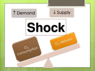
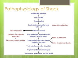
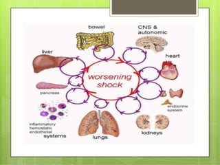
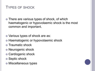
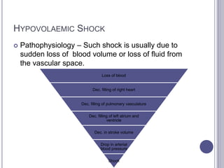
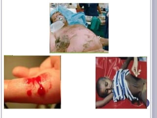
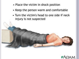
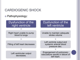
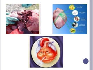
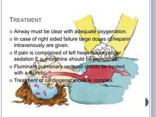
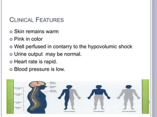
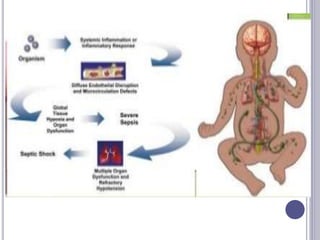
This seminar covers different types of shock including definitions, pathophysiology, clinical features, investigations, and treatment. The main types discussed are hypovolaemic shock, traumatic shock, cardiogenic shock, neurogenic shock, septic shock, and crush syndrome. Hypovolaemic shock is the most common and results from sudden loss of blood or fluid volume. Treatment focuses on fluid resuscitation and controlling bleeding. Septic shock has a high mortality and is usually caused by gram-negative bacteria. Crush syndrome occurs after body portions are compressed by heavy weights.
Subject: Medical Surgical Nursing / Adult Health Nursing
Title: Shock
Prepared by: Misfa Khatun, Nursing tutor
Content:
- Introduction
- Definition of Shock
- Classify Shock
- Stages of Shock
- Enumerate the Causes of shock
- Pathophysiology of Shock
- Identify the Signs and symptoms of Shock
- First ais management of Shock
- Treatment of Shock
- Management of Shock
- Nursing management of Shock
Cardiogenic shock is a condition of diminished cardiac output that severely impairs cardiac perfusion. In this condition in which the heart suddenly can't pump enough blood to meet the body's needs.
Simple medical student presentation about distributive shock, type and pathophysiology of each septic shock, anaphylactic shock, neurogenic shock
including management, prognosis and disposition of patient..
brief info of type of inotropes and when to start.
Ridge augmentation procedures /orthodontic courses by Indian dental academy Indian dental academy
The Indian Dental Academy is the Leader in continuing dental education , training dentists in all aspects of dentistry and
offering a wide range of dental certified courses in different formats.for more details please visit
www.indiandentalacademy.com
The Indian Dental Academy is the Leader in continuing dental education , training dentists in all aspects of dentistry and offering a wide range of dental certified courses in different formats.
Indian dental academy provides dental crown & Bridge,rotary endodontics,fixed orthodontics,
Dental implants courses.for details pls visit www.indiandentalacademy.com ,or call
00919248678078
Indian Dental Academy: will be one of the most relevant and exciting training center with best faculty and flexible training programs for dental professionals who wish to advance in their dental practice,Offers certified courses in Dental implants,Orthodontics,Endodontics,Cosmetic Dentistry, Prosthetic Dentistry, Periodontics and General Dentistry.
shock is the state of insufficient blood flow to the tissues of the body .it contains introduction, definition, stages of shock, types of shock, diagnostic evaluation, prognosis ,prevention, care for each stage.
Indian Dental Academy: will be one of the most relevant and exciting training center with best faculty and flexible training programs for dental professionals who wish to advance in their dental practice,Offers certified courses in Dental implants,Orthodontics,Endodontics,Cosmetic Dentistry, Prosthetic Dentistry, Periodontics and General Dentistry.
A very narrative discussion over Shock & Haemorrhage, Blood Transfusion, Blood Products which is presented in seminers. A concise guideline of a vast chapter.
Anti ulcer drugs and their Advance pharmacology ||
Anti-ulcer drugs are medications used to prevent and treat ulcers in the stomach and upper part of the small intestine (duodenal ulcers). These ulcers are often caused by an imbalance between stomach acid and the mucosal lining, which protects the stomach lining.
||Scope: Overview of various classes of anti-ulcer drugs, their mechanisms of action, indications, side effects, and clinical considerations.
TEST BANK for Operations Management, 14th Edition by William J. Stevenson, Ve...kevinkariuki227
TEST BANK for Operations Management, 14th Edition by William J. Stevenson, Verified Chapters 1 - 19, Complete Newest Version.pdf
Lung Cancer: Artificial Intelligence, Synergetics, Complex System Analysis, S...Oleg Kshivets
RESULTS: Overall life span (LS) was 2252.1±1742.5 days and cumulative 5-year survival (5YS) reached 73.2%, 10 years – 64.8%, 20 years – 42.5%. 513 LCP lived more than 5 years (LS=3124.6±1525.6 days), 148 LCP – more than 10 years (LS=5054.4±1504.1 days).199 LCP died because of LC (LS=562.7±374.5 days). 5YS of LCP after bi/lobectomies was significantly superior in comparison with LCP after pneumonectomies (78.1% vs.63.7%, P=0.00001 by log-rank test). AT significantly improved 5YS (66.3% vs. 34.8%) (P=0.00000 by log-rank test) only for LCP with N1-2. Cox modeling displayed that 5YS of LCP significantly depended on: phase transition (PT) early-invasive LC in terms of synergetics, PT N0—N12, cell ratio factors (ratio between cancer cells- CC and blood cells subpopulations), G1-3, histology, glucose, AT, blood cell circuit, prothrombin index, heparin tolerance, recalcification time (P=0.000-0.038). Neural networks, genetic algorithm selection and bootstrap simulation revealed relationships between 5YS and PT early-invasive LC (rank=1), PT N0—N12 (rank=2), thrombocytes/CC (3), erythrocytes/CC (4), eosinophils/CC (5), healthy cells/CC (6), lymphocytes/CC (7), segmented neutrophils/CC (8), stick neutrophils/CC (9), monocytes/CC (10); leucocytes/CC (11). Correct prediction of 5YS was 100% by neural networks computing (area under ROC curve=1.0; error=0.0).
CONCLUSIONS: 5YS of LCP after radical procedures significantly depended on: 1) PT early-invasive cancer; 2) PT N0--N12; 3) cell ratio factors; 4) blood cell circuit; 5) biochemical factors; 6) hemostasis system; 7) AT; 8) LC characteristics; 9) LC cell dynamics; 10) surgery type: lobectomy/pneumonectomy; 11) anthropometric data. Optimal diagnosis and treatment strategies for LC are: 1) screening and early detection of LC; 2) availability of experienced thoracic surgeons because of complexity of radical procedures; 3) aggressive en block surgery and adequate lymph node dissection for completeness; 4) precise prediction; 5) adjuvant chemoimmunoradiotherapy for LCP with unfavorable prognosis.
micro teaching on communication m.sc nursing.pdfAnurag Sharma
Microteaching is a unique model of practice teaching. It is a viable instrument for the. desired change in the teaching behavior or the behavior potential which, in specified types of real. classroom situations, tends to facilitate the achievement of specified types of objectives.
Factory Supply Best Quality Pmk Oil CAS 28578–16–7 PMK Powder in Stockrebeccabio
Factory Supply Best Quality Pmk Oil CAS 28578–16–7 PMK Powder in Stock
Telegram: bmksupplier
signal: +85264872720
threema: TUD4A6YC
You can contact me on Telegram or Threema
Communicate promptly and reply
Free of customs clearance, Double Clearance 100% pass delivery to USA, Canada, Spain, Germany, Netherland, Poland, Italy, Sweden, UK, Czech Republic, Australia, Mexico, Russia, Ukraine, Kazakhstan.Door to door service
Hot Selling Organic intermediates
MANAGEMENT OF ATRIOVENTRICULAR CONDUCTION BLOCK.pdfJim Jacob Roy
Cardiac conduction defects can occur due to various causes.
Atrioventricular conduction blocks ( AV blocks ) are classified into 3 types.
This document describes the acute management of AV block.
These lecture slides, by Dr Sidra Arshad, offer a quick overview of physiological basis of a normal electrocardiogram.
Learning objectives:
1. Define an electrocardiogram (ECG) and electrocardiography
2. Describe how dipoles generated by the heart produce the waveforms of the ECG
3. Describe the components of a normal electrocardiogram of a typical bipolar leads (limb II)
4. Differentiate between intervals and segments
5. Enlist some common indications for obtaining an ECG
Study Resources:
1. Chapter 11, Guyton and Hall Textbook of Medical Physiology, 14th edition
2. Chapter 9, Human Physiology - From Cells to Systems, Lauralee Sherwood, 9th edition
3. Chapter 29, Ganong’s Review of Medical Physiology, 26th edition
4. Electrocardiogram, StatPearls - https://www.ncbi.nlm.nih.gov/books/NBK549803/
5. ECG in Medical Practice by ABM Abdullah, 4th edition
6. ECG Basics, http://www.nataliescasebook.com/tag/e-c-g-basics
New Directions in Targeted Therapeutic Approaches for Older Adults With Mantl...i3 Health
i3 Health is pleased to make the speaker slides from this activity available for use as a non-accredited self-study or teaching resource.
This slide deck presented by Dr. Kami Maddocks, Professor-Clinical in the Division of Hematology and
Associate Division Director for Ambulatory Operations
The Ohio State University Comprehensive Cancer Center, will provide insight into new directions in targeted therapeutic approaches for older adults with mantle cell lymphoma.
STATEMENT OF NEED
Mantle cell lymphoma (MCL) is a rare, aggressive B-cell non-Hodgkin lymphoma (NHL) accounting for 5% to 7% of all lymphomas. Its prognosis ranges from indolent disease that does not require treatment for years to very aggressive disease, which is associated with poor survival (Silkenstedt et al, 2021). Typically, MCL is diagnosed at advanced stage and in older patients who cannot tolerate intensive therapy (NCCN, 2022). Although recent advances have slightly increased remission rates, recurrence and relapse remain very common, leading to a median overall survival between 3 and 6 years (LLS, 2021). Though there are several effective options, progress is still needed towards establishing an accepted frontline approach for MCL (Castellino et al, 2022). Treatment selection and management of MCL are complicated by the heterogeneity of prognosis, advanced age and comorbidities of patients, and lack of an established standard approach for treatment, making it vital that clinicians be familiar with the latest research and advances in this area. In this activity chaired by Michael Wang, MD, Professor in the Department of Lymphoma & Myeloma at MD Anderson Cancer Center, expert faculty will discuss prognostic factors informing treatment, the promising results of recent trials in new therapeutic approaches, and the implications of treatment resistance in therapeutic selection for MCL.
Target Audience
Hematology/oncology fellows, attending faculty, and other health care professionals involved in the treatment of patients with mantle cell lymphoma (MCL).
Learning Objectives
1.) Identify clinical and biological prognostic factors that can guide treatment decision making for older adults with MCL
2.) Evaluate emerging data on targeted therapeutic approaches for treatment-naive and relapsed/refractory MCL and their applicability to older adults
3.) Assess mechanisms of resistance to targeted therapies for MCL and their implications for treatment selection
Title: Sense of Smell
Presenter: Dr. Faiza, Assistant Professor of Physiology
Qualifications:
MBBS (Best Graduate, AIMC Lahore)
FCPS Physiology
ICMT, CHPE, DHPE (STMU)
MPH (GC University, Faisalabad)
MBA (Virtual University of Pakistan)
Learning Objectives:
Describe the primary categories of smells and the concept of odor blindness.
Explain the structure and location of the olfactory membrane and mucosa, including the types and roles of cells involved in olfaction.
Describe the pathway and mechanisms of olfactory signal transmission from the olfactory receptors to the brain.
Illustrate the biochemical cascade triggered by odorant binding to olfactory receptors, including the role of G-proteins and second messengers in generating an action potential.
Identify different types of olfactory disorders such as anosmia, hyposmia, hyperosmia, and dysosmia, including their potential causes.
Key Topics:
Olfactory Genes:
3% of the human genome accounts for olfactory genes.
400 genes for odorant receptors.
Olfactory Membrane:
Located in the superior part of the nasal cavity.
Medially: Folds downward along the superior septum.
Laterally: Folds over the superior turbinate and upper surface of the middle turbinate.
Total surface area: 5-10 square centimeters.
Olfactory Mucosa:
Olfactory Cells: Bipolar nerve cells derived from the CNS (100 million), with 4-25 olfactory cilia per cell.
Sustentacular Cells: Produce mucus and maintain ionic and molecular environment.
Basal Cells: Replace worn-out olfactory cells with an average lifespan of 1-2 months.
Bowman’s Gland: Secretes mucus.
Stimulation of Olfactory Cells:
Odorant dissolves in mucus and attaches to receptors on olfactory cilia.
Involves a cascade effect through G-proteins and second messengers, leading to depolarization and action potential generation in the olfactory nerve.
Quality of a Good Odorant:
Small (3-20 Carbon atoms), volatile, water-soluble, and lipid-soluble.
Facilitated by odorant-binding proteins in mucus.
Membrane Potential and Action Potential:
Resting membrane potential: -55mV.
Action potential frequency in the olfactory nerve increases with odorant strength.
Adaptation Towards the Sense of Smell:
Rapid adaptation within the first second, with further slow adaptation.
Psychological adaptation greater than receptor adaptation, involving feedback inhibition from the central nervous system.
Primary Sensations of Smell:
Camphoraceous, Musky, Floral, Pepperminty, Ethereal, Pungent, Putrid.
Odor Detection Threshold:
Examples: Hydrogen sulfide (0.0005 ppm), Methyl-mercaptan (0.002 ppm).
Some toxic substances are odorless at lethal concentrations.
Characteristics of Smell:
Odor blindness for single substances due to lack of appropriate receptor protein.
Behavioral and emotional influences of smell.
Transmission of Olfactory Signals:
From olfactory cells to glomeruli in the olfactory bulb, involving lateral inhibition.
Primitive, less old, and new olfactory systems with different path
Couples presenting to the infertility clinic- Do they really have infertility...Sujoy Dasgupta
Dr Sujoy Dasgupta presented the study on "Couples presenting to the infertility clinic- Do they really have infertility? – The unexplored stories of non-consummation" in the 13th Congress of the Asia Pacific Initiative on Reproduction (ASPIRE 2024) at Manila on 24 May, 2024.
3. DEFINITION
It is difficult to define shock in one sentence.
However,
It may be defined as a condition in which circulation
fails to meet the nutritional needs of the cells and at
the same time fails to remove the metabolic waste
products.
4.
5.
6.
7.
8. TYPES OF SHOCK
There are various types of shock, of which
haematogenic or hypovolaemic shock is the most
common and important.
Various types of shock are as:
Haematogenic or hypovolaemic shock
Traumatic shock
Neurogenic shock
Cardiogenic shock
Septic shock
Miscellaneous types
9.
10. HYPOVOLAEMIC SHOCK
Pathophysiology – Such shock is usually due to
sudden loss of blood volume or loss of fluid from
the vascular space.
Loss of blood
Dec. filling of right heart
Dec. filling of pulmonary vasculature
Dec. filling of left atrium and
ventricle
Dec. in stroke volume
Drop in arterial
blood pressure
Shock
11. COMPENSATORY MECHANISMS
After hemorrahge this mechanism follows:
1. Adrenergic discharge
2. Hyperventilation
3. Release of vasoactive hormones
4. Collapse
5. Resorption of fluid from the interstitial space
6. Resorption of fluid from the intercellular to the
extracellular space
7. Renal conservation of body water and electroytes.
12. CLINICAL FEATURES
Depends on the degree of loss of blood volume and
on the duration of shock.
MILD SHOCK: loss of less than 20% of blood
voluyme is included in this category.
Feet become pale and cool
Sweating
Urinary output pulse rate and blood pressure remai
normal
Thirst and cold.
13.
14. Moderate shock: Loss of bloos volume from 20 –
40% cause this shock.
Oliguria
Pulse rate increased to 100 beats / min.
Normal blood pressure.
Severe Shock: Loss of blood volume more than
40% usually causes severe shock.
Paller occurs
Low urinary output
Rapid pulse rate
Low blood pressure.
15. INVESTIGATIONS
Monitoring a shock includes:
Blood pressure- measurement of blood pressure is very
essential in shock
Respiration: hyperventilation is and important indicator
of shock.
Urine- urine is affected quite early even in moderate
shock. It is a good index of adequacy of replacement
therapy.
CVP: Measurement of CVP is quite important in
assessing shock.
In hypovolumic shock there is dec. in the blood volume
ans so is the CVP.
ECG: in severe shock it may show signs of myocardial
ischemia
SWAN-GANG CATHETER:
16. TREATMENT
1 Resuscitation: This should begin immediately as the
patient is admitted with hypovolaemic shock.
Establish clear airway
Maintain adequate ventilation and oxugenation.
Lowering of head to prevent pheripheral pooling of blood
and odema.
Increases cerebral circulation.
2 Immediate control of bleeding is highly important in
case of heamorrhagic shock.
Raise the footend of the bed and by compression bandage
to tamponade external haemorrhage
Bleeding can be stoppped surgically as well.
17. 3 Extracellular fluid replacement is probably the
most important point in the treatment of
hypovolaemic shock.
Ringer’s lactate, ringer’s acetate or normal saline
can be administered.
A few points to be remembered in case of
extracellular fluid replacement:
The I.V fluid particularly the crystalloid, ehich
should be given first, is administered with rapidly so
that replacemnet is done as quickly as possible
B.P, pulse rate, urine output, CVP and other
laboratory tests should be performed.
If there is blood loss, it is replaced by blood
18.
19. DRUGS: A few drugs are commonly used different
shock.
Sedatives: Morphine is quite good in this respect
and sholud be given IV.
Injection pethidine can be also be used
intramuscularly.
Chronotropic Agents: Atropine is the most widely
used in this group Followed by isoproterenol.
Inotropic agents: Most commonly used drugs in this
cateogory are the dopamine and dobutamine these
should be used in low dosages.
Othr used agents are vasodialators, vasoconstrictors,
betablockers, Diuretics.
20. 654 × 668 - esicm.org
TRAUMATIC SHOCK
Pathophysiology:
The pecularity of this shock is that traumatised
tissues activate the coagulation system and release
microthrombi into the circulation. These may
occlude or constrict parts of pulmonary
microvasculature to increse pulmonary vascular
resistance. This inc. right ventricular diastolic and
right atrial pressures. Inc. in capillary permeability.
Loss of plasma depletion of the vascular volume to
a great extent.
21. CLINICAL FEATURE
Presence of peripheral and pulmonary oedema in
this type of shock
Infusion of large volumes of fluid whih may be
adequate for pure hypovolaemic shock is usually
inadequate for traumatic shock.
22. TREATMENT
Resuscitation: In this type of shock shock
mechanical ventilatory support is more needed.
Local treatment of trauma and control of bleeding:
This is almost similar to hypovolaemic shock.
Surgical debridement of ischaemic and dead
tissues and immobilisation of fractures may be
required.
Fluid replacement: adquate fluid replacemnet
should be done with a dose of 10,000 units of
heparin seemes to be effective.
23.
24. CARDIOGENIC SHOCK
Pathophysiology:
Dysfunction of the
right ventricle
Right heart unable to pump
blood to lungs
Filling of left heart decreases
Left ventricular output
decreases.
Dysfunction of the
left ventricle
Unable to maintain adequate
stroke volume
Left ventricle output and
systemic arterial blood
pressure dec.
Engorgement of pulmonary
vasculature results in failure of
left heart.
25. CARDIAC COMPRESSIVE SHOCK
This arises when the heart is compressed enough from
outside to decrease cardiac output.
Causes: Tension pneumothorax, pericardial tamponade and
diaphragmatic rupture with herniation of the bowel into chest.
Clinical features: In the beginning the skin is pale and cool
and the urine output is low.
Pulse becomes rapid
Arterial blood flow becomes low
Left ventricular dysfuctional the patient has broncheal rales
and a third heart sound is heard.
Gradually the heart becomes enlarged and when the right
ventricle also fails distended neck veins will be visible.
26.
27. TREATMENT
Airway must be clear with adequate oxygenation.
In case of right sided failure large doses of heparin
intravenously are given.
If pain is complained of left heart failure proper
sedation E.g. morphine should be prescribed.
Fluminant pulmonary oedema should be treated
with a diuretic.
Treatment of cardiogenic shock is complex.
28. NEUROGENIC SHOCK
Pathophysiology:
Dialatation of systemic vasculature
Dec. arterial pressure
Blood pools in the systemic venules and small veins
Right heart filling and stroke volume dec.
Dec. in pulmonary blood volume and left
heart filling
Left ventricular output dec.
Compensatory
mechanisms of
adrenergic nervous
system fail.
shock
29. CLINICAL FEATURES
Skin remains warm
Pink in color
Well perfused in contarry to the hypovolumic shock
Urine output may be normal.
Heart rate is rapid.
Blood pressure is low.
30. TREATMENT
Elevation of the legs is effective in treating patients
with neurogenic shock.
Assumption of Trendelenburg position displaces
blood from the systemic venules and small veins
into the right heart and thus inc. cardiac output.
Administration of fluid is important though not so as
in hypovolaemic shock. This increase filling of the
right heart which in its increases cardiac output.
Neurogenic shock is probably the only form of
shock that can be safely treated with a
vasocaonstrictor drug.
31. SEPTIC SHOCK
Pathophysiology: High mortality rate
Most frequently causitive organisms are gram
positive and gram negative bacteria,
Because of effective antibiotic treatment available
for most gram positive infections, the majority of
cases of septic shock are now caused by gram
negative bacteria.
32. COMMON ORGANISMS CONCERNED WITH
SEPTIC SHOCK
E.Coli
Klebsiells aerobacter
Proteus
Pseudomonas
Bacteriods
Gram- positive sepsis and shock: Clostridium tetani
or Clostridium perfringes, streptococcus,
staphylococcus, pseudomonas.
Gram- negative sepsis and shock: Genitourinary
system associated with instrumentation.
33. CLINICAL FEATURE:
Recognized by development of chills and elevated
temperature above 100 F.
Two types:
Early warm shock
Late cold shock
In early warm shock there is cutaneous vasodialation.
Inc. in body temperature.
Cutaneous dilation.
Pulse rate high
Intermittent spikes of fever alternating with bouts of
chills.
34.
35. Late cold Shock: inc. permeability due to liberation
of toxic products into the center circulation.
Clinically difficult to differentiate b/w hemorrahgic ,
traumatic shock.
Only guide is the knowlegde of septic focus.
Treatment:
Treatment can be classified into two groups :
Treatment of the infection by early surgical
debridement or drainage and by use of appropriate
antibiotics
Treatment of shock which includes fluid
replacement , steroid administration and use of
vasoactive drugs.
36. Cephalothin (6-8gm/day I.V in 4 -6 divided doses)
Gentamicin (5mg/kg/day)
Clindamycin (bacteroides)
Chloromycetin
Fluid replacement is of great importance.
Mechanical ventilation along with endotracheal
intubation.
Steriods can be used sometimes.
Use of vasoactive drugs with mixed alpha and beta
adrenergic effects may be indicated.
37. CRUSH SYNDROME
It is a symptom complex in which a portion of the body
becomes crushed due to a heavy weight falls on that potion of
the body and is kept there for sometime to crush all the
tissues in that portion of the body.
This type of injury is come across after earthquakes, mine
injuries, air raids, collapse of a building or use of tourniquiet
for longer period.
In this syndrome oligaemic shock occurs due to extravasation
of blood into the muscle in the affceted portion of the body.
Sweeling of the muscles, myohaemoglobin enter the
circulation
At this stage the limbs fills tense and the patient complains of
severe pain in the limb.
Urine output will be obviously reduced if uraemia supervenes,
the patient may show restlessness, mild delirium.
38. TREATMENT
As a first aid measure application of tourniquet to
the affected limb above the crush injury is a good
method to reduce admission of deleterious
substances into the general circulation.
Parallel incisions ,ay be applied to relieve temsion,
through which muscles protruded.
Low molecular weight dextran (40000)or
Rheomacrodex is effective in this condition as it
prevents sludging of red cells in small blood vessels
and maintain circulation to the kidneys.
.
39. Mannitol is also very effective in this condition
1gm/kg body weight of mannitol is given IV as 20%
solution in 12 hours.
Catheterisation of the bladder should be performed
before instituting mannitol.
Tourniquet should be removed as scheduled, blood
transfusion should be done.
Haemodialysis should be used as life saving
procedure in grave conditions.