Download to read offline


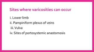


Varicose veins are dilated, tortuous veins caused by faulty valves that allow blood to flow backwards. The great saphenous and small saphenous veins are commonly affected. Symptoms include heavy legs, aching, night cramps, and skin changes. Conservative treatments include compression stockings and elevation while surgical options are available for more severe cases.


