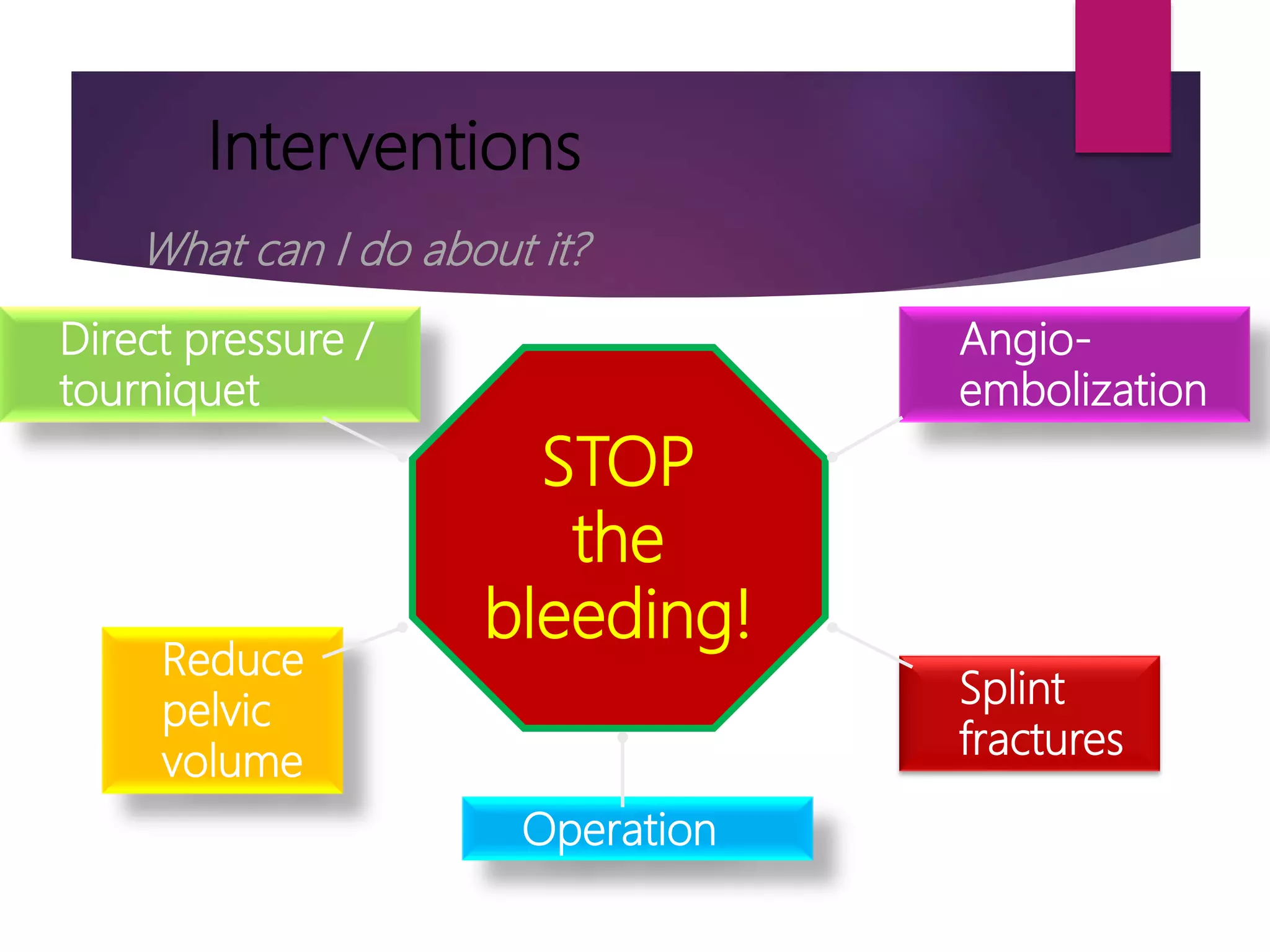
Shock in Polytrauma
DR. LALA ROBIN
DEPT. OF GENERAL SURGERY
CMC LUDHIANA
Definition of shock
 Shock is a systemic state of low tissue
perfusion which is inadequate for normal
cellular respiration.
Inadequate
Cellular
Oxygen
Delivery
Anaerobic
Metabolism
Inadequate
Energy
Production
Metabolic
Failure
Lactic
Acid
Production
Metabolic
AcidosisCELL
DEATH
Ultimate Effects
of Anaerobic
Metabolism
Initial Patient Assessment
 Two important questions in a patient presenting to
ER
 1. Is the patient in shock?
 2. What is the cause of shock?
RECOGNITION OF SHOCK
 Airway, Breathing ensured
 Circulatory status evaluated carefully to identify the early
manifestations of shock, including tachycardia and
cutaneous vasoconstriction
 Any injured patient who is cool and has tachycardia is
considered to be in shock until proven otherwise
 pulse rate, pulse character, respiratory rate, skin
circulation, and pulse pressure
 > 160 - Infants
 > 140 -Preschool
child
 > 120 -School age
to puberty
 > 100 - Adult
pitfalls
 Reliance solely on systolic blood pressure delays diagnosis of
shock
 Compensatory mechanisms can preclude a measurable fall in
systolic pressure until up to 30% of the patient’s blood
volume.
 Elderly patients may not exhibit tachycardia
 A narrowed pulse pressure suggests significant blood loss
and involvement of compensatory mechanisms
 Laboratory values for hematocrit or haemoglobin
concentration may be unreliable.
 Serial measurement of these parameters may be used to
monitor a patient’s response to therapy
CLINICAL DIFFERENTIATION OF
CAUSE OF SHOCK
 Hemorrhage (most common cause)
 Cardiogenic
 Neurogenic
 Tension pneumothorax
 Sepsis
 The response to initial treatment coupled with the finding
during the primary and secondary patient surveys, usually
provides sufficient information to determine the cause of
the shock state.
Hemorrhagic Shock
 Assessment of hemorrhagic includes a rapid
determination of the site of blood loss.
 Sources of potential blood loss—chest, abdomen, pelvis,
retroperitoneum, extremities, and external bleeding
 Chest x-ray, pelvic x-ray, abdominal assessment with
either focused assessment sonography in trauma (FAST)
or diagnostic peritoneal lavage (DPL), and bladder
catheterization may all be necessary to determine the
source of blood loss
on the floor plus four more
A) the chest; (B) the abdomen; (C) the pelvis; and (D) the
femur.
Nonhemorrhagic Shock
 Cardiogenic Shock - blunt cardiac injury, cardiac
tamponade, air embolus, or, rarely, a myocardial
infarction  constant electrocardiographic (ECG)
monitoring to detect injury patterns and dysrhythmias.
 Cardiac Tamponade
 penetrating thoracic trauma,
 Tachycardia, muffled heart sounds, and dilated,engorged
neck veins with hypotension resistant to fluid therapy
suggest cardiac tamponade
 Thoracotomy, pericardiocentesis.
Tension Pneumothorax
 acute respiratory distress, subcutaneous emphysema,
absent breath sounds, hyperresonance to percussion,
and tracheal shift
 Needs chest tube/ needle decompressoion immediately
 Do not wait for chest xray
Neurogenic Shock
 Isolated intracranial injuries do not cause shock.
 The classic picture of neurogenic shock is hypotension
without tachycardia or cutaneous vasoconstriction.
 Cervical or upper thoracic spinal cord injury can produce
hypotension due to loss of sympathetic tone
 often have concurrent torso trauma
 should be treated initially for hypovolemia. The failure of
fluid resuscitation to restore organ perfusion suggests
either continuing haemorrhage or neurogenic shock
 CVP monitoring may be helpful
Septic Shock
 Septic shock can occur in patients with penetrating
abdominal injuries and contamination of the peritoneal
cavity by intestinal contents
 difficult to distinguish from those in hypovolemic shock,
as both groups can manifest tachycardia, cutaneous
vasoconstriction, impaired urinary output, decreased
systolic pressure, and narrow pulse pressure
Hemorrhagic Shock
 Hemorrhage is the most common cause of shock in trauma
patients
 Hemorrhage is defined as an acute loss of circulating blood
volume
 Advanced Trauma Life Support (ATLS) manual describes four
classes of hemorrhage to emphasize the early signs of the shock
state.
 Normal Adult blood volume = Approximately- 7% of body
weight ( 70 kg= 5 L)
 Children= 8-9% of Body weight (80-90 ml/kg).
 Volume replacement is determined by patient’s response to
initial therapy.
 Hemorrhage control and balanced fluid resuscitation
must be initiated  when early signs and symptoms of
blood loss are apparent or suspected  not when the
blood pressure is falling or absent
 Bleeding patients need blood!
 Definitive control of haemorrhage and restoration of
adequate circulating volume are the goals of treatment
of hemorrhagic shock
Question
 A 70-kg patient arrives at an ED or trauma center with
hypotension. What is the minimum amount of blood loss
in this patient?
 Class III haemorrhage  atleast 30% blood loss
 70 kg x 7% x 30% = 1.47 L, or 1470 mL
 Resuscitation will likely require crystalloid, pRBCs, and
blood products
 Nonresponse to fluid administration almost always
indicates persistent blood loss with the need for
operative or angiographic control.
FLUID CHANGES SECONDARY TO
SOFT TISSUE INJURYAND FRACTURES
 Blood is lost into the site of injury, particularly in cases of
major fractures
 fractured tibia or humerus  750 mL
 Fracture femur  1500 mL
 pelvic fracture  retroperitoneal hematoma  more than 3
L
 Edema that occurs in injured soft tissues
Initial Management of
Hemorrhagic Shock
 The diagnosis and treatment of shock must occur
almost,simultaneously
 The basic management principle is to stop the bleeding
and replace the volume loss.
PHYSICAL EXAMINATION
 Immediate diagnosis of life-threatening injuries and
includes assessment of the ABCDEs
 Airway and Breathing
 Circulation—Hemorrhage Control
 Disability—Neurologic Examination
 Exposure—Complete Examination
 Gastric Dilation—Decompression
 Urinary Catheterization
Interventions
Direct pressure /
tourniquet
STOP
the
bleeding!
Reduce
pelvic
volume
Angio-
embolization
Splint
fractures
Operation
What can I do about it?
VASCULAR ACCESS LINES
 inserting two large-caliber (minimum of 16-gauge in an
adult) peripheral intravenous catheters
 Forearms and antecubital veins
 The rate of flow is proportional to the fourth power of the
radius of the cannula and inversely related to its length
(Poiseuille’s law)
 Fluid warmers and rapid infusion pumps are used in the
presence of massive haemorrhage and severe hypotension.
 In children younger than 6 years - intraosseous needle
 Blood samples, ABG
INITIAL FLUID THERAPY
 Warmed isotonic electrolyte solutions, such as lactated
Ringer’s and normal saline
 1 to 2 L for adults
 20 mL/kg for pediatric patients
 assess the patient’s response to fluid resuscitation and
identify evidence of adequate end-organ perfusion and
oxygenation (urinary output, level of consciousness, and
peripheral perfusion)
 Persistent infusion of large volumes of fluid and blood in an
attempt to achieve a normal blood pressure is not a
substitute for definitive control of bleeding.
Blood Replacement
 Patients who are transient responders or
nonresponders—those with Class III or Class IV
haemorrhage will need pRBCs and blood products as an
early part of their resuscitation
 complete crossmatching process requires approximately
1 hour
 Type-specific blood can be provided by most blood
banks within 10 minutes
 type O negative packed cells are indicated for patients
with exsanguinating hemorrhage
 WARMING FLUIDS—PLASMA AND CRYSTALLOID
 heat the fluid to 39°C (102.2° F) before infusing it. This
can be accomplished by storing crystalloids in a warmer
 AUTOTRANSFUSION -Collection of shed blood for
autotransfusion should be considered for any patient
a major hemothorax.
 MASSIVE TRANSFUSION PROTOCOL -defined as >10
units of pRBCs within the first 24 hours of admission.
 balanced, hemostatic or damage control resuscitation.
 COAGULOPATHY
 present in up to 30% of severely injured patients on
admission
 Massive fluid resuscitation, with the resultant dilution of
platelets and clotting factors, along with the adverse
effect of hypothermia on platelet aggregation and the
clotting cascade
 platelets, cryoprecipitate, and fresh-frozen plasma
 CALCIUM ADMINISTRATION - guided by measurement
of ionized calcium
Special Considerations
 Equating blood pressure with cardiac output
 Advanced age
 Athletes
 Pregnancy
 Medications
 Hypothermia
 Presence of pacemaker
Conclusion
 Management of shock in trauma is a challenge
 Most of trauma deaths can be prevented by followings
ATLS protocols
 Early involvement of surgeon is mandatory in trauma
patients.
THANK YOU

Shock in

  • 1.
    Shock in Polytrauma DR.LALA ROBIN DEPT. OF GENERAL SURGERY CMC LUDHIANA
  • 2.
    Definition of shock Shock is a systemic state of low tissue perfusion which is inadequate for normal cellular respiration.
  • 3.
  • 4.
    Initial Patient Assessment Two important questions in a patient presenting to ER  1. Is the patient in shock?  2. What is the cause of shock?
  • 5.
    RECOGNITION OF SHOCK Airway, Breathing ensured  Circulatory status evaluated carefully to identify the early manifestations of shock, including tachycardia and cutaneous vasoconstriction  Any injured patient who is cool and has tachycardia is considered to be in shock until proven otherwise  pulse rate, pulse character, respiratory rate, skin circulation, and pulse pressure
  • 6.
     > 160- Infants  > 140 -Preschool child  > 120 -School age to puberty  > 100 - Adult
  • 7.
    pitfalls  Reliance solelyon systolic blood pressure delays diagnosis of shock  Compensatory mechanisms can preclude a measurable fall in systolic pressure until up to 30% of the patient’s blood volume.  Elderly patients may not exhibit tachycardia  A narrowed pulse pressure suggests significant blood loss and involvement of compensatory mechanisms  Laboratory values for hematocrit or haemoglobin concentration may be unreliable.  Serial measurement of these parameters may be used to monitor a patient’s response to therapy
  • 8.
    CLINICAL DIFFERENTIATION OF CAUSEOF SHOCK  Hemorrhage (most common cause)  Cardiogenic  Neurogenic  Tension pneumothorax  Sepsis
  • 9.
     The responseto initial treatment coupled with the finding during the primary and secondary patient surveys, usually provides sufficient information to determine the cause of the shock state.
  • 10.
    Hemorrhagic Shock  Assessmentof hemorrhagic includes a rapid determination of the site of blood loss.  Sources of potential blood loss—chest, abdomen, pelvis, retroperitoneum, extremities, and external bleeding  Chest x-ray, pelvic x-ray, abdominal assessment with either focused assessment sonography in trauma (FAST) or diagnostic peritoneal lavage (DPL), and bladder catheterization may all be necessary to determine the source of blood loss
  • 11.
    on the floorplus four more A) the chest; (B) the abdomen; (C) the pelvis; and (D) the femur.
  • 12.
    Nonhemorrhagic Shock  CardiogenicShock - blunt cardiac injury, cardiac tamponade, air embolus, or, rarely, a myocardial infarction  constant electrocardiographic (ECG) monitoring to detect injury patterns and dysrhythmias.  Cardiac Tamponade  penetrating thoracic trauma,  Tachycardia, muffled heart sounds, and dilated,engorged neck veins with hypotension resistant to fluid therapy suggest cardiac tamponade  Thoracotomy, pericardiocentesis.
  • 13.
    Tension Pneumothorax  acuterespiratory distress, subcutaneous emphysema, absent breath sounds, hyperresonance to percussion, and tracheal shift  Needs chest tube/ needle decompressoion immediately  Do not wait for chest xray
  • 14.
    Neurogenic Shock  Isolatedintracranial injuries do not cause shock.  The classic picture of neurogenic shock is hypotension without tachycardia or cutaneous vasoconstriction.  Cervical or upper thoracic spinal cord injury can produce hypotension due to loss of sympathetic tone  often have concurrent torso trauma  should be treated initially for hypovolemia. The failure of fluid resuscitation to restore organ perfusion suggests either continuing haemorrhage or neurogenic shock  CVP monitoring may be helpful
  • 15.
    Septic Shock  Septicshock can occur in patients with penetrating abdominal injuries and contamination of the peritoneal cavity by intestinal contents  difficult to distinguish from those in hypovolemic shock, as both groups can manifest tachycardia, cutaneous vasoconstriction, impaired urinary output, decreased systolic pressure, and narrow pulse pressure
  • 16.
    Hemorrhagic Shock  Hemorrhageis the most common cause of shock in trauma patients  Hemorrhage is defined as an acute loss of circulating blood volume  Advanced Trauma Life Support (ATLS) manual describes four classes of hemorrhage to emphasize the early signs of the shock state.  Normal Adult blood volume = Approximately- 7% of body weight ( 70 kg= 5 L)  Children= 8-9% of Body weight (80-90 ml/kg).  Volume replacement is determined by patient’s response to initial therapy.
  • 18.
     Hemorrhage controland balanced fluid resuscitation must be initiated  when early signs and symptoms of blood loss are apparent or suspected  not when the blood pressure is falling or absent  Bleeding patients need blood!  Definitive control of haemorrhage and restoration of adequate circulating volume are the goals of treatment of hemorrhagic shock
  • 19.
    Question  A 70-kgpatient arrives at an ED or trauma center with hypotension. What is the minimum amount of blood loss in this patient?  Class III haemorrhage  atleast 30% blood loss  70 kg x 7% x 30% = 1.47 L, or 1470 mL  Resuscitation will likely require crystalloid, pRBCs, and blood products  Nonresponse to fluid administration almost always indicates persistent blood loss with the need for operative or angiographic control.
  • 20.
    FLUID CHANGES SECONDARYTO SOFT TISSUE INJURYAND FRACTURES  Blood is lost into the site of injury, particularly in cases of major fractures  fractured tibia or humerus  750 mL  Fracture femur  1500 mL  pelvic fracture  retroperitoneal hematoma  more than 3 L  Edema that occurs in injured soft tissues
  • 21.
    Initial Management of HemorrhagicShock  The diagnosis and treatment of shock must occur almost,simultaneously  The basic management principle is to stop the bleeding and replace the volume loss.
  • 22.
    PHYSICAL EXAMINATION  Immediatediagnosis of life-threatening injuries and includes assessment of the ABCDEs  Airway and Breathing  Circulation—Hemorrhage Control  Disability—Neurologic Examination  Exposure—Complete Examination  Gastric Dilation—Decompression  Urinary Catheterization
  • 23.
  • 24.
    VASCULAR ACCESS LINES inserting two large-caliber (minimum of 16-gauge in an adult) peripheral intravenous catheters  Forearms and antecubital veins  The rate of flow is proportional to the fourth power of the radius of the cannula and inversely related to its length (Poiseuille’s law)  Fluid warmers and rapid infusion pumps are used in the presence of massive haemorrhage and severe hypotension.  In children younger than 6 years - intraosseous needle  Blood samples, ABG
  • 25.
    INITIAL FLUID THERAPY Warmed isotonic electrolyte solutions, such as lactated Ringer’s and normal saline  1 to 2 L for adults  20 mL/kg for pediatric patients  assess the patient’s response to fluid resuscitation and identify evidence of adequate end-organ perfusion and oxygenation (urinary output, level of consciousness, and peripheral perfusion)  Persistent infusion of large volumes of fluid and blood in an attempt to achieve a normal blood pressure is not a substitute for definitive control of bleeding.
  • 27.
    Blood Replacement  Patientswho are transient responders or nonresponders—those with Class III or Class IV haemorrhage will need pRBCs and blood products as an early part of their resuscitation  complete crossmatching process requires approximately 1 hour  Type-specific blood can be provided by most blood banks within 10 minutes  type O negative packed cells are indicated for patients with exsanguinating hemorrhage
  • 28.
     WARMING FLUIDS—PLASMAAND CRYSTALLOID  heat the fluid to 39°C (102.2° F) before infusing it. This can be accomplished by storing crystalloids in a warmer  AUTOTRANSFUSION -Collection of shed blood for autotransfusion should be considered for any patient a major hemothorax.  MASSIVE TRANSFUSION PROTOCOL -defined as >10 units of pRBCs within the first 24 hours of admission.  balanced, hemostatic or damage control resuscitation.
  • 29.
     COAGULOPATHY  presentin up to 30% of severely injured patients on admission  Massive fluid resuscitation, with the resultant dilution of platelets and clotting factors, along with the adverse effect of hypothermia on platelet aggregation and the clotting cascade  platelets, cryoprecipitate, and fresh-frozen plasma  CALCIUM ADMINISTRATION - guided by measurement of ionized calcium
  • 30.
    Special Considerations  Equatingblood pressure with cardiac output  Advanced age  Athletes  Pregnancy  Medications  Hypothermia  Presence of pacemaker
  • 31.
    Conclusion  Management ofshock in trauma is a challenge  Most of trauma deaths can be prevented by followings ATLS protocols  Early involvement of surgeon is mandatory in trauma patients.
  • 32.