Downloaded 243 times

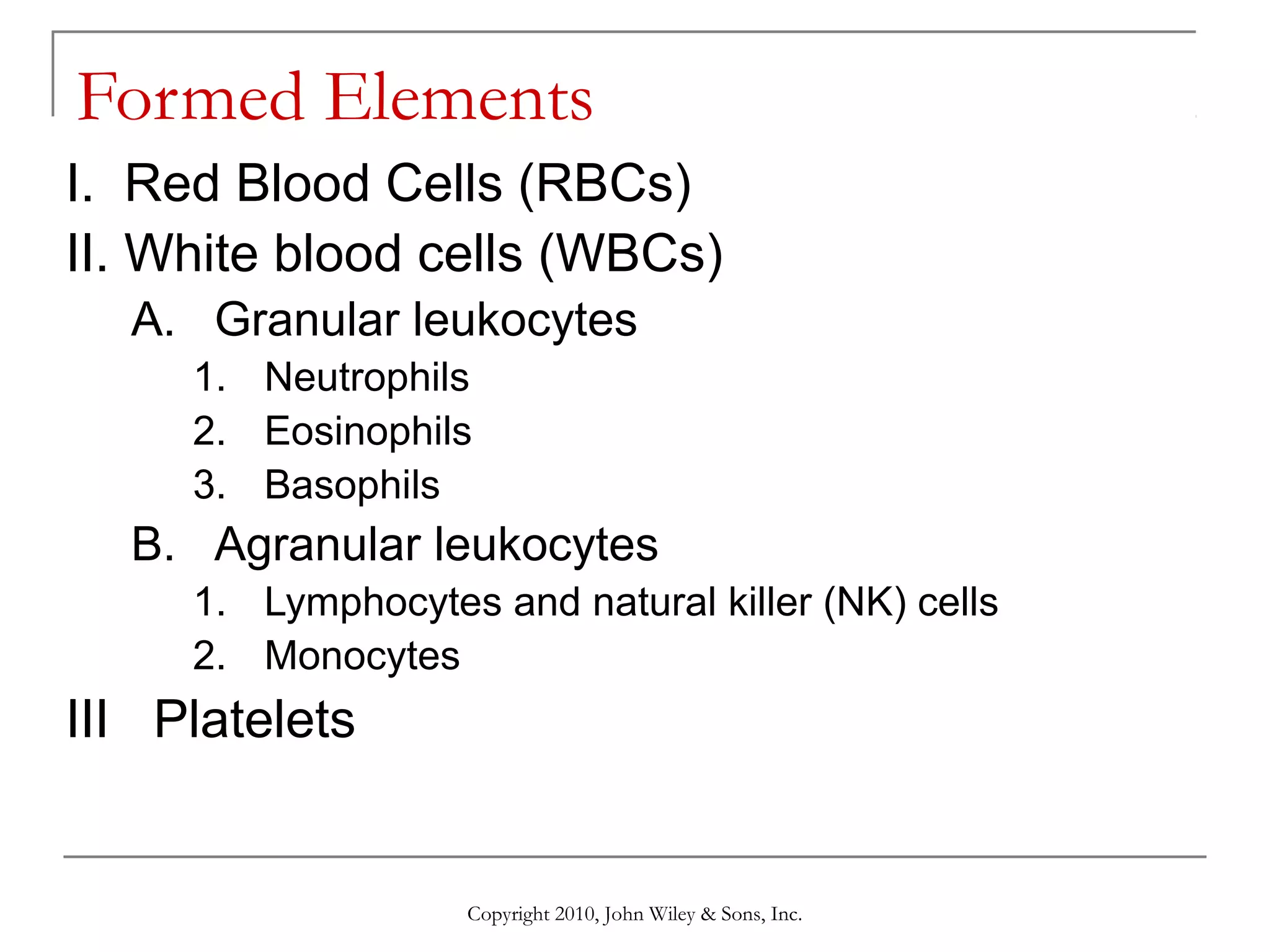
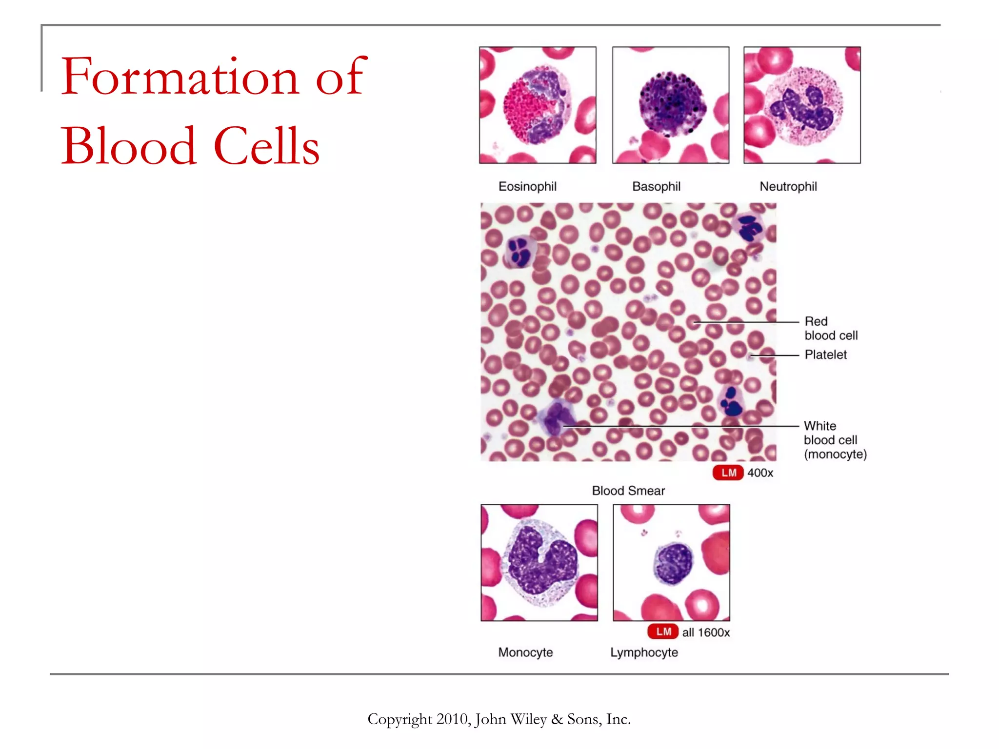
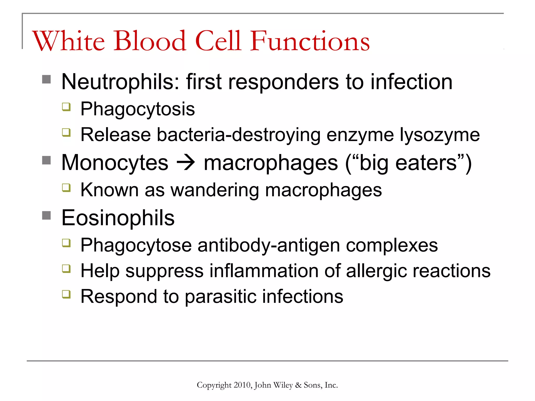
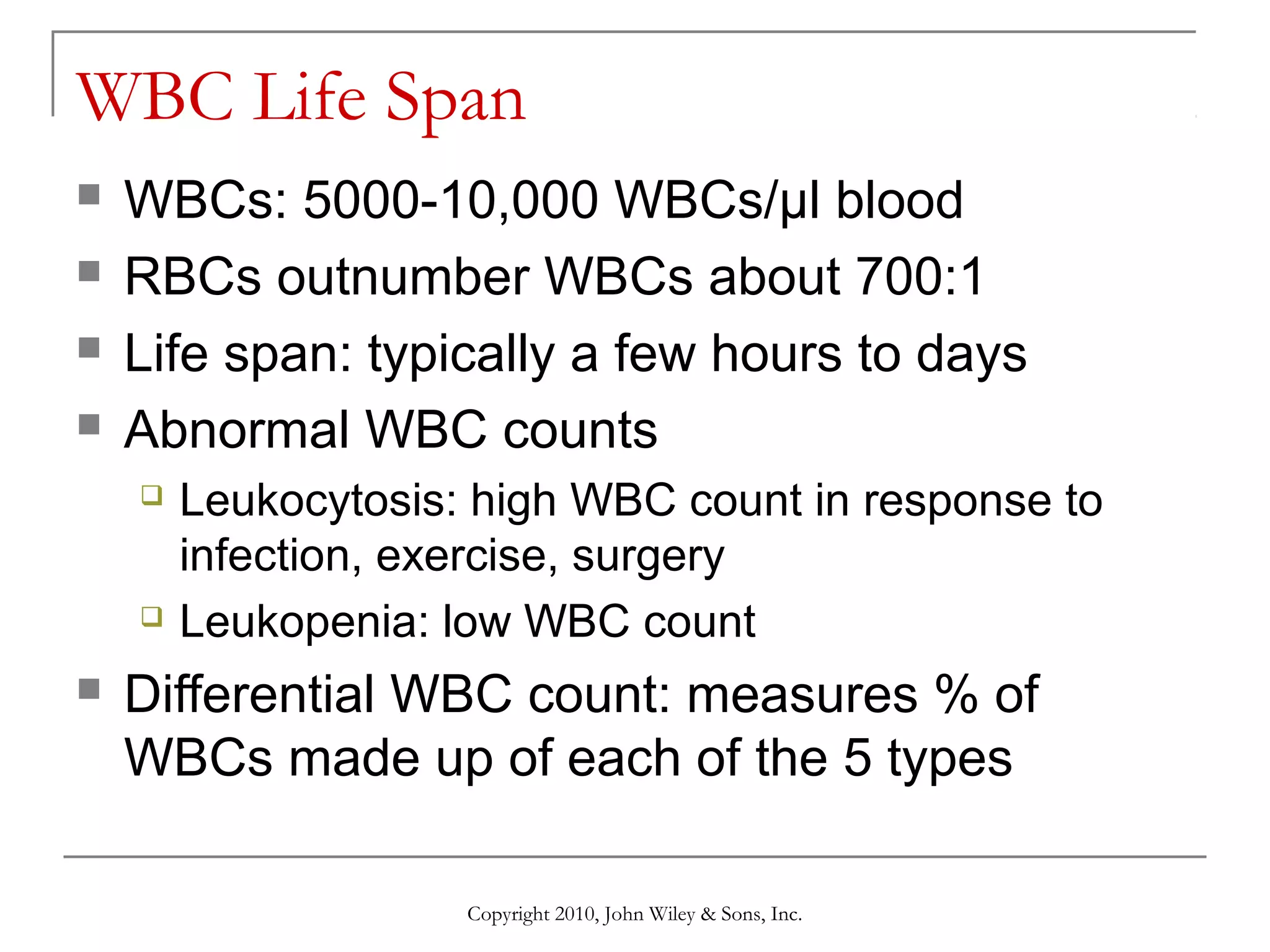
![White Blood Cells (WBCs or Leukocytes)
Appear white because lack hemoglobin
Normal WBC count: 5,000-10,000/µl
WBC count usually increases in infection
Two major classes based on presence or
absence of granules (vesicles) in them]
Granular: neutrophils, eosinophils, basophils
Neutrophils usually make up 2/3 of all WBCs
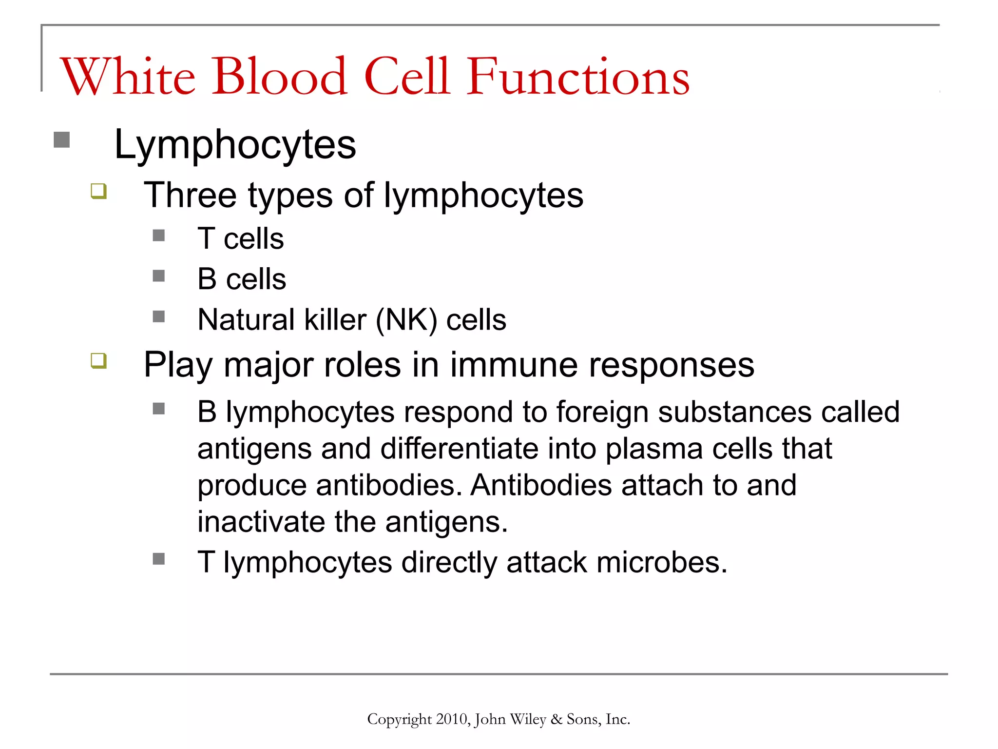
Agranular: lymphocytes, monocytes
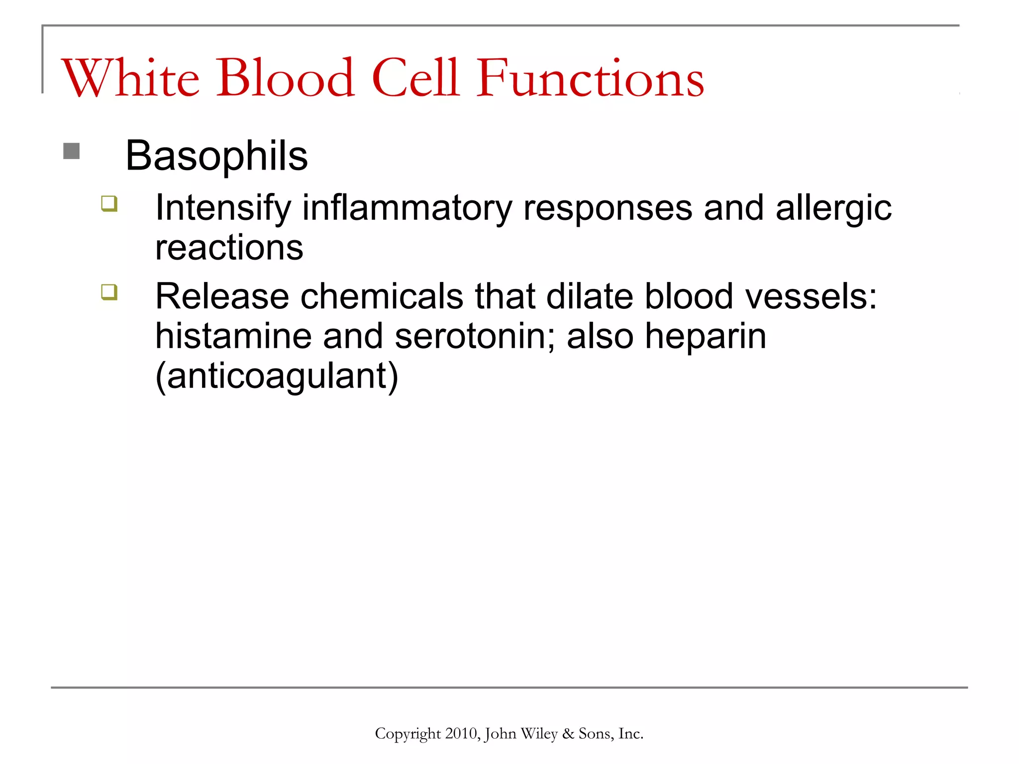
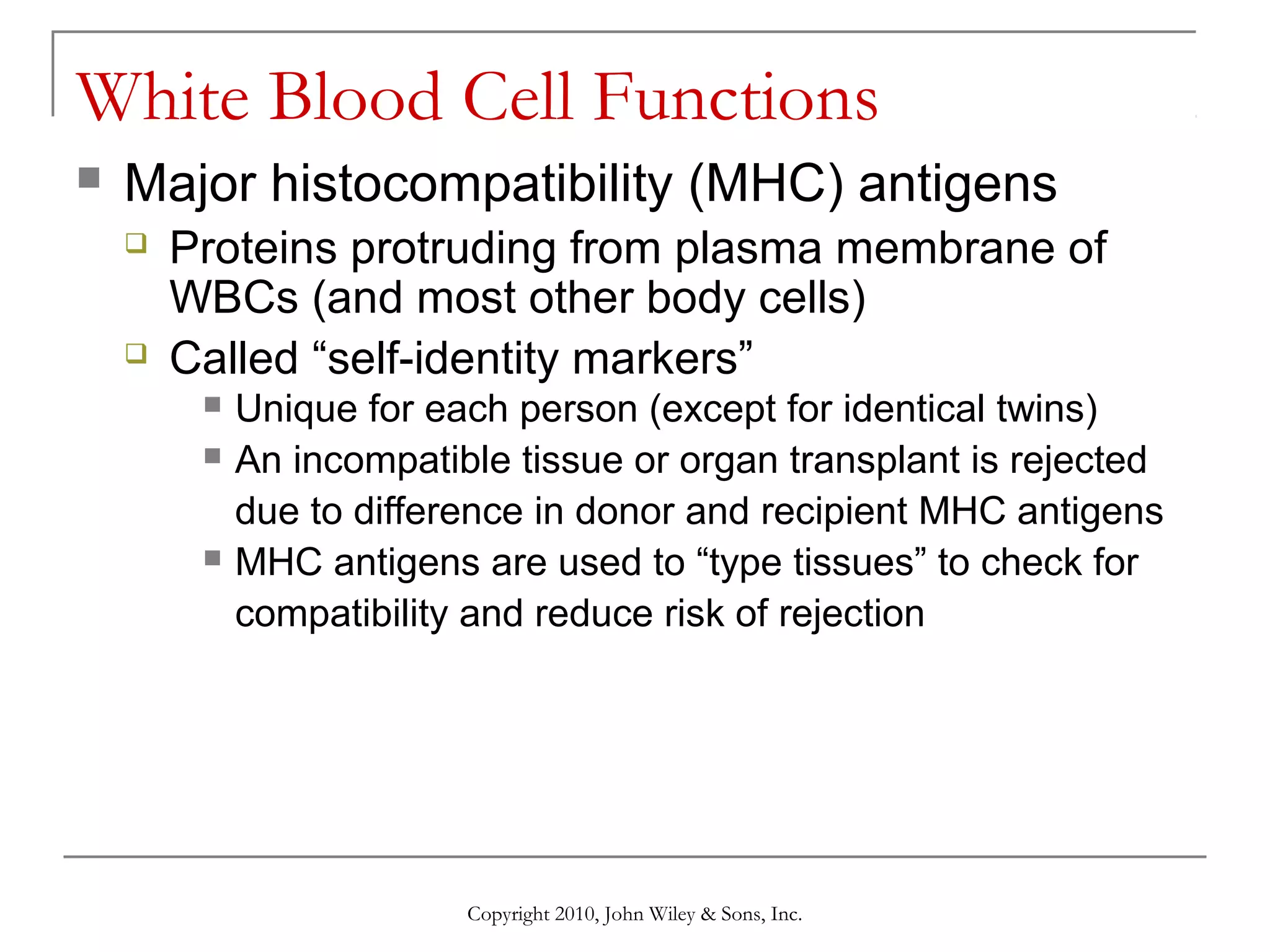
Major function: defense against
Infection and inflammation
Antigen-antibody (allergic) reactions
Copyright 2010, John Wiley & Sons, Inc.](https://image.slidesharecdn.com/lecture6thecardiovascularsystemblood-140306160029-phpapp01/75/Lecture-6-the-cardiovascular-system-blood-18-2048.jpg)

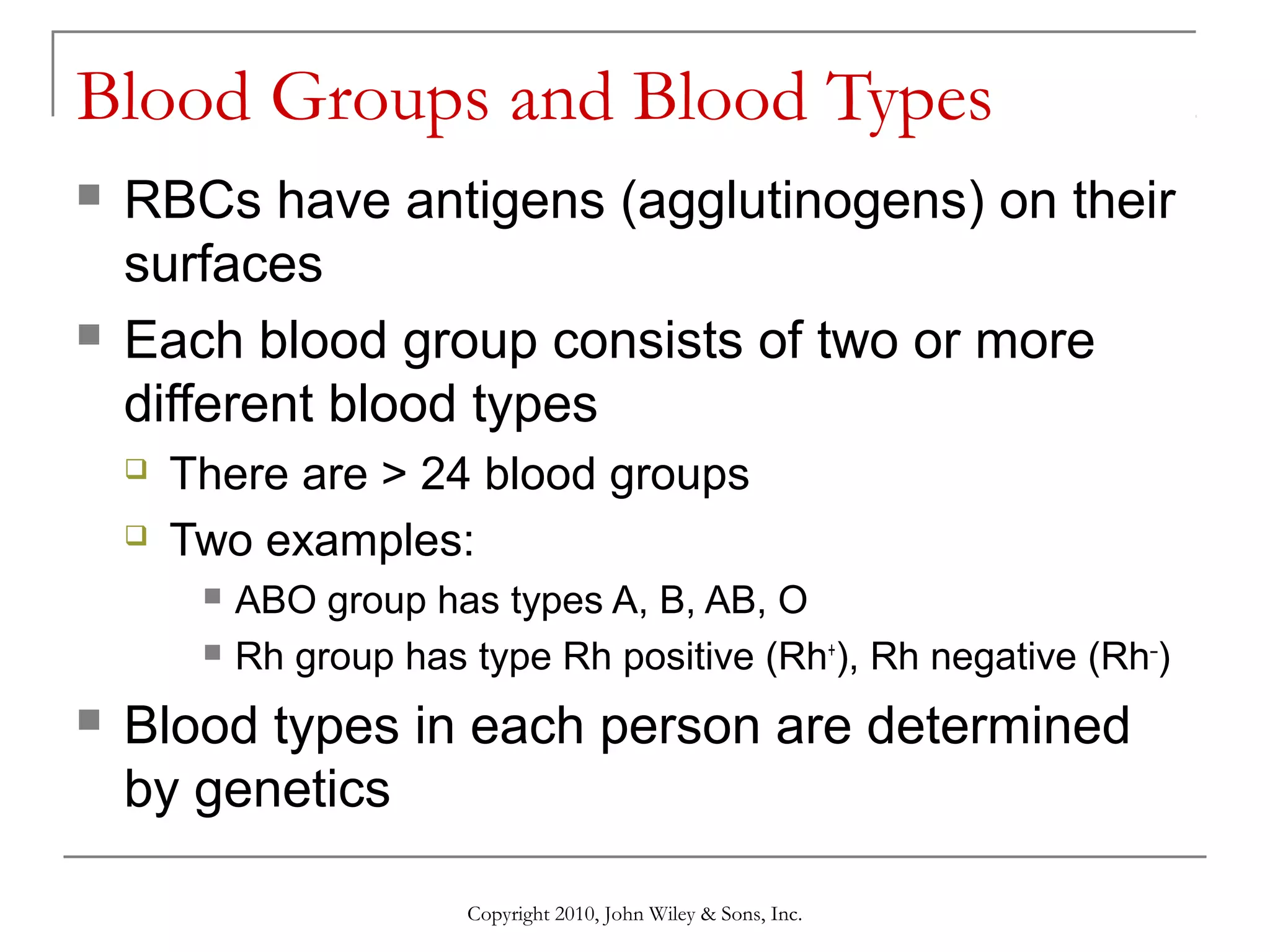
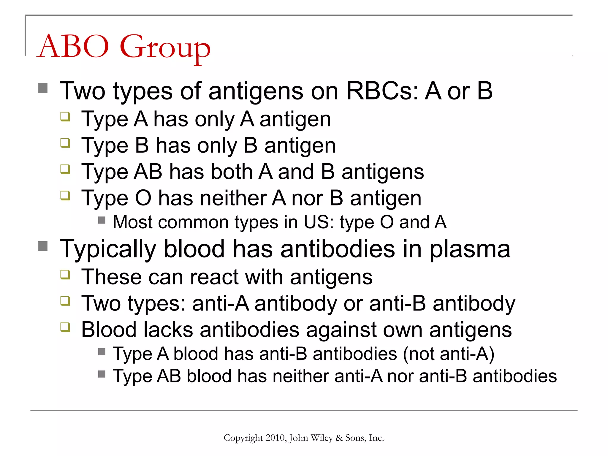
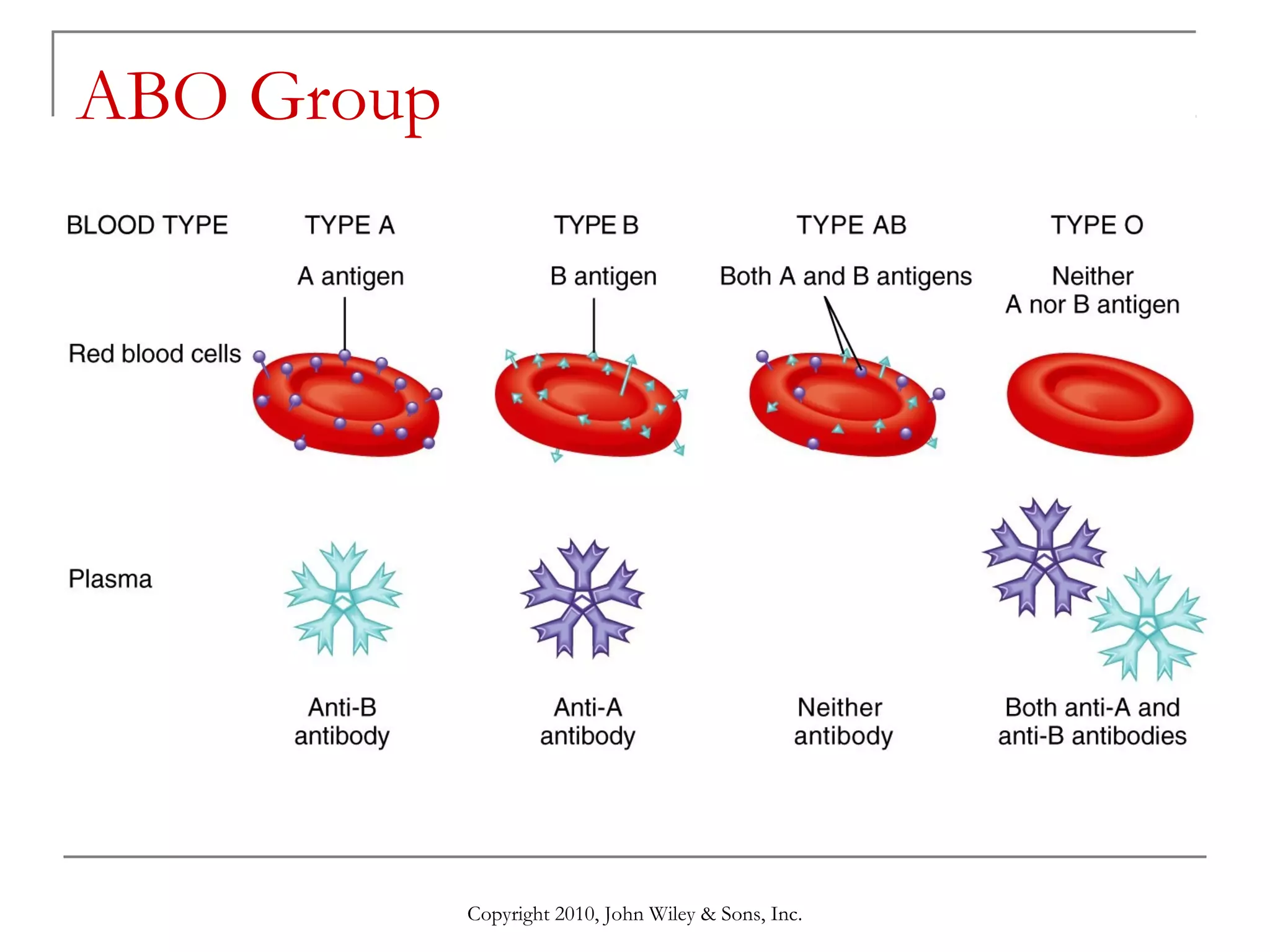

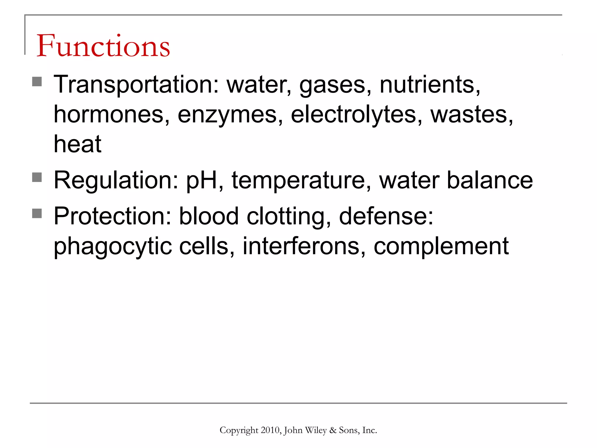
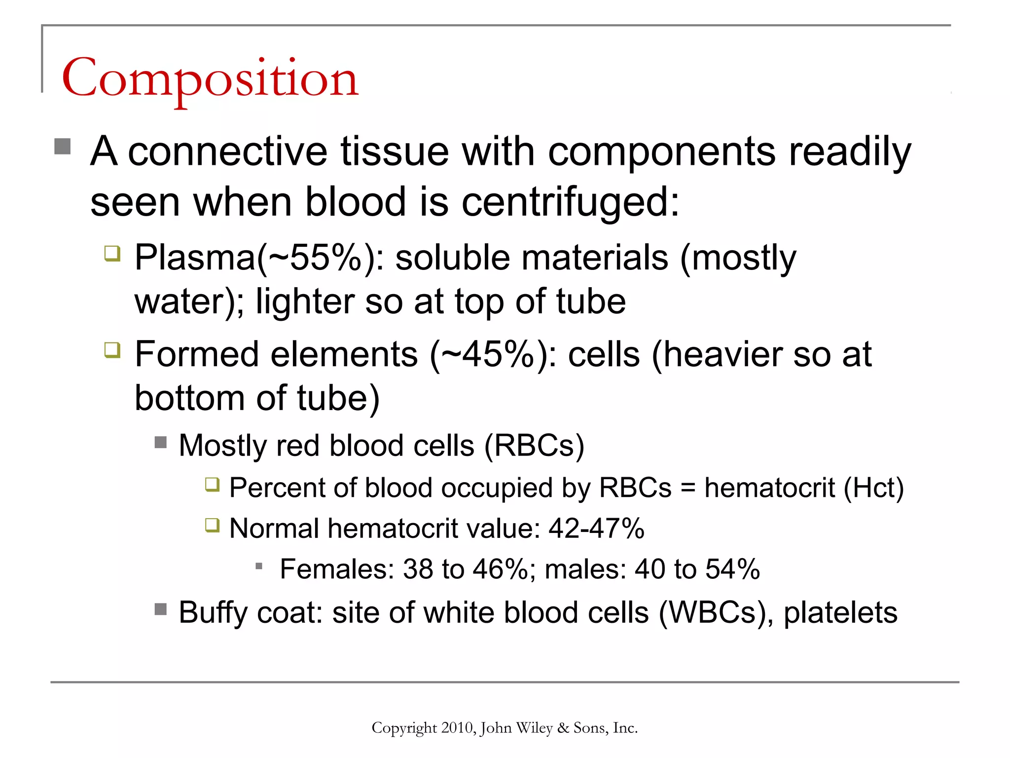
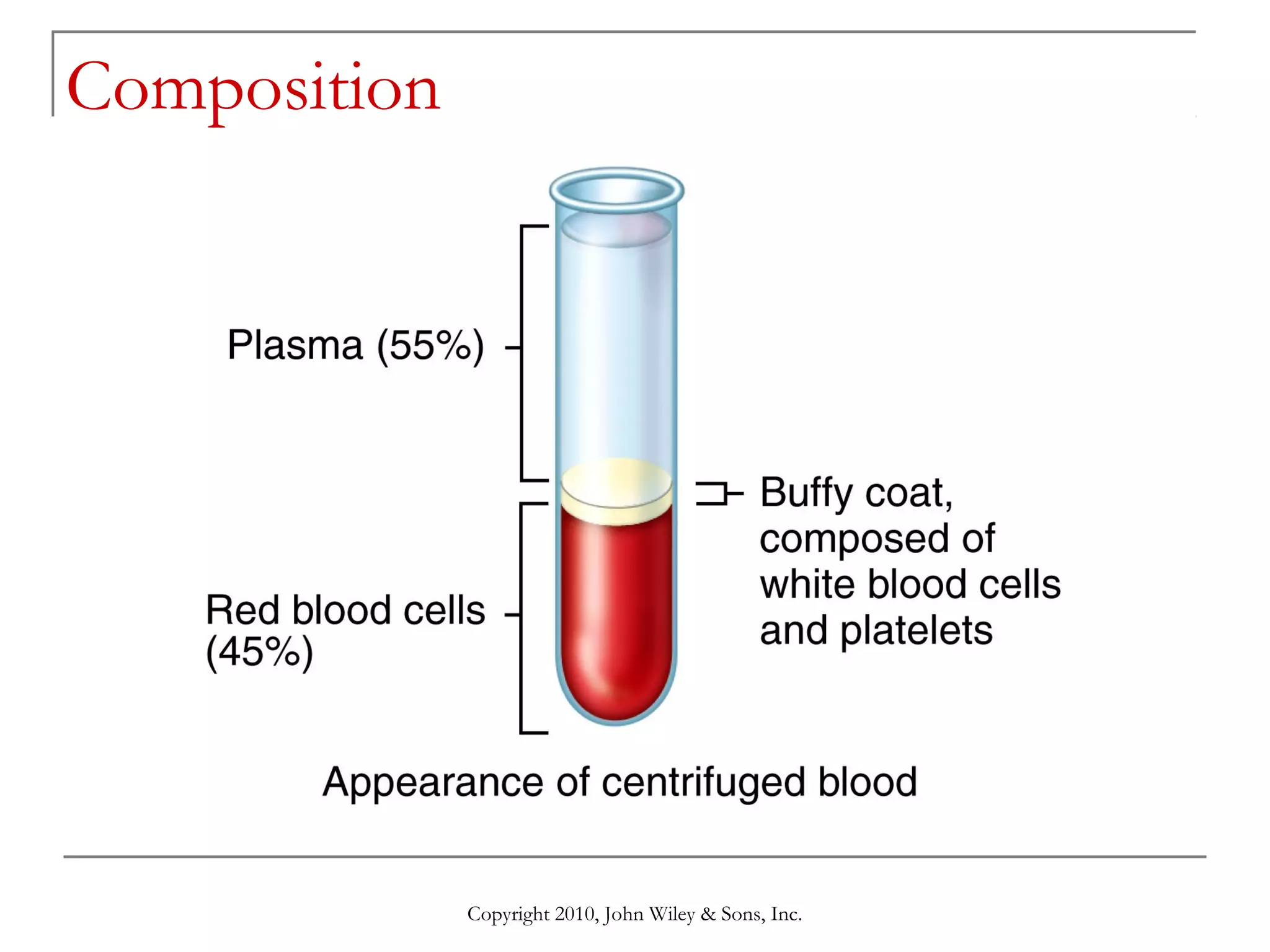
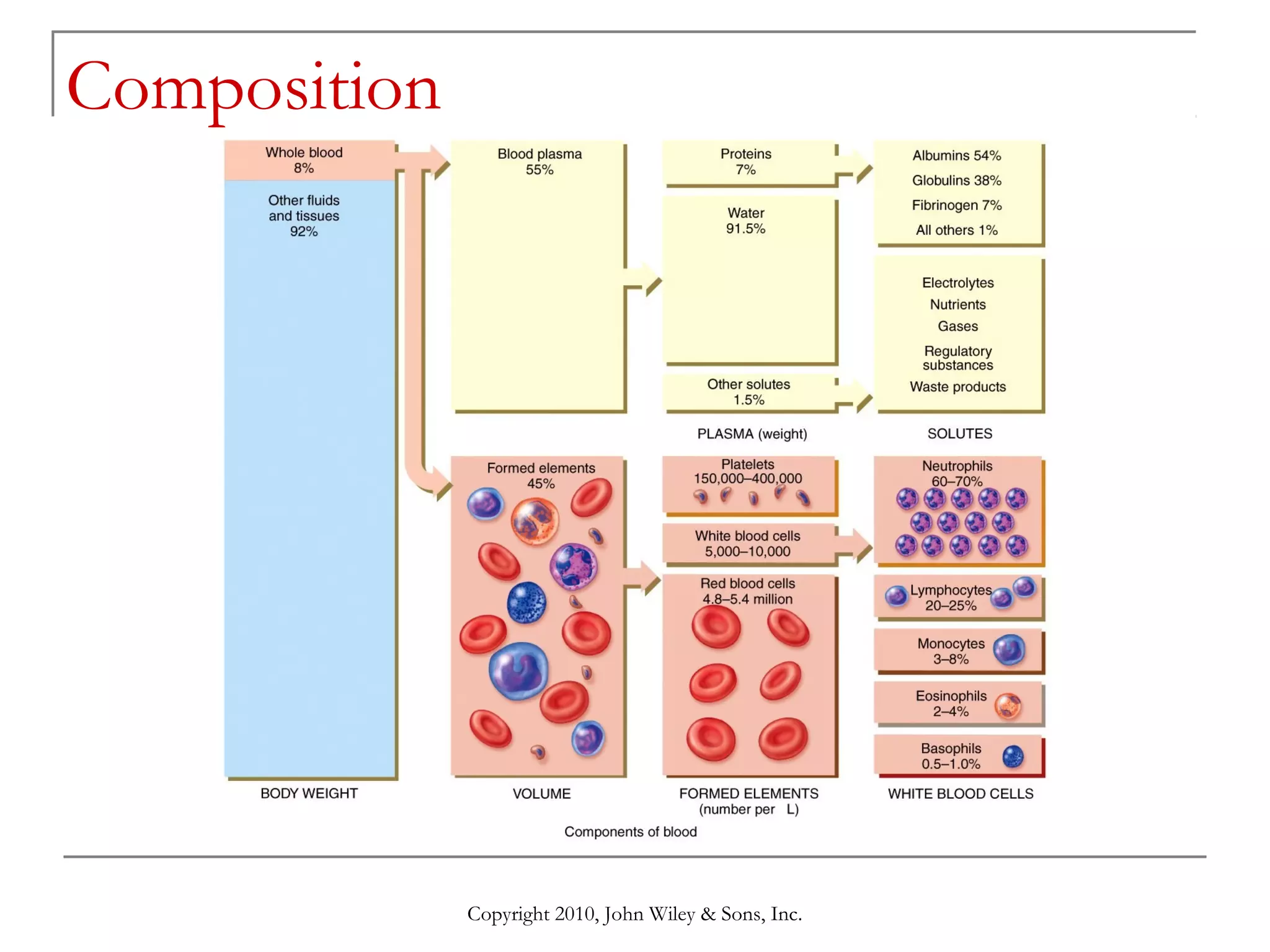
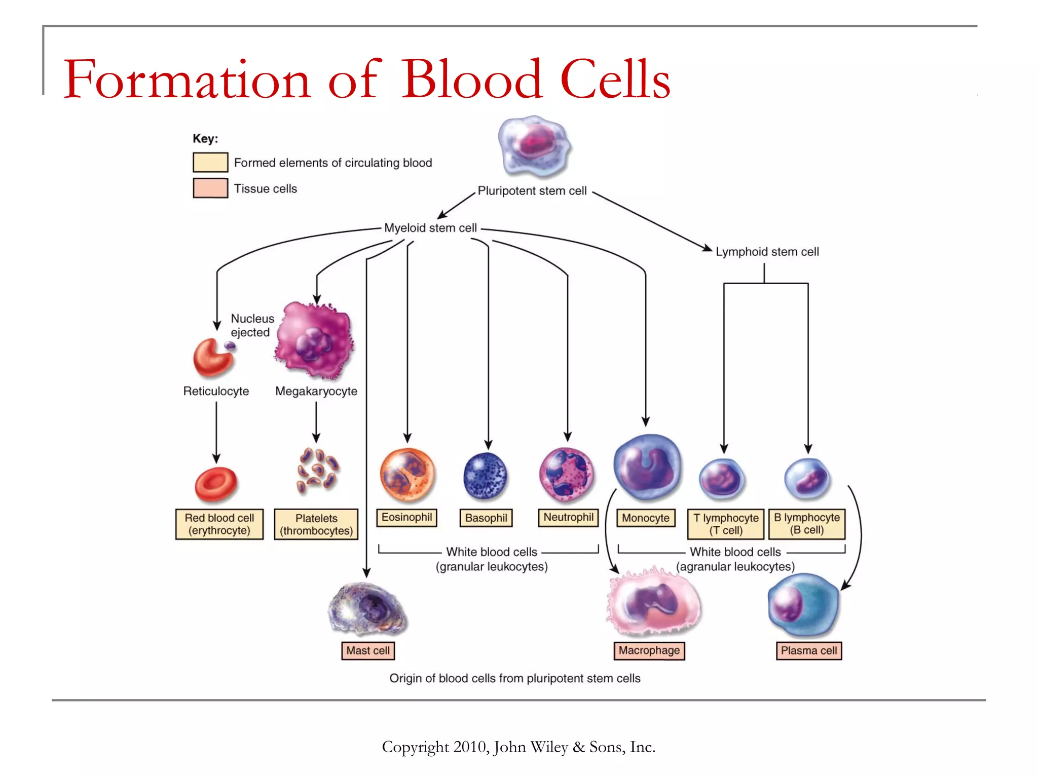
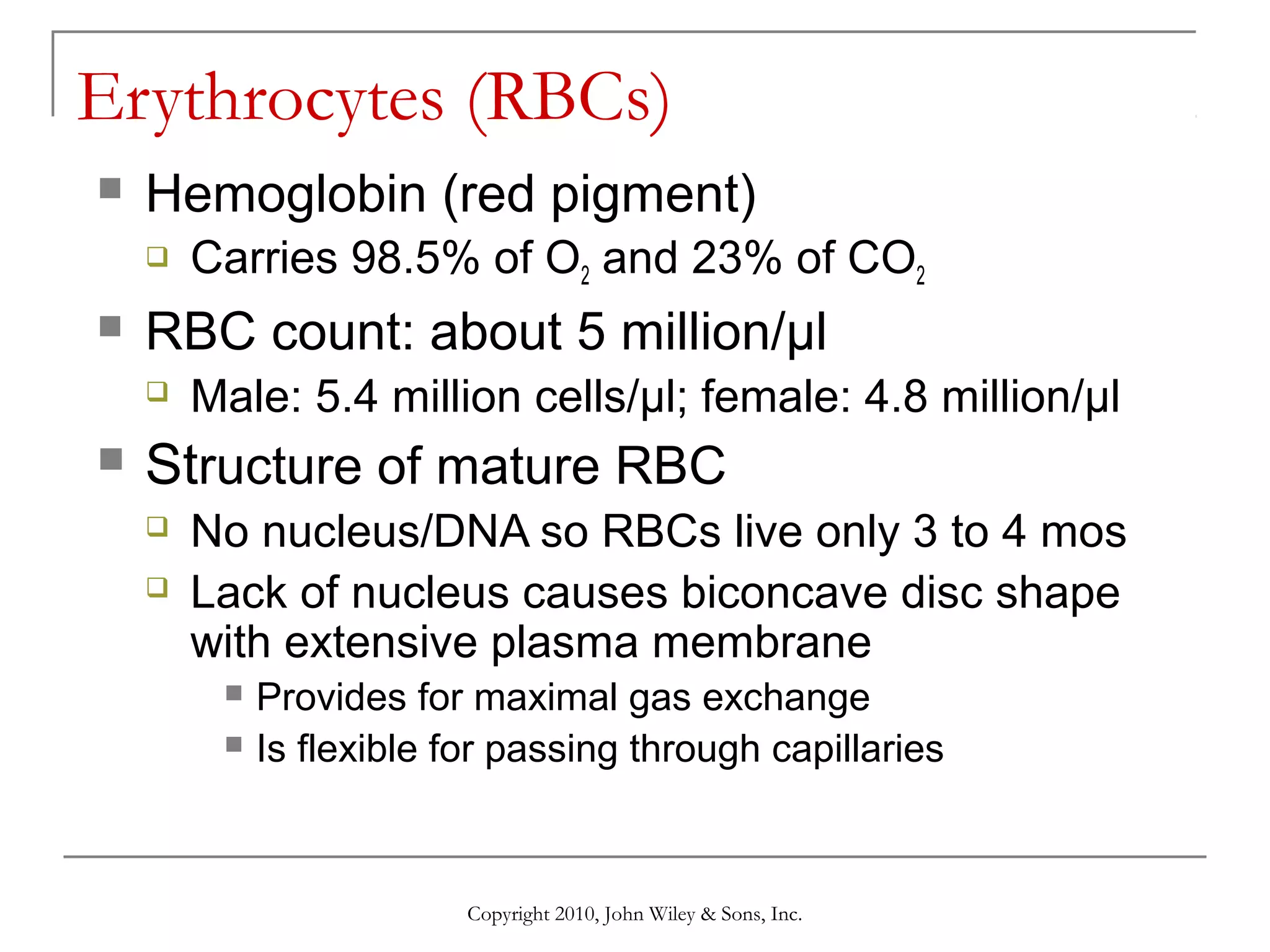
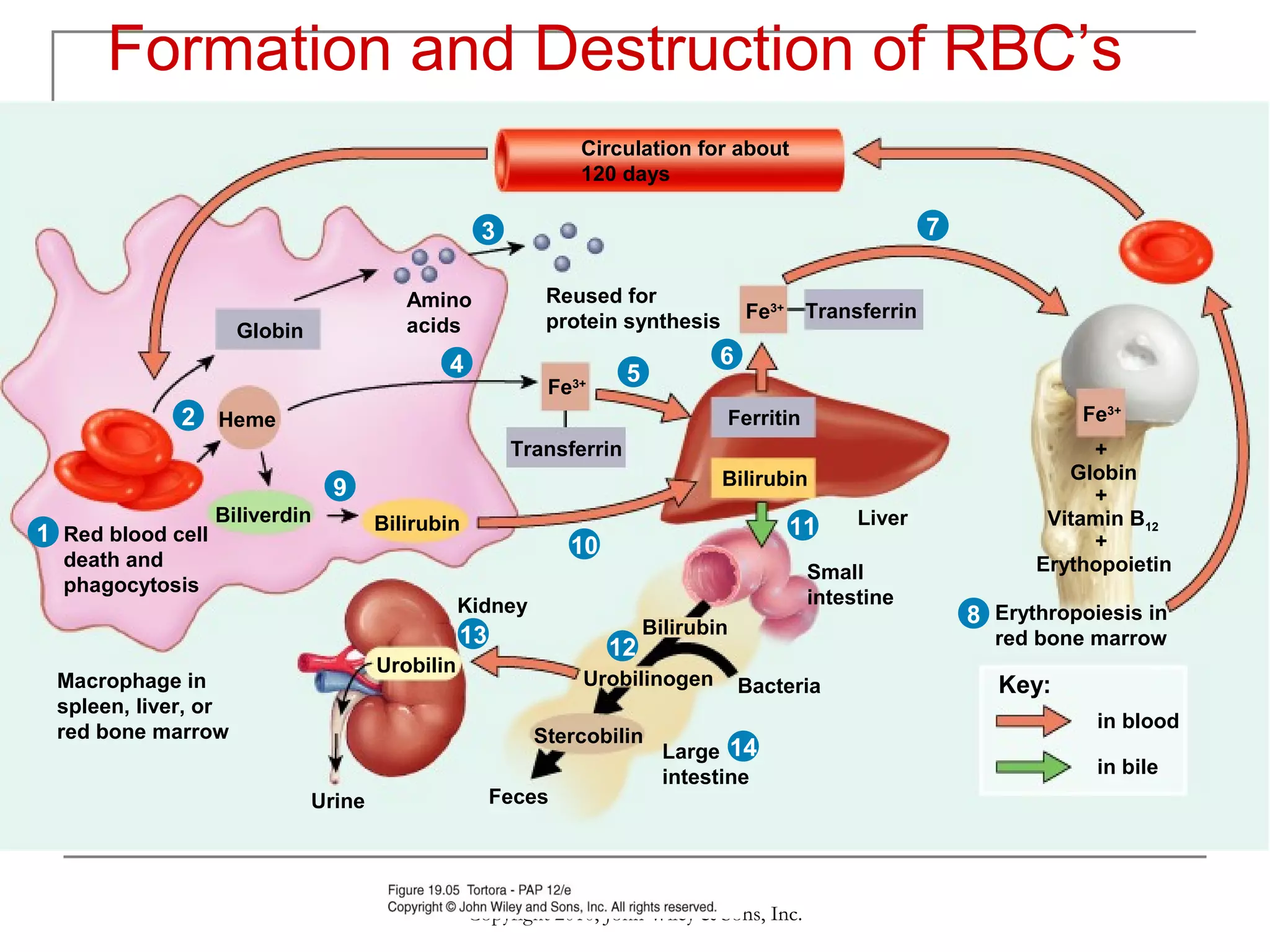
The cardiovascular system chapter discusses the functions, composition, and formation of blood. Blood functions include transportation, regulation, and protection. Blood composition includes plasma and formed elements such as red blood cells, white blood cells, and platelets. Red blood cells are formed through erythropoiesis and contain hemoglobin, while white blood cells help defend the body against infection and disease. Blood also contains different blood groups defined by antigens on red blood cells.


![26 [chapter 26 the urinary system]](https://cdn.slidesharecdn.com/ss_thumbnails/26chapter26theurinarysystem-170828044011-thumbnail.jpg?width=640&height=640&fit=bounds)

![12 [chapter 12 nervous tissue]](https://cdn.slidesharecdn.com/ss_thumbnails/12chapter12nervoustissue-170828041102-thumbnail.jpg?width=640&height=640&fit=bounds)


![20 [chapter 20 the cardiovascular system the heart]](https://cdn.slidesharecdn.com/ss_thumbnails/20chapter20thecardiovascularsystem-theheart-170828133506-thumbnail.jpg?width=640&height=640&fit=bounds)

![21 [chapter 21 the cardiovascular system blood vessels and hemodynamics][11e]](https://cdn.slidesharecdn.com/ss_thumbnails/21chapter21thecardiovascularsystem-bloodvesselsandhemodynamics11e-170828043342-thumbnail.jpg?width=640&height=640&fit=bounds)
![19 [chapter 19 the cardiovascular system the blood]](https://cdn.slidesharecdn.com/ss_thumbnails/19chapter19thecardiovascularsystem-theblood-170828042033-thumbnail.jpg?width=640&height=640&fit=bounds)

![18 [chapter 18 the endocrine system]](https://cdn.slidesharecdn.com/ss_thumbnails/18chapter18theendocrinesystem-170828042016-thumbnail.jpg?width=640&height=640&fit=bounds)