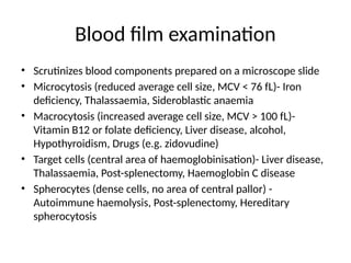
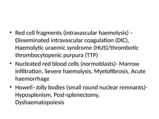
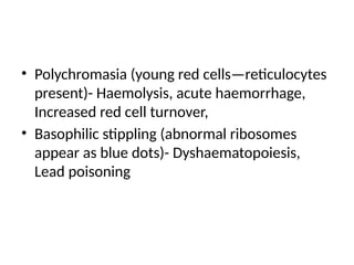
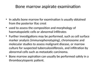
HEMATOLOGY
BY
NJIHIA MARTHA
FUNCTIONAL ANATOMY AND
PHYSIOLOGY
Introduction
• Blood flows throughout the body in the vascular
system, and consists of plasma and three cellular
components:
red cells, which transport oxygen from the lungs
to the tissues
white cells, which protect against infection
platelets, which interact with blood vessels and
clotting factors to maintain vascular integrity and
prevent bleeding.
Hematopoiesis
• Haematopoiesis describes the formation of blood cells
• During development, haematopoiesis occurs in the liverand
spleen and subsequently in red bone marrow in the
medullary cavity of all bones
• In childhood, red marrow is progressively replaced by fat
(yellow marrow), so that in adults normal haematopoiesis is
restricted to the vertebrae, pelvis, sternum, ribs, clavicles,
skull, upper humerus and proximal femur
• Bone marrow contains a range of immature haematopoietic
precursor cells and a storage pool of mature cells for release
at times of increased demand.
Red blood cells
• Red cell precursors formed in the bone marrow from the
erythroid (CFU–E) progenitor cells are called erythroblasts or
normoblasts
• They divide and acquire haemoglobin which turns the cytoplasm
pink; the nucleus condenses and is extruded from the cell.
• The first non-nucleated red cell is a reticulocyte which still
contains ribosomal material in the cytoplasm, giving these large
cells a faint blue tinge (‘polychromasia’).
• Reticulocytes lose their ribosomal material and mature over 3
days, during which time they are released into the circulation.
• Proliferation and differentiation of red cell precursors is
stimulated by erythropoietin, a polypeptide hormone produced
by renal tubular cells in response to hypoxia.
• Mature red cells circulate for about 120 days
Red blood cells
• They are biconcave discs lacking a nucleus but filled
with haemoglobin, which delivers oxygen to the
tissues from the lungs.
• In order to pass through the smallest capillaries the
red cell membrane is adapted to be deformable
• The ABO and Rhesus systems are the most
commonly recognised antigens inserted into the
lipid bilayer , but over 400 blood group antigens
have been described
Hemoglobin
• Haemoglobin is a protein specially adapted for
oxygen transport.
• It is composed of four globin chains, each
surrounding an iron-containing porphyrin
pigment molecule termed haem
• Haemoglobin A (αα/ββ) represents over 90%
of adult haemoglobin, whereas haemoglobin F
(αα/γ γ) is the predominant type in the fetus
Destruction
• Red cells at the end of their lifespan of approximately 120
days are phagocytosed by the reticulo-endothelial system.
• Amino acids from globin chains are recycled and iron is
removed from haem for reuse in haemoglobin synthesis.
• The remnant haem structure is degraded to bilirubin and
conjugated with glucuronic acid before being excreted in
bile.
• Free intravascular haemoglobin is toxic and is normally
bound by haptoglobins, which are plasma proteins
produced by the liver
White blood cells
• White cells or leucocytes in the blood consist of
granulocytes (neutrophils, eosinophils and basophils),
monocytes and lymphocytes
• Granulocytes and monocytes are formed from bone
marrow
• The first recognisable granulocyte in the marrow is the
myeloblast.
• As the cells divide and mature, the nucleus segments
and the cytoplasm acquires specific neutrophilic,
eosinophilic or basophilic granules
Neutrophils
• Neutrophils, the most common white blood cells in the
blood of adults.
• Have a multilobular nucleus
• Their main function is to recognise, ingest and destroy
foreign particles and microorganisms
• Neutrophils spend 6–10 hours in the circulation before being
 removed principally by the spleen or
 Alternatively, they pass into the tissues and either are
consumed in the inflammatory process or undergo
apoptotic cell death and phagocytosis by macrophages
Eosinophils
• Eosinophils represent 1–6% of the circulating white cells.
• They are a similar size to neutrophils but have a bilobed
nucleus and prominent orange granules on Romanowsky
staining.
• Eosinophils are phagocytic and their granules contain a
peroxidase capable of generating reactive oxygen species
and proteins involved in the intracellular killing of
protozoa and helminths
• They are also involved in allergic reactions (e.g. atopic
asthma,
Basophils
• These cells are less common than eosinophils,
representing less than 1% of circulating white
cells.
• They contain
• dense black granules which obscure the nucleus.
• Mast cells resemble basophils but are only found
in the tissues.
• These cells are involved in hypersensitivity
reactions
Monocytes
• Monocytes are the largest of the white cells
• These cells circulate for a few hours and then
migrate into the tissue where they become
macrophages, Kupffer cells or antigen-
presenting dendritic cells.
• The former phagocytose debris, apoptotic
cells and microorganisms
Lymphocytes
• Lymphocytes are derived from pluripotent haematopoietic stem cells in
the bone marrow.
• There are two main types:
 T cells (which mediate cellular immunity) and
 B cells (which mediate humoral immunity)
• Lymphoid cells which migrate to the thymus develop into T cells, whereas
B cells develop in the bone marrow.
• The majority of lymphocytes (approximately 80%) in the circulation are T
cells.
• Lymphocytes are heterogeneous, the smallest cells being the size of red
cells and the largest being the size of neutrophils.
• Small lymphocytes are circular with scanty cytoplasm but the larger cells
are more irregular with abundant blue cytoplasm.
• Lymphocyte subpopulations can be defined with specific functions and
their lifespan can vary from a few days to many years
Hemostasis
• Blood must be maintained in a fluid state in order to function as
a transport system, but must be able to solidify to form a clot
following vascular injury to prevent excessive bleeding, a
process known as haemostasis.
• Successful haemostasis is localised to the area of tissue damage
and is followed by removal of the clot and tissue repair.
• This is achieved by complex interactions between the vascular
endothelium, platelets, coagulation factors, natural
anticoagulants and fibrinolytic enzymes.
• Dysfunction of any of these components may result in
haemorrhage or thrombosis
Platelets
• Platelets are formed in the bone marrow from megakaryocytes.
• Megakaryocytic stem cells divide to form megakaryoblasts,
which undergo a process called ‘endomitotic reduplication’, in
which there is division of the nucleus but not the cell.
• This creates mature megakaryocytes, large cells with several
nuclei and cytoplasm containing platelet granules.
• Up to 3000 platelets then fragment off from each
megakaryocyte into the circulation in the marrow sinusoids.
• The formation and maturation of megakaryocytes are
stimulated by thrombopoietin produced in the liver.
• Platelets circulate for 8–10 days before they are destroyed in
the reticuloendothelial system
• Drugs which inhibit platelet function and thrombosis include
aspirin
Clotting factors
• Clotting factors are synthesised by the liver,
although factor V is also produced by platelets
and endothelial cells.
• Congenital (e.g. haemophilia) and acquired
(e.g. liver failure) causes of coagulation factor
deficiency are associated with bleeding
Physical examination
Introduction
 Observation
• General well-being
• Colour: pallor, plethora
• Breathlessness
 Pulse
• Rate
 Conjunctivae
• Pallor
• Jaundice
 Fundi
• Haemorrhage
• Hyperviscosity
• Engorged veins
• Papilloedema
• Haemorrhage
Introduction
Mouth
• Lips: angular stomatitis,
• telangiectasia
• Gum hypertrophy
• Tongue: colour,
• smoothness
• Buccal mucosa:
• petechiae
• Tonsils: size
Introduction
Lymph nodes-lymphadenopathy
Skin- Purpura, Bruising
Abdomen
• Masses
• Ascites
• Hepatomegaly
• Splenomegaly
• Inguinal and femoral
• nodes
Introduction
 Hands
• Perfusion
• Telangiectasia
• Skin crease pallor
• Koilonychia
 Joints
• Deformity
• Swelling- hemophilia
• Restricted movement
 Feet
• Peripheral circulation
• Toes: gangrene
Investigations
The full blood count (FBC)
• Measures the haematological parameters. These include
 numbers of circulating cells and platelets
 the proportion of whole blood volume occupied by red cells
(the haematocrit, Hct)
 the red cell indices which give information about the size of
red cells (mean cell volume, MCV)
 the amount of haemoglobin present in the red cells (mean
cell haemoglobin, MCH).
 types of white blood cell and give automated counts of
neutrophils, lymphocytes, monocytes, eosinophils and
basophils.
Blood film examination
• Scrutinizes blood components prepared on a microscope slide
• Microcytosis (reduced average cell size, MCV < 76 fL)- Iron
deficiency, Thalassaemia, Sideroblastic anaemia
• Macrocytosis (increased average cell size, MCV > 100 fL)-
Vitamin B12 or folate deficiency, Liver disease, alcohol,
Hypothyroidism, Drugs (e.g. zidovudine)
• Target cells (central area of haemoglobinisation)- Liver disease,
Thalassaemia, Post-splenectomy, Haemoglobin C disease
• Spherocytes (dense cells, no area of central pallor) -
Autoimmune haemolysis, Post-splenectomy, Hereditary
spherocytosis
• Red cell fragments (intravascular haemolysis) –
Disseminated intravascular coagulation (DIC),
Haemolytic uraemic syndrome (HUS)/thrombotic
thrombocytopenic purpura (TTP)
• Nucleated red blood cells (normoblasts)- Marrow
infiltration, Severe haemolysis, Myelofibrosis, Acute
haemorrhage
• Howell–Jolly bodies (small round nuclear remnants)-
Hyposplenism, Post-splenectomy,
Dyshaematopoiesis
• Polychromasia (young red cells—reticulocytes
present)- Haemolysis, acute haemorrhage,
Increased red cell turnover,
• Basophilic stippling (abnormal ribosomes
appear as blue dots)- Dyshaematopoiesis,
Lead poisoning
Bone marrow aspirate examination
• In adults bone marrow for examination is usually obtained
from the posterior iliac crest
• used to assess the composition and morphology of
haematopoietic cells or abnormal infiltrates
• Further investigations may be performed, such as cell surface
marker analysis (immunophenotyping), chromosome and
molecular studies to assess malignant disease, or marrow
culture for suspected tuberculosisfibrosis, and infiltration by
abnormal cells such as metastatic carcinoma.
• Bone marrow aspiration can usually be performed safely in a
thrombocytopenic patient.
Investigation of coagulation
• Prothrombin time (PT)- Assesses the extrinsic
pathway by the and
• Activated partial thromboplastin time (APTT)-asseses
the intrinsic pathway
• The international normalised ratio (INR) is validated
only to assess the therapeutic effect of coumarin
anticoagulants,including warfarin. INR is the ratio of
the patient’s prothrombin time to that of a normal
control, raised to the power of the international
sensitivity index of the thromboplastin used in the test

HEMATOLOGY presentation(internal medicine)

  • 1.
  • 2.
  • 3.
    Introduction • Blood flowsthroughout the body in the vascular system, and consists of plasma and three cellular components: red cells, which transport oxygen from the lungs to the tissues white cells, which protect against infection platelets, which interact with blood vessels and clotting factors to maintain vascular integrity and prevent bleeding.
  • 4.
    Hematopoiesis • Haematopoiesis describesthe formation of blood cells • During development, haematopoiesis occurs in the liverand spleen and subsequently in red bone marrow in the medullary cavity of all bones • In childhood, red marrow is progressively replaced by fat (yellow marrow), so that in adults normal haematopoiesis is restricted to the vertebrae, pelvis, sternum, ribs, clavicles, skull, upper humerus and proximal femur • Bone marrow contains a range of immature haematopoietic precursor cells and a storage pool of mature cells for release at times of increased demand.
  • 5.
    Red blood cells •Red cell precursors formed in the bone marrow from the erythroid (CFU–E) progenitor cells are called erythroblasts or normoblasts • They divide and acquire haemoglobin which turns the cytoplasm pink; the nucleus condenses and is extruded from the cell. • The first non-nucleated red cell is a reticulocyte which still contains ribosomal material in the cytoplasm, giving these large cells a faint blue tinge (‘polychromasia’). • Reticulocytes lose their ribosomal material and mature over 3 days, during which time they are released into the circulation. • Proliferation and differentiation of red cell precursors is stimulated by erythropoietin, a polypeptide hormone produced by renal tubular cells in response to hypoxia. • Mature red cells circulate for about 120 days
  • 6.
    Red blood cells •They are biconcave discs lacking a nucleus but filled with haemoglobin, which delivers oxygen to the tissues from the lungs. • In order to pass through the smallest capillaries the red cell membrane is adapted to be deformable • The ABO and Rhesus systems are the most commonly recognised antigens inserted into the lipid bilayer , but over 400 blood group antigens have been described
  • 7.
    Hemoglobin • Haemoglobin isa protein specially adapted for oxygen transport. • It is composed of four globin chains, each surrounding an iron-containing porphyrin pigment molecule termed haem • Haemoglobin A (αα/ββ) represents over 90% of adult haemoglobin, whereas haemoglobin F (αα/γ γ) is the predominant type in the fetus
  • 8.
    Destruction • Red cellsat the end of their lifespan of approximately 120 days are phagocytosed by the reticulo-endothelial system. • Amino acids from globin chains are recycled and iron is removed from haem for reuse in haemoglobin synthesis. • The remnant haem structure is degraded to bilirubin and conjugated with glucuronic acid before being excreted in bile. • Free intravascular haemoglobin is toxic and is normally bound by haptoglobins, which are plasma proteins produced by the liver
  • 9.
    White blood cells •White cells or leucocytes in the blood consist of granulocytes (neutrophils, eosinophils and basophils), monocytes and lymphocytes • Granulocytes and monocytes are formed from bone marrow • The first recognisable granulocyte in the marrow is the myeloblast. • As the cells divide and mature, the nucleus segments and the cytoplasm acquires specific neutrophilic, eosinophilic or basophilic granules
  • 10.
    Neutrophils • Neutrophils, themost common white blood cells in the blood of adults. • Have a multilobular nucleus • Their main function is to recognise, ingest and destroy foreign particles and microorganisms • Neutrophils spend 6–10 hours in the circulation before being  removed principally by the spleen or  Alternatively, they pass into the tissues and either are consumed in the inflammatory process or undergo apoptotic cell death and phagocytosis by macrophages
  • 11.
    Eosinophils • Eosinophils represent1–6% of the circulating white cells. • They are a similar size to neutrophils but have a bilobed nucleus and prominent orange granules on Romanowsky staining. • Eosinophils are phagocytic and their granules contain a peroxidase capable of generating reactive oxygen species and proteins involved in the intracellular killing of protozoa and helminths • They are also involved in allergic reactions (e.g. atopic asthma,
  • 12.
    Basophils • These cellsare less common than eosinophils, representing less than 1% of circulating white cells. • They contain • dense black granules which obscure the nucleus. • Mast cells resemble basophils but are only found in the tissues. • These cells are involved in hypersensitivity reactions
  • 13.
    Monocytes • Monocytes arethe largest of the white cells • These cells circulate for a few hours and then migrate into the tissue where they become macrophages, Kupffer cells or antigen- presenting dendritic cells. • The former phagocytose debris, apoptotic cells and microorganisms
  • 14.
    Lymphocytes • Lymphocytes arederived from pluripotent haematopoietic stem cells in the bone marrow. • There are two main types:  T cells (which mediate cellular immunity) and  B cells (which mediate humoral immunity) • Lymphoid cells which migrate to the thymus develop into T cells, whereas B cells develop in the bone marrow. • The majority of lymphocytes (approximately 80%) in the circulation are T cells. • Lymphocytes are heterogeneous, the smallest cells being the size of red cells and the largest being the size of neutrophils. • Small lymphocytes are circular with scanty cytoplasm but the larger cells are more irregular with abundant blue cytoplasm. • Lymphocyte subpopulations can be defined with specific functions and their lifespan can vary from a few days to many years
  • 15.
    Hemostasis • Blood mustbe maintained in a fluid state in order to function as a transport system, but must be able to solidify to form a clot following vascular injury to prevent excessive bleeding, a process known as haemostasis. • Successful haemostasis is localised to the area of tissue damage and is followed by removal of the clot and tissue repair. • This is achieved by complex interactions between the vascular endothelium, platelets, coagulation factors, natural anticoagulants and fibrinolytic enzymes. • Dysfunction of any of these components may result in haemorrhage or thrombosis
  • 16.
    Platelets • Platelets areformed in the bone marrow from megakaryocytes. • Megakaryocytic stem cells divide to form megakaryoblasts, which undergo a process called ‘endomitotic reduplication’, in which there is division of the nucleus but not the cell. • This creates mature megakaryocytes, large cells with several nuclei and cytoplasm containing platelet granules. • Up to 3000 platelets then fragment off from each megakaryocyte into the circulation in the marrow sinusoids. • The formation and maturation of megakaryocytes are stimulated by thrombopoietin produced in the liver. • Platelets circulate for 8–10 days before they are destroyed in the reticuloendothelial system • Drugs which inhibit platelet function and thrombosis include aspirin
  • 17.
    Clotting factors • Clottingfactors are synthesised by the liver, although factor V is also produced by platelets and endothelial cells. • Congenital (e.g. haemophilia) and acquired (e.g. liver failure) causes of coagulation factor deficiency are associated with bleeding
  • 18.
  • 19.
    Introduction  Observation • Generalwell-being • Colour: pallor, plethora • Breathlessness  Pulse • Rate  Conjunctivae • Pallor • Jaundice  Fundi • Haemorrhage • Hyperviscosity • Engorged veins • Papilloedema • Haemorrhage
  • 20.
    Introduction Mouth • Lips: angularstomatitis, • telangiectasia • Gum hypertrophy • Tongue: colour, • smoothness • Buccal mucosa: • petechiae • Tonsils: size
  • 21.
    Introduction Lymph nodes-lymphadenopathy Skin- Purpura,Bruising Abdomen • Masses • Ascites • Hepatomegaly • Splenomegaly • Inguinal and femoral • nodes
  • 22.
    Introduction  Hands • Perfusion •Telangiectasia • Skin crease pallor • Koilonychia  Joints • Deformity • Swelling- hemophilia • Restricted movement  Feet • Peripheral circulation • Toes: gangrene
  • 23.
  • 24.
    The full bloodcount (FBC) • Measures the haematological parameters. These include  numbers of circulating cells and platelets  the proportion of whole blood volume occupied by red cells (the haematocrit, Hct)  the red cell indices which give information about the size of red cells (mean cell volume, MCV)  the amount of haemoglobin present in the red cells (mean cell haemoglobin, MCH).  types of white blood cell and give automated counts of neutrophils, lymphocytes, monocytes, eosinophils and basophils.
  • 25.
    Blood film examination •Scrutinizes blood components prepared on a microscope slide • Microcytosis (reduced average cell size, MCV < 76 fL)- Iron deficiency, Thalassaemia, Sideroblastic anaemia • Macrocytosis (increased average cell size, MCV > 100 fL)- Vitamin B12 or folate deficiency, Liver disease, alcohol, Hypothyroidism, Drugs (e.g. zidovudine) • Target cells (central area of haemoglobinisation)- Liver disease, Thalassaemia, Post-splenectomy, Haemoglobin C disease • Spherocytes (dense cells, no area of central pallor) - Autoimmune haemolysis, Post-splenectomy, Hereditary spherocytosis
  • 26.
    • Red cellfragments (intravascular haemolysis) – Disseminated intravascular coagulation (DIC), Haemolytic uraemic syndrome (HUS)/thrombotic thrombocytopenic purpura (TTP) • Nucleated red blood cells (normoblasts)- Marrow infiltration, Severe haemolysis, Myelofibrosis, Acute haemorrhage • Howell–Jolly bodies (small round nuclear remnants)- Hyposplenism, Post-splenectomy, Dyshaematopoiesis
  • 27.
    • Polychromasia (youngred cells—reticulocytes present)- Haemolysis, acute haemorrhage, Increased red cell turnover, • Basophilic stippling (abnormal ribosomes appear as blue dots)- Dyshaematopoiesis, Lead poisoning
  • 28.
    Bone marrow aspirateexamination • In adults bone marrow for examination is usually obtained from the posterior iliac crest • used to assess the composition and morphology of haematopoietic cells or abnormal infiltrates • Further investigations may be performed, such as cell surface marker analysis (immunophenotyping), chromosome and molecular studies to assess malignant disease, or marrow culture for suspected tuberculosisfibrosis, and infiltration by abnormal cells such as metastatic carcinoma. • Bone marrow aspiration can usually be performed safely in a thrombocytopenic patient.
  • 29.
    Investigation of coagulation •Prothrombin time (PT)- Assesses the extrinsic pathway by the and • Activated partial thromboplastin time (APTT)-asseses the intrinsic pathway • The international normalised ratio (INR) is validated only to assess the therapeutic effect of coumarin anticoagulants,including warfarin. INR is the ratio of the patient’s prothrombin time to that of a normal control, raised to the power of the international sensitivity index of the thromboplastin used in the test