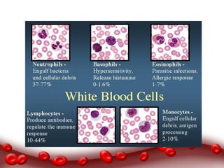
Cardiovascular System
Chapter 14 Blood
Functions of Blood
• Blood is the only liquid connective tissue.
• Blood has three main functions.
• 1. Transportation- blood transports oxygen from the
lungs to the cells throughout the body.
• 2. Regulation- blood helps regular pH of body fluids
and also the temperature of the body.
• 3. Protection- blood clots protect against excessive
loss of blood. White blood cells patrol the body and
protect it against disease.
Components of Whole Blood
• Blood is denser and stickier than water.
• Blood is kept at a temperature of about
100.4 F and has a slightly alkaline pH.
• Blood constitutes about 8% of the total
body weight.
• The average male human contains 5-6 liters
of blood with the average female human
containing 4-5 liters of blood.
• Whole blood is composed of two parts: blood
plasma and formed elements.
• Blood is 45% formed elements and 55%
plasma.
• 99% of the formed elements are red blood
cells (RBC) and less than 1% of the blood is
white blood cells and platelets.
• When blood is centrifuged, the formed
elements like the RBC’s sink to the bottom of
the tube and the liquid plasma floats on top.
Blood Plasma
• When formed elements are removed, a straw colored
liquid called blood plasma is left.
• Plasma is 91.5% water, 7% proteins, and 1.5% other
dissolved solutes.
• Proteins in the blood are synthesized by the liver.
• Some proteins include:
• Albumins-most plentiful, (egg whites)
• Globulins- antibodies and immunoglobulins
• Fibrinogen- blood clots
Formed Elements
This includes the following:
Red blood cells (RBC)
Platelets
White Blood Cells (WBC)
Granular leukocytes
Neutrophils
Eosinophils
Basophils
Agranular Leukocytes
T and B lymphocytes and natural killer
cells
Monocytes
Formation of Blood Cells
• The process in which the formed elements of blood develop
is called hemopoiesis.
• Before birth, this occurs in the yolk sac of the embryo and
later in the liver, spleen, thymus and lymph nodes of the
fetus.
• In the last three months before birth and the rest of human
life, red bone marrow becomes the primary site for blood
formation.
Red Bone Marrow
• High vascular connective tissue located
in spongy bone tissue.
• Red bone marrow cells are derived from
pluripotent stem cells.
• These cells have the capacity to develop
into several different types of cells.
• Pluripotent stems cells further develop
into myeloid stem cells and lymphoid
stem cells.
Red Blood Cells
• RBCs are also called erythrocytes.
• They contain the oxygen-carrying
protein hemoglobin which gives whole
blood its red color.
• A healthy adult male typically has 5.4
million red blood cells per microliter of
blood and a healthy adult female has
about 4.8 million per microliter.
• To maintain normal numbers, new mature RBCs must
enter the bloodstream at 2 million per second; a pace
that balancing the RBC destruction.
• Mature RBC do not have a nucleus and cannot divide
or carry out metabolic activities.
• RBCs are a biconcave disk which increases the surface
area for gas diffusion.
RBC Life Cycle
• RBCs only live about 120 days because of wear and tear and
are removed from circulation.
• 1. Macrophages in the spleen, liver and red bone marrow
phagocytize ruptured and worn-out cells splitting the heme
and globin from each other.
• 2. Globin is broken down into amino acids and reused.
• 3. Iron is removed from the heme portion and hooks up
with the plasma protein transferrin which acts as a
transporter.
• 4. The iron-transferrin complex is carried to the red bone
marrow where it is used to create new hemoglobin.
Vitamin B-12 is also used.
• 5. Red blood cells are formed in the red bone
marrow.
• 6. When iron is removed from heme, the non-iron
portion of heme is converted to biliverdin and
bilirubin. Bilirubin enters the blood and transported
to the liver and secreted into bile.
• 7. In the large intestine, bacteria convert bilirubin
into urobilinogen. This can be absorbed back into
the blood, excreted as urine or excreted in feces to
give it the brown color.
• Free iron is damaging, transferrin acts a protective
protein escort and as a result, plasma has virtually
no free iron.
• Iron overload can result when the body has too
much buildup of iron.
• The body has no method to eliminate the excess
iron.
• This can cause diseases of the heart, liver pancreas
and gonads.
Blood plasma
Formed Elements
Hemopoiesis
Red bone
marrow
Plutipotent stem
cells
RBC or
erythrocytes
Hemoglobin
White Blood Cells
• Unlike red blood cells, WBCs or leukocytes, have nuclei
and do not contain hemoglobin.
• They can be classified as granular or agranular depending
if they contain vesicles.
• WBCs are only made visible by staining. They are
normally clear.
• WBCs are active in the immune response of the body.
Normal stained slide
SEM with all three componenets
Platelets
WBC Functions
• Some WBCs combat microbes via phagocytosis
while others types produce antibodies.
• Neutrophils response first by releasing enzymes
to destroy microbes and ingesting them.
• Monocytes take longer to reach the site of
infection but arrive in large numbers.
• Monocytes migrate into infected tissues and
become wandering macrophages that can ingest
many microbes and clean up cellular debris.
• Eosinophils leave the capillaries and enter interstitial
fluid to combat inflammation in allergic reactions.
• They phagocytize antigen-antibody complexes and
are effective against certain parasitic worms.
• A high eosinophil count indicates an allergic
condition or parasitic infection.
• Basophils are also involved in inflammatory and
allergic reactions.
• They leave the capillaries and enter the tissues and
help release histamine, heparin and serotonin which
intensify the inflammatory reaction.
• B cells, T Cells and natural killer cells are WBC that
are active in the immune system.
• B cells become plasma cells that produce antibodies
and inactivate toxins.
• T cells attack microbes directly.
• NKC attack microbes and tumor cells.
• White blood cells and other nucleated body
cells have MHC proteins.
• Red blood cells possess group antigens but
lack MHC antigens.
• Tissue transplant rejections happen in part
to MHC proteins being incompatible.
• Red blood cell antigens do make blood
donations difficult because of
incompatibilities but NOT due to MHC.
• Many WBC’s live only a few days and during
cycles of infection, some only live a few
hours.
• However, there are some WBCs that are in
the body for years to provide continued
protection as memory cells.
WBC Counts
• Leukocytosis is an increase in the number of WBCs as a
protective response to invading microbes, strenuous
exercise, anesthesia and surgery.
• Determining the percentage of each type of WBC in a
blood sample is useful for diagnosing a condition because
each type of WBC plays a different role in the immune
response.
• This test is called the differential white blood count.
• Leukopenia is an abnormally low level of WBCs and can be
caused by radiation, shock and other chemical exposure.
WBC or
leukocytes
Neutrophil
Monocytes
Eosinophils
Basophils
Differential
WBC count
Platelets
• Pluripotent cells also differentiate into platelets.
• They are disk shaped and have many vesicles but no
nucleus.
• When blood vessels are damaged, platelets help stop blood
loss by forming a platelet plug.
• The platelets also contain chemicals that promote blood
clotting.
• Platelets only live for 5-9 days.
Platelet Plug Formation
• 1. Platelets contact and stick to parts
of the damaged blood vessel. This is
called platelet adhesion.
• 2. The platelets become activated and
they extend many projections that
enable them to contact and interact
with each other. They release
chemicals contained in their vesicles
which decrease blood flow. This is
called platelet release reaction.
• 3. The chemicals released makes all the
platelets in the area sticky and causes them
to clump together. This is called platelet
aggregation. Eventually large numbers
form and a platelet plug forms and stops
blood leakage.
Blood Groups and Blood Types
• The surfaces of blood cells contain genetically
determined assortments of proteins and lipids.
• Based on the presence or absence of certain antigens,
blood is categorized into different blood groups.
• Within each blood group, there may be different blood
types.
• There are at least 24 blood groups and 100 or so
different antigens.
• The major ones are ABO and Rh blood groups.
Rh factor and Rh disease
• The Rh blood group is named for the rhesus
monkey in which it was first discovered.
• People whose RBC’s have the Rh antigen are Rh +
and people who lack the antigen are Rh-.
• Under normal conditions, blood plasma does NOT
contain anti-Rh antibodies.
• If an Rh negative person receives a transfusion of
Rh positive blood however, the immune system
starts to make anti-Rh antibodies that remain in
the blood.
• Rh disease or hemolytic disease of the
newborn (HDN) is a problem that results
from Rh incompatibility between a mother
and her fetus.
• Normally no direct contact occurs maternal and fetal
blood however if a small amount of fetal Rh+ blood leaks
into an Rh- mother, the mother will develop anti-Rh
antibodies.
• The greatest possibility of blood transfer occurs during
delivery so the first born child is typically un-affected.
• Subsequent children are at risk if the fetus happens to be
Rh+ since the mother has anti-Rh antibodies in her
system.
• These antibodies will attack the fetus causing life-
threatening hemolysis or rupture of RBC’s.
• If the mother and infant are Rh+, no complications
will result since Rh+ people cannot make anti-Rh
antibodies.
• Issues only arise when the baby and mother are
opposite and the child is NOT the first born child.
Platelets
Platelet adhesion
Platelet release
reaction
Platelet
aggregation
ABO blood groups
Rh factor
Rh disease
20 total cards

Cardiovascular notes blood

  • 1.
  • 2.
    Functions of Blood •Blood is the only liquid connective tissue. • Blood has three main functions. • 1. Transportation- blood transports oxygen from the lungs to the cells throughout the body. • 2. Regulation- blood helps regular pH of body fluids and also the temperature of the body. • 3. Protection- blood clots protect against excessive loss of blood. White blood cells patrol the body and protect it against disease.
  • 3.
    Components of WholeBlood • Blood is denser and stickier than water. • Blood is kept at a temperature of about 100.4 F and has a slightly alkaline pH. • Blood constitutes about 8% of the total body weight. • The average male human contains 5-6 liters of blood with the average female human containing 4-5 liters of blood.
  • 4.
    • Whole bloodis composed of two parts: blood plasma and formed elements. • Blood is 45% formed elements and 55% plasma. • 99% of the formed elements are red blood cells (RBC) and less than 1% of the blood is white blood cells and platelets. • When blood is centrifuged, the formed elements like the RBC’s sink to the bottom of the tube and the liquid plasma floats on top.
  • 6.
    Blood Plasma • Whenformed elements are removed, a straw colored liquid called blood plasma is left. • Plasma is 91.5% water, 7% proteins, and 1.5% other dissolved solutes. • Proteins in the blood are synthesized by the liver. • Some proteins include: • Albumins-most plentiful, (egg whites) • Globulins- antibodies and immunoglobulins • Fibrinogen- blood clots
  • 8.
    Formed Elements This includesthe following: Red blood cells (RBC) Platelets White Blood Cells (WBC) Granular leukocytes Neutrophils Eosinophils Basophils Agranular Leukocytes T and B lymphocytes and natural killer cells Monocytes
  • 9.
    Formation of BloodCells • The process in which the formed elements of blood develop is called hemopoiesis. • Before birth, this occurs in the yolk sac of the embryo and later in the liver, spleen, thymus and lymph nodes of the fetus. • In the last three months before birth and the rest of human life, red bone marrow becomes the primary site for blood formation.
  • 10.
    Red Bone Marrow •High vascular connective tissue located in spongy bone tissue. • Red bone marrow cells are derived from pluripotent stem cells. • These cells have the capacity to develop into several different types of cells. • Pluripotent stems cells further develop into myeloid stem cells and lymphoid stem cells.
  • 12.
    Red Blood Cells •RBCs are also called erythrocytes. • They contain the oxygen-carrying protein hemoglobin which gives whole blood its red color. • A healthy adult male typically has 5.4 million red blood cells per microliter of blood and a healthy adult female has about 4.8 million per microliter.
  • 14.
    • To maintainnormal numbers, new mature RBCs must enter the bloodstream at 2 million per second; a pace that balancing the RBC destruction. • Mature RBC do not have a nucleus and cannot divide or carry out metabolic activities. • RBCs are a biconcave disk which increases the surface area for gas diffusion.
  • 15.
    RBC Life Cycle •RBCs only live about 120 days because of wear and tear and are removed from circulation. • 1. Macrophages in the spleen, liver and red bone marrow phagocytize ruptured and worn-out cells splitting the heme and globin from each other. • 2. Globin is broken down into amino acids and reused. • 3. Iron is removed from the heme portion and hooks up with the plasma protein transferrin which acts as a transporter. • 4. The iron-transferrin complex is carried to the red bone marrow where it is used to create new hemoglobin. Vitamin B-12 is also used.
  • 16.
    • 5. Redblood cells are formed in the red bone marrow. • 6. When iron is removed from heme, the non-iron portion of heme is converted to biliverdin and bilirubin. Bilirubin enters the blood and transported to the liver and secreted into bile. • 7. In the large intestine, bacteria convert bilirubin into urobilinogen. This can be absorbed back into the blood, excreted as urine or excreted in feces to give it the brown color.
  • 18.
    • Free ironis damaging, transferrin acts a protective protein escort and as a result, plasma has virtually no free iron. • Iron overload can result when the body has too much buildup of iron. • The body has no method to eliminate the excess iron. • This can cause diseases of the heart, liver pancreas and gonads.
  • 19.
    Blood plasma Formed Elements Hemopoiesis Redbone marrow Plutipotent stem cells RBC or erythrocytes Hemoglobin
  • 20.
    White Blood Cells •Unlike red blood cells, WBCs or leukocytes, have nuclei and do not contain hemoglobin. • They can be classified as granular or agranular depending if they contain vesicles. • WBCs are only made visible by staining. They are normally clear. • WBCs are active in the immune response of the body.
  • 21.
    Normal stained slide SEMwith all three componenets Platelets
  • 22.
    WBC Functions • SomeWBCs combat microbes via phagocytosis while others types produce antibodies. • Neutrophils response first by releasing enzymes to destroy microbes and ingesting them. • Monocytes take longer to reach the site of infection but arrive in large numbers. • Monocytes migrate into infected tissues and become wandering macrophages that can ingest many microbes and clean up cellular debris.
  • 23.
    • Eosinophils leavethe capillaries and enter interstitial fluid to combat inflammation in allergic reactions. • They phagocytize antigen-antibody complexes and are effective against certain parasitic worms. • A high eosinophil count indicates an allergic condition or parasitic infection.
  • 24.
    • Basophils arealso involved in inflammatory and allergic reactions. • They leave the capillaries and enter the tissues and help release histamine, heparin and serotonin which intensify the inflammatory reaction.
  • 25.
    • B cells,T Cells and natural killer cells are WBC that are active in the immune system. • B cells become plasma cells that produce antibodies and inactivate toxins. • T cells attack microbes directly. • NKC attack microbes and tumor cells.
  • 27.
    • White bloodcells and other nucleated body cells have MHC proteins. • Red blood cells possess group antigens but lack MHC antigens. • Tissue transplant rejections happen in part to MHC proteins being incompatible.
  • 28.
    • Red bloodcell antigens do make blood donations difficult because of incompatibilities but NOT due to MHC. • Many WBC’s live only a few days and during cycles of infection, some only live a few hours. • However, there are some WBCs that are in the body for years to provide continued protection as memory cells.
  • 30.
    WBC Counts • Leukocytosisis an increase in the number of WBCs as a protective response to invading microbes, strenuous exercise, anesthesia and surgery. • Determining the percentage of each type of WBC in a blood sample is useful for diagnosing a condition because each type of WBC plays a different role in the immune response. • This test is called the differential white blood count. • Leukopenia is an abnormally low level of WBCs and can be caused by radiation, shock and other chemical exposure.
  • 31.
  • 32.
    Platelets • Pluripotent cellsalso differentiate into platelets. • They are disk shaped and have many vesicles but no nucleus. • When blood vessels are damaged, platelets help stop blood loss by forming a platelet plug. • The platelets also contain chemicals that promote blood clotting. • Platelets only live for 5-9 days.
  • 33.
    Platelet Plug Formation •1. Platelets contact and stick to parts of the damaged blood vessel. This is called platelet adhesion.
  • 34.
    • 2. Theplatelets become activated and they extend many projections that enable them to contact and interact with each other. They release chemicals contained in their vesicles which decrease blood flow. This is called platelet release reaction.
  • 35.
    • 3. Thechemicals released makes all the platelets in the area sticky and causes them to clump together. This is called platelet aggregation. Eventually large numbers form and a platelet plug forms and stops blood leakage.
  • 38.
    Blood Groups andBlood Types • The surfaces of blood cells contain genetically determined assortments of proteins and lipids. • Based on the presence or absence of certain antigens, blood is categorized into different blood groups. • Within each blood group, there may be different blood types. • There are at least 24 blood groups and 100 or so different antigens. • The major ones are ABO and Rh blood groups.
  • 41.
    Rh factor andRh disease • The Rh blood group is named for the rhesus monkey in which it was first discovered. • People whose RBC’s have the Rh antigen are Rh + and people who lack the antigen are Rh-. • Under normal conditions, blood plasma does NOT contain anti-Rh antibodies.
  • 42.
    • If anRh negative person receives a transfusion of Rh positive blood however, the immune system starts to make anti-Rh antibodies that remain in the blood. • Rh disease or hemolytic disease of the newborn (HDN) is a problem that results from Rh incompatibility between a mother and her fetus.
  • 43.
    • Normally nodirect contact occurs maternal and fetal blood however if a small amount of fetal Rh+ blood leaks into an Rh- mother, the mother will develop anti-Rh antibodies. • The greatest possibility of blood transfer occurs during delivery so the first born child is typically un-affected. • Subsequent children are at risk if the fetus happens to be Rh+ since the mother has anti-Rh antibodies in her system. • These antibodies will attack the fetus causing life- threatening hemolysis or rupture of RBC’s.
  • 44.
    • If themother and infant are Rh+, no complications will result since Rh+ people cannot make anti-Rh antibodies. • Issues only arise when the baby and mother are opposite and the child is NOT the first born child.
  • 45.