Downloaded 664 times
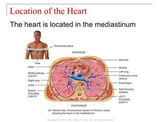
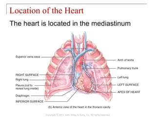
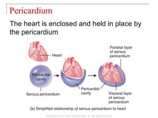
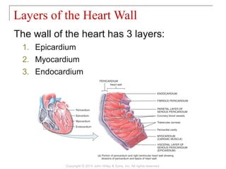
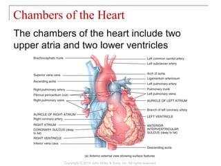
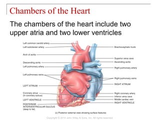
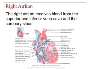
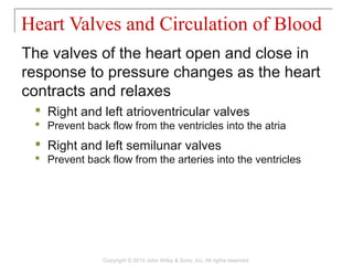
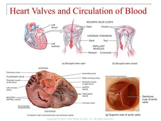
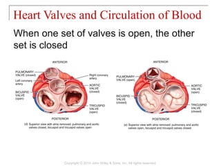
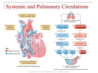
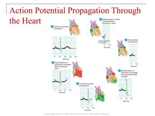
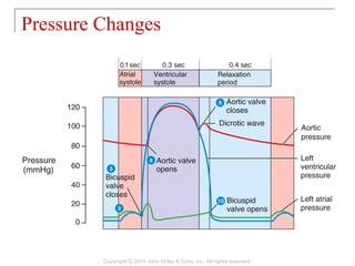

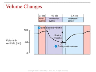
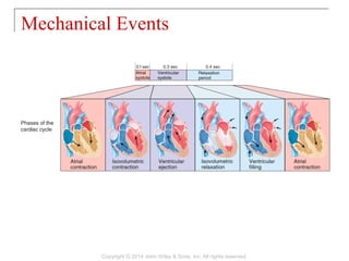
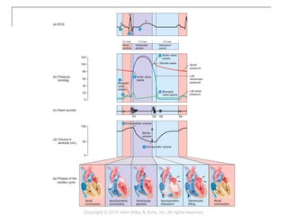
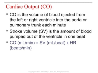
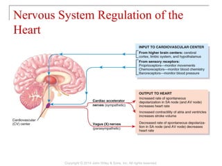
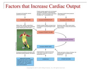
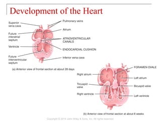


The document summarizes Chapter 20 from the textbook "Principles of Anatomy and Physiology". The chapter discusses the cardiovascular system with a focus on the anatomy and physiology of the heart. It describes the location of the heart in the mediastinum and its layers. It explains the cardiac cycle, conduction system, heart valves and chambers. Factors that influence heart rate and cardiac output are also summarized.
![19 [chapter 19 the cardiovascular system the blood]](https://cdn.slidesharecdn.com/ss_thumbnails/19chapter19thecardiovascularsystem-theblood-170828042033-thumbnail.jpg?width=640&height=640&fit=bounds)
![14 [chapter 14 the brain and cranial nerves]](https://cdn.slidesharecdn.com/ss_thumbnails/14chapter14thebrainandcranialnerves-170828133437-thumbnail.jpg?width=640&height=640&fit=bounds)
![10 [chapter 10 muscular tissue]](https://cdn.slidesharecdn.com/ss_thumbnails/10chapter10musculartissue-170828040153-thumbnail.jpg?width=640&height=640&fit=bounds)
![25 [chapter 25 metabolism and nutrition]](https://cdn.slidesharecdn.com/ss_thumbnails/25chapter25metabolismandnutrition-170828145139-thumbnail.jpg?width=640&height=640&fit=bounds)
![22 [chapter 22 the lymphatic system and immunity]](https://cdn.slidesharecdn.com/ss_thumbnails/22chapter22thelymphaticsystemandimmunity-170828153258-thumbnail.jpg?width=640&height=640&fit=bounds)
![06 [chapter 6 the skeletal system bone tissue]](https://cdn.slidesharecdn.com/ss_thumbnails/06chapter6theskeletalsystem-bonetissue-170828035633-thumbnail.jpg?width=640&height=640&fit=bounds)
![24 [chapter 24 the digestive system][11e]](https://cdn.slidesharecdn.com/ss_thumbnails/24chapter24thedigestivesystem11e-170828043714-thumbnail.jpg?width=640&height=640&fit=bounds)
![08 [chapter 8 the skeletal system appendicular skeleton]](https://cdn.slidesharecdn.com/ss_thumbnails/08chapter8theskeletalsystem-appendicularskeleton-170828041008-thumbnail.jpg?width=640&height=640&fit=bounds)
![23 [chapter 23 the respiratory system]](https://cdn.slidesharecdn.com/ss_thumbnails/23chapter23therespiratorysystem-170828043650-thumbnail.jpg?width=640&height=640&fit=bounds)
![11 [chapter 11 the muscular system]](https://cdn.slidesharecdn.com/ss_thumbnails/11chapter11themuscularsystem-170828041038-thumbnail.jpg?width=640&height=640&fit=bounds)
![26 [chapter 26 the urinary system]](https://cdn.slidesharecdn.com/ss_thumbnails/26chapter26theurinarysystem-170828044011-thumbnail.jpg?width=640&height=640&fit=bounds)
![05 [chapter 5 the integumentary system]](https://cdn.slidesharecdn.com/ss_thumbnails/05chapter5theintegumentarysystem-170828035624-thumbnail.jpg?width=640&height=640&fit=bounds)
![18 [chapter 18 the endocrine system]](https://cdn.slidesharecdn.com/ss_thumbnails/18chapter18theendocrinesystem-170828042016-thumbnail.jpg?width=640&height=640&fit=bounds)
![17 [chapter 17 the special senses]](https://cdn.slidesharecdn.com/ss_thumbnails/17chapter17thespecialsenses-170828041636-thumbnail.jpg?width=640&height=640&fit=bounds)
![28 [chapter 28 the reproductive system]](https://cdn.slidesharecdn.com/ss_thumbnails/28chapter28thereproductivesystem-170828134050-thumbnail.jpg?width=640&height=640&fit=bounds)
![01 [chapter 1 an introduction to the human body]](https://cdn.slidesharecdn.com/ss_thumbnails/01chapter1anintroductiontothehumanbody-170828035545-thumbnail.jpg?width=640&height=640&fit=bounds)
![13 [chapter 13 the spinal cord and spinal nerves]](https://cdn.slidesharecdn.com/ss_thumbnails/13chapter13thespinalcordandspinalnerves-170828040950-thumbnail.jpg?width=640&height=640&fit=bounds)
![03 [chapter 3 the cellular level of organization]](https://cdn.slidesharecdn.com/ss_thumbnails/03chapter3thecellularleveloforganization-170828035521-thumbnail.jpg?width=640&height=640&fit=bounds)
![07 [chapter 7 the skeletal system the axial skeleton]](https://cdn.slidesharecdn.com/ss_thumbnails/07chapter7theskeletalsystem-theaxialskeleton-170828035650-thumbnail.jpg?width=640&height=640&fit=bounds)
![15 [chapter 15 the autonomic nervous system]](https://cdn.slidesharecdn.com/ss_thumbnails/15chapter15theautonomicnervoussystem-170828041929-thumbnail.jpg?width=640&height=640&fit=bounds)
![12 [chapter 12 nervous tissue]](https://cdn.slidesharecdn.com/ss_thumbnails/12chapter12nervoustissue-170828041102-thumbnail.jpg?width=640&height=640&fit=bounds)
![02 [chapter 2 the chemical level of organization]](https://cdn.slidesharecdn.com/ss_thumbnails/02chapter2thechemicalleveloforganization-170828035601-thumbnail.jpg?width=640&height=640&fit=bounds)
![09 [chapter 9 joints]](https://cdn.slidesharecdn.com/ss_thumbnails/09chapter9joints-170828041032-thumbnail.jpg?width=640&height=640&fit=bounds)

![04 [chapter 4 the tissue level of organization][11e]](https://cdn.slidesharecdn.com/ss_thumbnails/04chapter4thetissueleveloforganization11e-170828035609-thumbnail.jpg?width=640&height=640&fit=bounds)
![11 [chapter 11 the muscular system][11e]](https://cdn.slidesharecdn.com/ss_thumbnails/11chapter11themuscularsystem11e-170828040427-thumbnail.jpg?width=640&height=640&fit=bounds)


