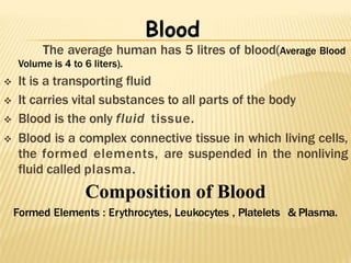
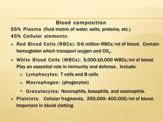
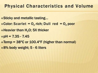
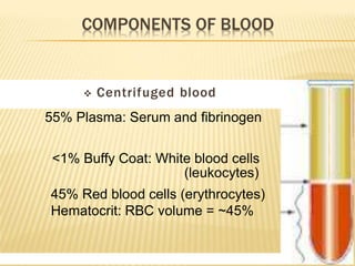
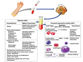
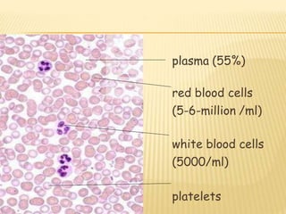
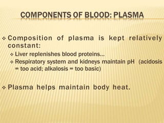
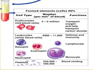
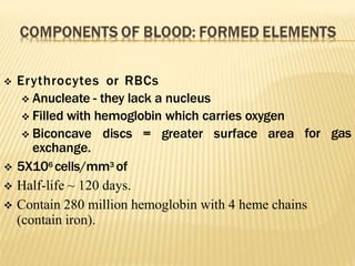
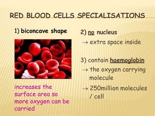
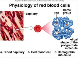
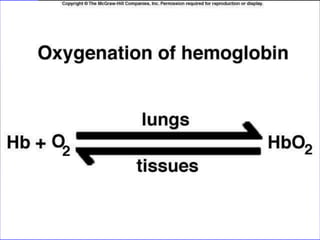
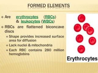
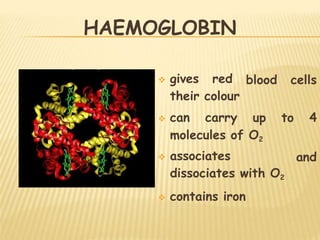
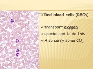
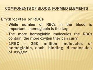
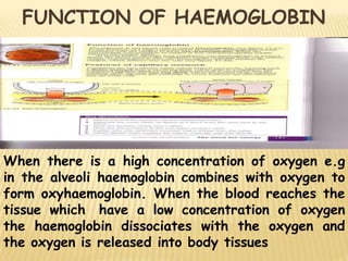
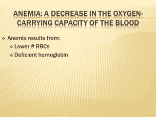
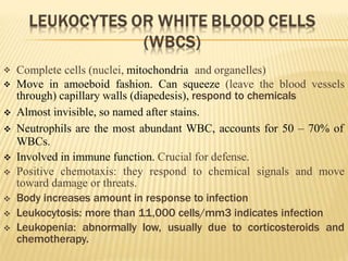
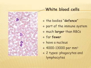
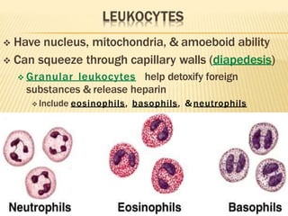
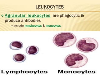
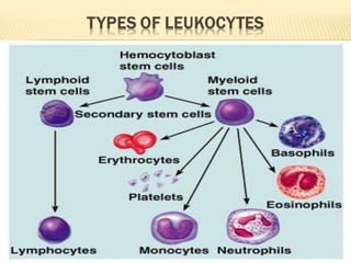
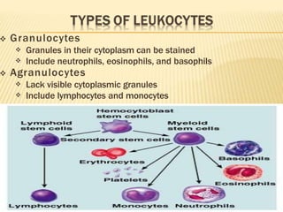
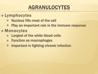
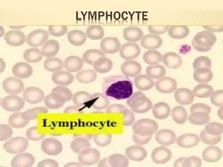
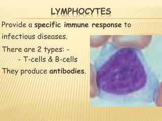
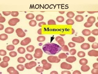
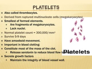
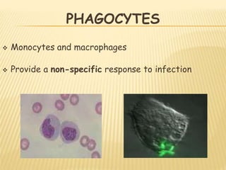
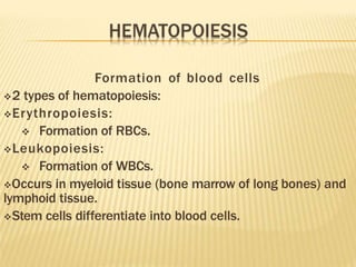
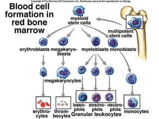
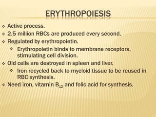
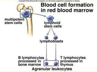
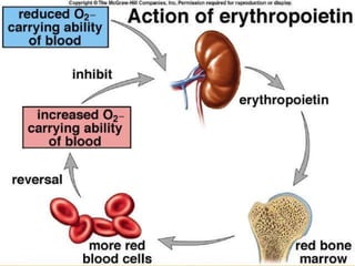
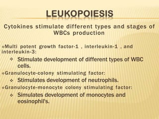
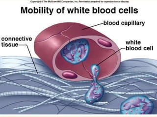
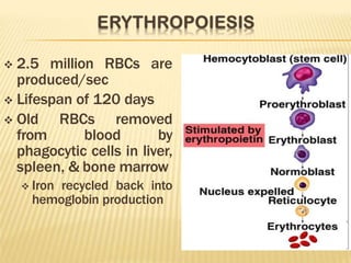
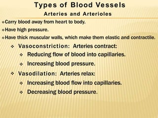
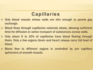
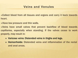
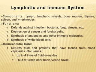
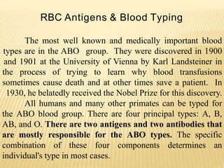
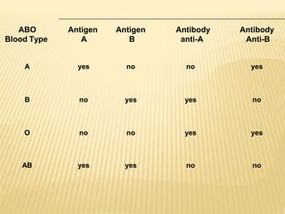
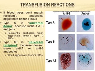
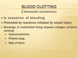
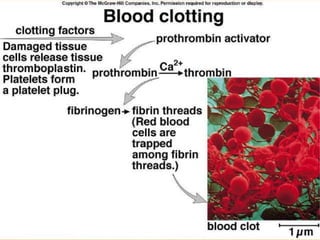
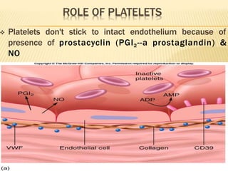
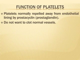
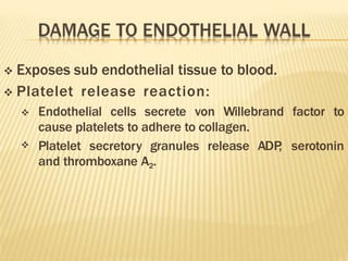
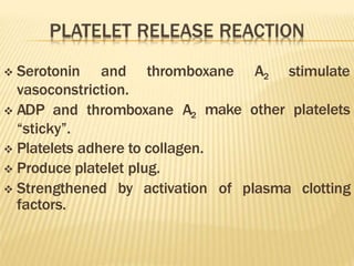
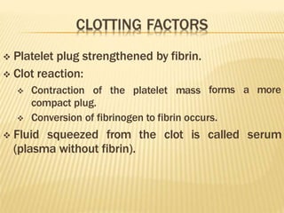
The average human has 5 litres of blood(Average Blood
Volume is 4 to 6 liters).
 It is a transporting fluid
 It carries vital substances to all parts of the body
 Blood is the only fluid tissue.
 Blood is a complex connective tissue in which living cells,
the formed elements, are suspended in the nonliving
fluid called plasma.
Composition of Blood
Formed Elements : Erythrocytes, Leukocytes , Platelets & Plasma.
Blood
Blood composition
55% Plasma (fluid matrix of water, salts, proteins, etc.)
45% Cellular elements:

 Red Blood Cells (RBCs): 5-6 million RBCs/ml of blood. Contain
hemoglobin which transport oxygen and CO2.
White Blood Cells (WBCs): 5,000-10,000 WBCs/ml of blood.
Play an essential role in immunity and defense. Include:


 Lymphocytes: T cells and B cells
Macrophages: (phagocytes)
Granulocytes: Neutrophils, basophils, and eosinophils.
 Platelets: Cellular fragments, 250,000- 400,000/ml of blood.
Important in blood clotting.
HUMAN BLOOD SMEAR
Physical Characteristics and Volume
Sticky and metallic tasting…
Color: Scarlet = O2 rich; Dull red = O2 poor
Heavier than H2O; 5X thicker
pH = 7.35 - 7.45
Temp = 38ºC or 100.4ºF (higher than normal)
8% body weight; 5 - 6 liters
 Centrifuged blood
55% Plasma: Serum and fibrinogen
<1% Buffy Coat: White blood cells
(leukocytes)
45% Red blood cells (erythrocytes)
Hematocrit: RBC volume = ~45%
plasma (55%)
red blood cells
(5-6-million /ml)
white blood cells
(5000/ml)
platelets
Plasma
Straw-colored liquid.
Consists of H20 and dissolved
solutes.
Ions, metabolites, hormones,
antibodies.
Na+ is the major solute of the
plasma.
liquid part of blood
Plasma transports
soluble food molecules
waste products
hormones
antibodies
 90% of plasma is water:
 >100 other substances in plasma: salts (electrolytes),
nutrients, gases, hormones, plasma proteins, various
wastes and products of cell metabolism.
 Plasma proteins:
 Constitute 7-9% of plasma.
 Provide the colloid osmotic pressure needed to draw H20
from interstitial fluid to capillaries.




Maintain blood pressure.
Albumin:. Maintains osmotic pressure of blood.
Clotting proteins
Antibodies


Constitute 7-9% of plasma
Three types of plasma proteins: albumins, globulins, & fibrinogen

 Albumin accounts for 60-80, plasma protein made by the liver,
Creates colloid osmotic pressure that draws H20 from interstitial
fluid into capillaries to maintain blood volume & pressure
Globulins carry lipids
  globulin: Transport lipids and fat soluble vitamins.
  globulin: Transport lipids and fat soluble vitamins.
  globulin:Antibodies that function in immunity.

Gamma globulins are antibodies
Fibrinogen Constitutes 4% of plasma proteins. Important clotting
factor. Converted into fibrin during the clotting process.
Serum is fluid left when
blood clots
 Composition of plasma is kept relatively
constant:
 Liver replenishes blood proteins…
 Respiratory system and kidneys maintain pH (acidosis
= too acid; alkalosis = too basic)
 Plasma helps maintain body heat.
for gas
 Erythrocytes or RBCs
 Anucleate - they lack a nucleus
 Filled with hemoglobin which carries oxygen
 Biconcave discs = greater surface area
exchange.
 5X10⁶ cells/mm3 of
 Half-life ~ 120 days.
 Contain 280 million hemoglobin with 4 heme chains
(contain iron).
RED BLOOD CELLS SPECIALISATIONS
2) no nucleus
 extra space inside
3) contain haemoglobin
 the oxygen carrying
molecule
 250million molecules
/ cell
1) biconcave shape
increases the
surface area so
more oxygen can be
carried
 Are erythrocytes (RBCs)
& leukocytes (WBCs)
 RBCs are flattened biconcave
discs
 Shape provides increased surface
area for diffusion
 Lack nuclei & mitochondria
 Each RBC contains 280 million
hemoglobins
HAEMOGLOBIN
blood cells
 gives red
their colour
 can carry up to 4
molecules of O2
and
 associates
dissociates with O2
 contains iron
 Red blood cells (RBCs)
 transport oxygen
 specialised to do this
 Also carry some CO2
Erythrocytes or RBCs
While number of RBCs in the blood is
important….hemoglobin is the key.
The more hemoglobin molecules the RBCs
contain, the more oxygen they can carry.
1RBC - 250 million molecules of
hemoglobin, each binding 4 molecules
of oxygen.
When there is a high concentration of oxygen e.g
in the alveoli haemoglobin combines with oxygen to
form oxyhaemoglobin. When the blood reaches the
tissue which have a low concentration of oxygen
the haemoglobin dissociates with the oxygen and
the oxygen is released into body tissues
FUNCTION OF HAEMOGLOBIN
 Anemia results from:
 Lower # RBCs
 Deficient hemoglobin
 Hemorrhagic anemia: sudden hemorrhage
 Hemolytic anemia: lysis of RBCs from bact. infection
 Pernicious anemia: lack of B12
 Aplastic anemia: depression/destruction of bone
marrow by cancer, radiation, meds.
 Sickle Cell Anemia:
 Deficient hemoglobin - a point mutation changes a
single amino acid.
 Polycythemia - abnormal increase in erythrocytes,
caused by:
 Bone marrow cancer
 High altitudes
 Thickens and slows blood, impairs
circulation


Complete cells (nuclei, mitochondria and organelles)
Move in amoeboid fashion. Can squeeze (leave the blood vessels
through) capillary walls (diapedesis), respond to chemicals
 Almost invisible, so named after stains.
 Neutrophils are the most abundant WBC, accounts for 50 – 70% of
WBCs.
 Involved in immune function. Crucial for defense.
 Positive chemotaxis: they respond to chemical signals and move
toward damage or threats.
 Body increases amount in response to infection
 Leukocytosis: more than 11,000 cells/mm3 indicates infection
 Leukopenia: abnormally low, usually due to corticosteroids and
chemotherapy.
White blood cells
 the bodies “defence”
 part of the immune system
 much larger than RBCs
 far fewer
 have a nucleus
 4000-13000 per mm3
 2 types: phagocytes and
lymphocytes
 Have nucleus, mitochondria, & amoeboid ability
 Can squeeze through capillary walls (diapedesis)
 Granular leukocytes help detoxify foreign
substances & release heparin
 Include eosinophils, basophils, & neutrophils
 Agranular leukocytes are phagocytic &
produce antibodies
 Include lymphocytes & monocytes
 Granulocytes


Granules in their cytoplasm can be stained
Include neutrophils, eosinophils, and basophils
 Agranulocytes


Lack visible cytoplasmic granules
Include lymphocytes and monocytes
 Neutrophils


Multi-lobed nucleus with fine granules
Act as phagocytes at active sites of infection
 Eosinophils


Large brick-red cytoplasmic granules
Found in response to allergies and parasitic worms
 Basophils


Have histamine-containing granules
Initiate inflammation
 Lymphocytes


Nucleus fills most of the cell
Play an important role in the immune response
 Monocytes



Largest of the white blood cells
Function as macrophages
Important in fighting chronic infection
Provide a specific immune response to
infectious diseases.
There are 2 types: -
- T-cells & B-cells
They produce antibodies.
MONOCYTES
 Also called thrombocytes.
 Derived from ruptured multinucleate cells (megakaryocytes)
 Smallest of formed elements.


Are fragments of megakaryocytes.
Lack nuclei.


Normal platelet count = 300,000/mm3
Survive 5-9 days
 Have amoeboid movement.
 Important in blood clotting:
 Constitute most of the mass of the clot.
 Release serotonin to reduce blood flow to area.
 Secrete growth factors
 Maintain the integrity of blood vessel wall.
PHAGOCYTES
 Monocytes and macrophages
 Provide a non-specific response to infection
HEMATOPOIESIS
 Is formation of blood cells from stem cells in
marrow (myeloid tissue) & lymphoid tissue
 Erythropoiesis is formation of RBCs
 Stimulated by erythropoietin (EPO) from kidney
 Leukopoiesis is formation of WBCs
 Stimulated by variety of cytokines
 = autocrine regulators secreted by immune system
Formation of blood cells
2 types of hematopoiesis:
Erythropoiesis:
 Formation of RBCs.
Leukopoiesis:
 Formation of WBCs.
Occurs in myeloid tissue (bone marrow of long bones) and
lymphoid tissue.
Stem cells differentiate into blood cells.
 Active process.
 2.5 million RBCs are produced every second.
 Regulated by erythropoietin.
 Erythropoietin binds to membrane receptors,
stimulating cell division.
 Old cells are destroyed in spleen and liver.
 Iron recycled back to myeloid tissue to be reused in
RBC synthesis.
 Need iron, vitamin B12 and folic acid for synthesis.
Cytokines stimulate different types and stages of
WBCs production
Multi potent growth factor-1 , interleukin-1 , and
interleukin-3:
 Stimulate development of different types of WBC
cells.
Granulocyte-colony stimulating factor:
 Stimulates development of neutrophils.
Granulocyte-monocyte colony stimulating factor:
 Simulates development of monocytes and
eosinophil's.
 2.5 million RBCs are
produced/sec
 Lifespan of 120 days
 Old RBCs removed
from blood by
phagocytic cells in liver,
spleen, & bone marrow
 Iron recycled back into
hemoglobin production
Types of Blood Vessels
Arteries and Arterioles
Carry blood away from heart to body.
Have high pressure.
Have thick muscular walls, which make them elastic and contractile.
 Vasoconstriction: Arteries contract:

 Reducing flow of blood into capillaries.
Increasing blood pressure.
 Vasodilation: Arteries relax:
 Increasing blood flow into capillaries.
 Decreasing blood pressure.
Capillaries




Only blood vessels whose walls are thin enough to permit gas
exchange.
Blood flows through capillaries relatively slowly, allowing sufficient
time for diffusion or active transport of substances across walls.
Only about 5 to 10% of capillaries have blood flowing through
them. Only a few organs (brain and heart) always carry full load of
blood.
Blood flow to different organs is controlled by pre capillary
sphincters of smooth muscle.
Veins and Venules
Collect blood from all tissues and organs and carry it back towards
heart.
Have low pressure and thin walls.
Veins have small valves that prevent backflow of blood towards
capillaries, especially when standing. If the valves cease to work
properly, may result in:

 Varicose veins: Distended veins in thighs and legs.
Hemorrhoids: Distended veins and inflammation of the rectal
and anal areas.
Lymphatic and Immune System
Components: Lymph, lymphatic vessels, bone marrow, thymus,
spleen, and lymph nodes.
Functions:




Defends against infection: bacteria, fungi, viruses, etc.
Destruction of cancer and foreign cells.
Synthesis of antibodies and other immune molecules.
Synthesis of white blood cells.
Homeostatic Role:
 Returns fluid and proteins that have leaked from blood
capillaries into tissues.


Up to 4 liters of fluid every day.
Fluid returned near heart/venae cavae.
RBC Antigens & Blood Typing
The most well known and medically important blood
types are in the ABO group. They were discovered in 1900
and 1901 at the University of Vienna by Karl Landsteiner in
the process of trying to learn why blood transfusions
sometimes cause death and at other times save a patient. In
1930, he belatedly received the Nobel Prize for this discovery.
All humans and many other primates can be typed for
the ABO blood group. There are four principal types: A, B,
AB, and O. There are two antigens and two antibodies that
are mostly responsible for the ABO types. The specific
combination of these four components determines an
individual's type in most cases.
ABO
Blood Type
Antigen
A
Antigen
B
Antibody
anti-A
Antibody
Anti-B
A yes no no yes
B no yes yes no
O no no yes yes
AB yes yes no no
People with type A blood will have the A antigen on the surface of their
red cells . As a result, anti-A antibodies will not be produced by them
because they would cause the destruction of their own blood. However,
if B type blood is injected into their systems, anti-B antibodies in their
plasma will recognize it as alien and burst or agglutinate the introduced
red cells in order to cleanse the blood of alien protein.
Individuals with type O blood do not produce ABO antigens.
Therefore, their blood normally will not be rejected when it is given to
others with different ABO types. As a result, type O people are
universal donors for transfusions, but they can receive only type O
blood themselves.
ABO antibodies.
other ABO type.
Those who have type AB blood do not make any
Their blood does not discriminate against any
Consequently, they are universal receivers for
transfusions, but their blood will be agglutinated when given to people
with every other type because they produce both kinds of antigens.
It is easy and inexpensive to determine an individual's ABO
type from a few drops of blood. A serum containing anti-A
antibodies is mixed with some of the blood. Another serum with
anti-B antibodies is mixed with the remaining sample. Whether or
not agglutination occurs in either sample indicates the ABO type. It
is a simple process of elimination of the possibilities. For instance, if
an individual's blood sample is agglutinated by the anti-A antibody,
but not the anti-B antibody, it means that the A antigen is present
but not the B antigen. Therefore, the blood type is A.
 ABO system:
 Major group of antigens of RBCs.
 Type A:
 Only A antigens present.
 Type B:
 Only B antigens present.
 Type AB:
 Both A and B antigens present.
 Type O:
 Neither A or B antigens present.
 Each person inherits 2 genes that control the
production of ABO groups.
 Type A:


May have inherited A gene from each parent.
May have inherited A gene from 1 parent and O gene
from the other.
 Type B:


May have inherited B gene from each parent.
May have inherited B gene from 1 parent and O gene
from the other parent.
 Type AB:
 Inherited the A gene from one parent and the B gene
from the other parent.
 Type O:
 Inherited O gene from each parent.
People with blood group 0 Rh - are called "universal donors" and
people with blood group AB Rh+ are called "universal receivers."
Rh+ blood can never be given to someone with Rh - blood, but the
other way around works. For example, 0 Rh+ blood can not be given
to someone with the blood type AB Rh -.
 People with Type A blood
make antibodies to Type B
RBCs, but not to Type A




Type B blood has antibodies to
Type A RBCs but not to Type B
Type AB blood doesn’t have
antibodies to A or B
Type O has antibodies to both
Type A & B
If different blood types are
mixed, antibodies will cause
mixture to agglutinate
 If blood types do not match, the recipient’s antibodies
attach to donor’s RBCs and agglutinate.
 Type O


Universal donor.
Recipient’s antibodies cannot agglutinate the donor’s
RBCs.
 Type AB
Universal recipient


Lack the anti-A and anti-B antibodies.
Cannot agglutinate donor’s RBCs.
 Another group of antigens found on RBCs.
 Rh positive:
 Have these antigens.
 Rh negative:
 Do not have these antigens.
 Significant when Rh negative mother give birth
to Rh positive baby.

 At birth, mother may become exposed to Rh positive
blood of fetus.
Mother at subsequent pregnancies may produce
antibodies against the Rh factor.
 If blood types don't match,
recipient’s antibodies
agglutinate donor’s RBCs
 Type O is “universal
donor” because lacks A & B
antigens
antibodies won’t
donor’s Type O
 Recipient’s
agglutinate
RBCs
 Type AB is “universal
recipient” because doesn’t
make anti-A or anti-B
antibodies
 Won’t agglutinate donor’s RBCs
🞭 Insert fig. 13.6
 Is cessation of bleeding
 Promoted by reactions initiated by vessel injury
 Breakage of endothelial lining exposes collagen proteins
causing:



Vasoconstriction.
Platelet plug.
Web of fibrin.
 Platelets don't stick to intact endothelium because of
presence of prostacyclin (PGI2--a prostaglandin) &
NO
 Keep clots from forming & are vasodilators
 Damage to endothelium
exposed collagen
allows platelets to bind to
increases bond by binding to both
 von Willebrand factor
collagen & platelets
 Platelets stick to collagen & release ADP, serotonin, &
thromboxane A2
 = platelet release reaction
 Serotonin & thromboxane A2 stimulate
vasoconstriction, reducing blood flow to wound
thromboxane A2 cause other platelets to
sticky & attach & undergo platelet release
 ADP &
become
reaction
 This continues until platelet plug is formed
 Platelets normally repelled away from endothelial
lining by prostacyclin (prostaglandin).
 Do not want to clot normal vessels.
 Exposes sub endothelial tissue to blood.
 Platelet release reaction:

 Endothelial cells secrete von Willebrand factor to
cause platelets to adhere to collagen.
Platelet secretory granules release ADP
, serotonin
and thromboxane A2.
 Serotonin and thromboxane A2 stimulate
make other platelets
vasoconstriction.
 ADP and thromboxane A2
“sticky”.
of plasma clotting
 Platelets adhere to collagen.
 Produce platelet plug.
 Strengthened by activation
factors.
forms a more
 Platelet plug strengthened by fibrin.
 Clot reaction:
 Contraction of the platelet mass
compact plug.
 Conversion of fibrinogen to fibrin occurs.
 Fluid squeezed from the clot is called serum
(plasma without fibrin).
Intrinsic Pathway





Initiated by exposure of blood to a negatively charged
surface (collagen).
This activates Factor XII (protease), which activates
other clotting factors.
Ca++ and phospholipids convert prothrombin to
thrombin.
Thrombin converts fibrinogen to fibrin.
Produces meshwork of insoluble fibrin polymers.
 Thromboplastin is not a part of the blood, so
called extrinsic pathway.
 Damaged tissue release a thromboplastin.
 Thromboplastin initiates a short cut to formation
of fibrin.
 Activated factor XII converts an inactive molecule
into the active form (kallikrein).
 Kallikrein converts plasminogen to plasmin.
 Plasmin is an enzyme that digests the fibrin.
 Clot dissolution occurs.
 When damage is repaired, activated factor XII
causes activation of kallikrein
 Kallikrein converts plasminogen to plasmin
 Plasmin digests fibrin, dissolving clot
 Clotting can be prevented by Ca+2 chelators (e.g.
sodium citrate or EDTA)
 or heparin which activates antithrombin III
(blocks thrombin)
 Coumarin blocks clotting by inhibiting
activation of Vit K
 Vit K works indirectly by reducing Ca+2 availability

blooditsfunctions-copy-110325121157-phpapp02.pptx

  • 2.
    The average humanhas 5 litres of blood(Average Blood Volume is 4 to 6 liters).  It is a transporting fluid  It carries vital substances to all parts of the body  Blood is the only fluid tissue.  Blood is a complex connective tissue in which living cells, the formed elements, are suspended in the nonliving fluid called plasma. Composition of Blood Formed Elements : Erythrocytes, Leukocytes , Platelets & Plasma. Blood
  • 3.
    Blood composition 55% Plasma(fluid matrix of water, salts, proteins, etc.) 45% Cellular elements:   Red Blood Cells (RBCs): 5-6 million RBCs/ml of blood. Contain hemoglobin which transport oxygen and CO2. White Blood Cells (WBCs): 5,000-10,000 WBCs/ml of blood. Play an essential role in immunity and defense. Include:    Lymphocytes: T cells and B cells Macrophages: (phagocytes) Granulocytes: Neutrophils, basophils, and eosinophils.  Platelets: Cellular fragments, 250,000- 400,000/ml of blood. Important in blood clotting.
  • 4.
  • 5.
    Physical Characteristics andVolume Sticky and metallic tasting… Color: Scarlet = O2 rich; Dull red = O2 poor Heavier than H2O; 5X thicker pH = 7.35 - 7.45 Temp = 38ºC or 100.4ºF (higher than normal) 8% body weight; 5 - 6 liters
  • 6.
     Centrifuged blood 55%Plasma: Serum and fibrinogen <1% Buffy Coat: White blood cells (leukocytes) 45% Red blood cells (erythrocytes) Hematocrit: RBC volume = ~45%
  • 8.
    plasma (55%) red bloodcells (5-6-million /ml) white blood cells (5000/ml) platelets
  • 9.
    Plasma Straw-colored liquid. Consists ofH20 and dissolved solutes. Ions, metabolites, hormones, antibodies. Na+ is the major solute of the plasma. liquid part of blood Plasma transports soluble food molecules waste products hormones antibodies
  • 10.
     90% ofplasma is water:  >100 other substances in plasma: salts (electrolytes), nutrients, gases, hormones, plasma proteins, various wastes and products of cell metabolism.  Plasma proteins:  Constitute 7-9% of plasma.  Provide the colloid osmotic pressure needed to draw H20 from interstitial fluid to capillaries.     Maintain blood pressure. Albumin:. Maintains osmotic pressure of blood. Clotting proteins Antibodies
  • 11.
      Constitute 7-9% ofplasma Three types of plasma proteins: albumins, globulins, & fibrinogen   Albumin accounts for 60-80, plasma protein made by the liver, Creates colloid osmotic pressure that draws H20 from interstitial fluid into capillaries to maintain blood volume & pressure Globulins carry lipids   globulin: Transport lipids and fat soluble vitamins.   globulin: Transport lipids and fat soluble vitamins.   globulin:Antibodies that function in immunity.  Gamma globulins are antibodies Fibrinogen Constitutes 4% of plasma proteins. Important clotting factor. Converted into fibrin during the clotting process. Serum is fluid left when blood clots
  • 13.
     Composition ofplasma is kept relatively constant:  Liver replenishes blood proteins…  Respiratory system and kidneys maintain pH (acidosis = too acid; alkalosis = too basic)  Plasma helps maintain body heat.
  • 15.
    for gas  Erythrocytesor RBCs  Anucleate - they lack a nucleus  Filled with hemoglobin which carries oxygen  Biconcave discs = greater surface area exchange.  5X10⁶ cells/mm3 of  Half-life ~ 120 days.  Contain 280 million hemoglobin with 4 heme chains (contain iron).
  • 16.
    RED BLOOD CELLSSPECIALISATIONS 2) no nucleus  extra space inside 3) contain haemoglobin  the oxygen carrying molecule  250million molecules / cell 1) biconcave shape increases the surface area so more oxygen can be carried
  • 19.
     Are erythrocytes(RBCs) & leukocytes (WBCs)  RBCs are flattened biconcave discs  Shape provides increased surface area for diffusion  Lack nuclei & mitochondria  Each RBC contains 280 million hemoglobins
  • 20.
    HAEMOGLOBIN blood cells  givesred their colour  can carry up to 4 molecules of O2 and  associates dissociates with O2  contains iron
  • 21.
     Red bloodcells (RBCs)  transport oxygen  specialised to do this  Also carry some CO2
  • 22.
    Erythrocytes or RBCs Whilenumber of RBCs in the blood is important….hemoglobin is the key. The more hemoglobin molecules the RBCs contain, the more oxygen they can carry. 1RBC - 250 million molecules of hemoglobin, each binding 4 molecules of oxygen.
  • 23.
    When there isa high concentration of oxygen e.g in the alveoli haemoglobin combines with oxygen to form oxyhaemoglobin. When the blood reaches the tissue which have a low concentration of oxygen the haemoglobin dissociates with the oxygen and the oxygen is released into body tissues FUNCTION OF HAEMOGLOBIN
  • 24.
     Anemia resultsfrom:  Lower # RBCs  Deficient hemoglobin
  • 25.
     Hemorrhagic anemia:sudden hemorrhage  Hemolytic anemia: lysis of RBCs from bact. infection  Pernicious anemia: lack of B12  Aplastic anemia: depression/destruction of bone marrow by cancer, radiation, meds.
  • 26.
     Sickle CellAnemia:  Deficient hemoglobin - a point mutation changes a single amino acid.
  • 27.
     Polycythemia -abnormal increase in erythrocytes, caused by:  Bone marrow cancer  High altitudes  Thickens and slows blood, impairs circulation
  • 28.
      Complete cells (nuclei,mitochondria and organelles) Move in amoeboid fashion. Can squeeze (leave the blood vessels through) capillary walls (diapedesis), respond to chemicals  Almost invisible, so named after stains.  Neutrophils are the most abundant WBC, accounts for 50 – 70% of WBCs.  Involved in immune function. Crucial for defense.  Positive chemotaxis: they respond to chemical signals and move toward damage or threats.  Body increases amount in response to infection  Leukocytosis: more than 11,000 cells/mm3 indicates infection  Leukopenia: abnormally low, usually due to corticosteroids and chemotherapy.
  • 29.
    White blood cells the bodies “defence”  part of the immune system  much larger than RBCs  far fewer  have a nucleus  4000-13000 per mm3  2 types: phagocytes and lymphocytes
  • 30.
     Have nucleus,mitochondria, & amoeboid ability  Can squeeze through capillary walls (diapedesis)  Granular leukocytes help detoxify foreign substances & release heparin  Include eosinophils, basophils, & neutrophils
  • 31.
     Agranular leukocytesare phagocytic & produce antibodies  Include lymphocytes & monocytes
  • 33.
     Granulocytes   Granules intheir cytoplasm can be stained Include neutrophils, eosinophils, and basophils  Agranulocytes   Lack visible cytoplasmic granules Include lymphocytes and monocytes
  • 34.
     Neutrophils   Multi-lobed nucleuswith fine granules Act as phagocytes at active sites of infection  Eosinophils   Large brick-red cytoplasmic granules Found in response to allergies and parasitic worms  Basophils   Have histamine-containing granules Initiate inflammation
  • 35.
     Lymphocytes   Nucleus fillsmost of the cell Play an important role in the immune response  Monocytes    Largest of the white blood cells Function as macrophages Important in fighting chronic infection
  • 37.
    Provide a specificimmune response to infectious diseases. There are 2 types: - - T-cells & B-cells They produce antibodies.
  • 38.
  • 39.
     Also calledthrombocytes.  Derived from ruptured multinucleate cells (megakaryocytes)  Smallest of formed elements.   Are fragments of megakaryocytes. Lack nuclei.   Normal platelet count = 300,000/mm3 Survive 5-9 days  Have amoeboid movement.  Important in blood clotting:  Constitute most of the mass of the clot.  Release serotonin to reduce blood flow to area.  Secrete growth factors  Maintain the integrity of blood vessel wall.
  • 40.
    PHAGOCYTES  Monocytes andmacrophages  Provide a non-specific response to infection
  • 41.
    HEMATOPOIESIS  Is formationof blood cells from stem cells in marrow (myeloid tissue) & lymphoid tissue  Erythropoiesis is formation of RBCs  Stimulated by erythropoietin (EPO) from kidney  Leukopoiesis is formation of WBCs  Stimulated by variety of cytokines  = autocrine regulators secreted by immune system
  • 42.
    Formation of bloodcells 2 types of hematopoiesis: Erythropoiesis:  Formation of RBCs. Leukopoiesis:  Formation of WBCs. Occurs in myeloid tissue (bone marrow of long bones) and lymphoid tissue. Stem cells differentiate into blood cells.
  • 44.
     Active process. 2.5 million RBCs are produced every second.  Regulated by erythropoietin.  Erythropoietin binds to membrane receptors, stimulating cell division.  Old cells are destroyed in spleen and liver.  Iron recycled back to myeloid tissue to be reused in RBC synthesis.  Need iron, vitamin B12 and folic acid for synthesis.
  • 47.
    Cytokines stimulate differenttypes and stages of WBCs production Multi potent growth factor-1 , interleukin-1 , and interleukin-3:  Stimulate development of different types of WBC cells. Granulocyte-colony stimulating factor:  Stimulates development of neutrophils. Granulocyte-monocyte colony stimulating factor:  Simulates development of monocytes and eosinophil's.
  • 49.
     2.5 millionRBCs are produced/sec  Lifespan of 120 days  Old RBCs removed from blood by phagocytic cells in liver, spleen, & bone marrow  Iron recycled back into hemoglobin production
  • 50.
    Types of BloodVessels Arteries and Arterioles Carry blood away from heart to body. Have high pressure. Have thick muscular walls, which make them elastic and contractile.  Vasoconstriction: Arteries contract:   Reducing flow of blood into capillaries. Increasing blood pressure.  Vasodilation: Arteries relax:  Increasing blood flow into capillaries.  Decreasing blood pressure.
  • 51.
    Capillaries     Only blood vesselswhose walls are thin enough to permit gas exchange. Blood flows through capillaries relatively slowly, allowing sufficient time for diffusion or active transport of substances across walls. Only about 5 to 10% of capillaries have blood flowing through them. Only a few organs (brain and heart) always carry full load of blood. Blood flow to different organs is controlled by pre capillary sphincters of smooth muscle.
  • 52.
    Veins and Venules Collectblood from all tissues and organs and carry it back towards heart. Have low pressure and thin walls. Veins have small valves that prevent backflow of blood towards capillaries, especially when standing. If the valves cease to work properly, may result in:   Varicose veins: Distended veins in thighs and legs. Hemorrhoids: Distended veins and inflammation of the rectal and anal areas.
  • 54.
    Lymphatic and ImmuneSystem Components: Lymph, lymphatic vessels, bone marrow, thymus, spleen, and lymph nodes. Functions:     Defends against infection: bacteria, fungi, viruses, etc. Destruction of cancer and foreign cells. Synthesis of antibodies and other immune molecules. Synthesis of white blood cells. Homeostatic Role:  Returns fluid and proteins that have leaked from blood capillaries into tissues.   Up to 4 liters of fluid every day. Fluid returned near heart/venae cavae.
  • 56.
    RBC Antigens &Blood Typing The most well known and medically important blood types are in the ABO group. They were discovered in 1900 and 1901 at the University of Vienna by Karl Landsteiner in the process of trying to learn why blood transfusions sometimes cause death and at other times save a patient. In 1930, he belatedly received the Nobel Prize for this discovery. All humans and many other primates can be typed for the ABO blood group. There are four principal types: A, B, AB, and O. There are two antigens and two antibodies that are mostly responsible for the ABO types. The specific combination of these four components determines an individual's type in most cases.
  • 57.
    ABO Blood Type Antigen A Antigen B Antibody anti-A Antibody Anti-B A yesno no yes B no yes yes no O no no yes yes AB yes yes no no
  • 58.
    People with typeA blood will have the A antigen on the surface of their red cells . As a result, anti-A antibodies will not be produced by them because they would cause the destruction of their own blood. However, if B type blood is injected into their systems, anti-B antibodies in their plasma will recognize it as alien and burst or agglutinate the introduced red cells in order to cleanse the blood of alien protein.
  • 59.
    Individuals with typeO blood do not produce ABO antigens. Therefore, their blood normally will not be rejected when it is given to others with different ABO types. As a result, type O people are universal donors for transfusions, but they can receive only type O blood themselves. ABO antibodies. other ABO type. Those who have type AB blood do not make any Their blood does not discriminate against any Consequently, they are universal receivers for transfusions, but their blood will be agglutinated when given to people with every other type because they produce both kinds of antigens. It is easy and inexpensive to determine an individual's ABO type from a few drops of blood. A serum containing anti-A antibodies is mixed with some of the blood. Another serum with anti-B antibodies is mixed with the remaining sample. Whether or not agglutination occurs in either sample indicates the ABO type. It is a simple process of elimination of the possibilities. For instance, if an individual's blood sample is agglutinated by the anti-A antibody, but not the anti-B antibody, it means that the A antigen is present but not the B antigen. Therefore, the blood type is A.
  • 61.
     ABO system: Major group of antigens of RBCs.  Type A:  Only A antigens present.  Type B:  Only B antigens present.  Type AB:  Both A and B antigens present.  Type O:  Neither A or B antigens present.
  • 62.
     Each personinherits 2 genes that control the production of ABO groups.  Type A:   May have inherited A gene from each parent. May have inherited A gene from 1 parent and O gene from the other.
  • 63.
     Type B:   Mayhave inherited B gene from each parent. May have inherited B gene from 1 parent and O gene from the other parent.  Type AB:  Inherited the A gene from one parent and the B gene from the other parent.  Type O:  Inherited O gene from each parent.
  • 64.
    People with bloodgroup 0 Rh - are called "universal donors" and people with blood group AB Rh+ are called "universal receivers." Rh+ blood can never be given to someone with Rh - blood, but the other way around works. For example, 0 Rh+ blood can not be given to someone with the blood type AB Rh -.
  • 66.
     People withType A blood make antibodies to Type B RBCs, but not to Type A     Type B blood has antibodies to Type A RBCs but not to Type B Type AB blood doesn’t have antibodies to A or B Type O has antibodies to both Type A & B If different blood types are mixed, antibodies will cause mixture to agglutinate
  • 67.
     If bloodtypes do not match, the recipient’s antibodies attach to donor’s RBCs and agglutinate.  Type O   Universal donor. Recipient’s antibodies cannot agglutinate the donor’s RBCs.  Type AB Universal recipient   Lack the anti-A and anti-B antibodies. Cannot agglutinate donor’s RBCs.
  • 68.
     Another groupof antigens found on RBCs.  Rh positive:  Have these antigens.  Rh negative:  Do not have these antigens.  Significant when Rh negative mother give birth to Rh positive baby.   At birth, mother may become exposed to Rh positive blood of fetus. Mother at subsequent pregnancies may produce antibodies against the Rh factor.
  • 70.
     If bloodtypes don't match, recipient’s antibodies agglutinate donor’s RBCs  Type O is “universal donor” because lacks A & B antigens antibodies won’t donor’s Type O  Recipient’s agglutinate RBCs  Type AB is “universal recipient” because doesn’t make anti-A or anti-B antibodies  Won’t agglutinate donor’s RBCs 🞭 Insert fig. 13.6
  • 71.
     Is cessationof bleeding  Promoted by reactions initiated by vessel injury  Breakage of endothelial lining exposes collagen proteins causing:    Vasoconstriction. Platelet plug. Web of fibrin.
  • 73.
     Platelets don'tstick to intact endothelium because of presence of prostacyclin (PGI2--a prostaglandin) & NO  Keep clots from forming & are vasodilators
  • 74.
     Damage toendothelium exposed collagen allows platelets to bind to increases bond by binding to both  von Willebrand factor collagen & platelets  Platelets stick to collagen & release ADP, serotonin, & thromboxane A2  = platelet release reaction
  • 75.
     Serotonin &thromboxane A2 stimulate vasoconstriction, reducing blood flow to wound thromboxane A2 cause other platelets to sticky & attach & undergo platelet release  ADP & become reaction  This continues until platelet plug is formed
  • 78.
     Platelets normallyrepelled away from endothelial lining by prostacyclin (prostaglandin).  Do not want to clot normal vessels.
  • 79.
     Exposes subendothelial tissue to blood.  Platelet release reaction:   Endothelial cells secrete von Willebrand factor to cause platelets to adhere to collagen. Platelet secretory granules release ADP , serotonin and thromboxane A2.
  • 80.
     Serotonin andthromboxane A2 stimulate make other platelets vasoconstriction.  ADP and thromboxane A2 “sticky”. of plasma clotting  Platelets adhere to collagen.  Produce platelet plug.  Strengthened by activation factors.
  • 81.
    forms a more Platelet plug strengthened by fibrin.  Clot reaction:  Contraction of the platelet mass compact plug.  Conversion of fibrinogen to fibrin occurs.  Fluid squeezed from the clot is called serum (plasma without fibrin).
  • 82.
    Intrinsic Pathway      Initiated byexposure of blood to a negatively charged surface (collagen). This activates Factor XII (protease), which activates other clotting factors. Ca++ and phospholipids convert prothrombin to thrombin. Thrombin converts fibrinogen to fibrin. Produces meshwork of insoluble fibrin polymers.
  • 83.
     Thromboplastin isnot a part of the blood, so called extrinsic pathway.  Damaged tissue release a thromboplastin.  Thromboplastin initiates a short cut to formation of fibrin.
  • 84.
     Activated factorXII converts an inactive molecule into the active form (kallikrein).  Kallikrein converts plasminogen to plasmin.  Plasmin is an enzyme that digests the fibrin.  Clot dissolution occurs.
  • 85.
     When damageis repaired, activated factor XII causes activation of kallikrein  Kallikrein converts plasminogen to plasmin  Plasmin digests fibrin, dissolving clot
  • 86.
     Clotting canbe prevented by Ca+2 chelators (e.g. sodium citrate or EDTA)  or heparin which activates antithrombin III (blocks thrombin)  Coumarin blocks clotting by inhibiting activation of Vit K  Vit K works indirectly by reducing Ca+2 availability