Downloaded 22 times


![Composition of blood
Blood contain fluid called plasma, in which cellular elements of blood are
suspended.
1. Plasma: (55%)
a) Water: (91-92%)
b) Solids: (7-9%)
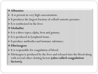
i. Plasma proteins: (7%) [Albumin, Globulin and Fibrinogen]
ii. Inorganic substance:(0.9%) [Na, K, Ca, Mg, Fe & Cu etc.]
iii. Organic substance: [Proteins, non-protein nitrogenous substance like
Urea, Uric acid,Amino acid, Glucose, Fat, Hormones & various enzymes
etc.]
2. Cell: (45%)
I. RBC
II. WBC
III. Platelets](https://image.slidesharecdn.com/blood-200718045931/85/Blood-4-320.jpg)


The document provides a comprehensive overview of blood as a specialized connective tissue, detailing its functions, composition, and types of blood cells (red blood cells, white blood cells, and platelets). It discusses the functions of blood, the processes of erythropoiesis and coagulation, as well as various blood disorders such as anemia, polycythemia, and leukocytosis. Additionally, it outlines blood group classifications, Rh factors, and potential complications like erythroblastosis fetalis.