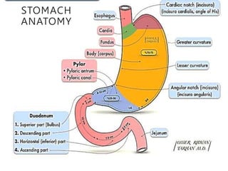
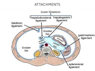
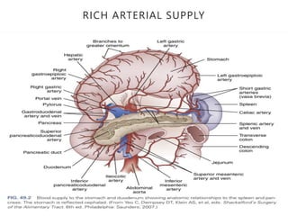
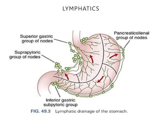
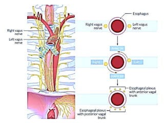
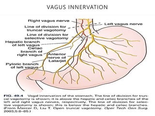
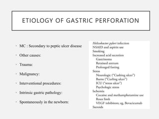
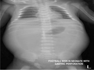
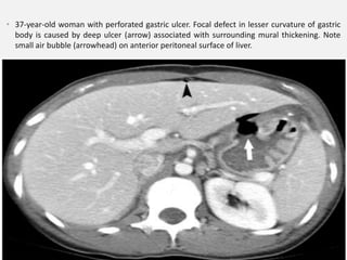
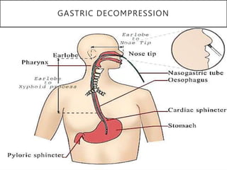
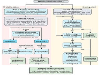
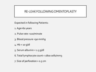
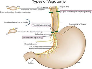
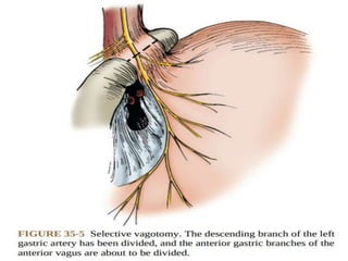
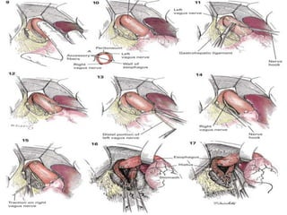
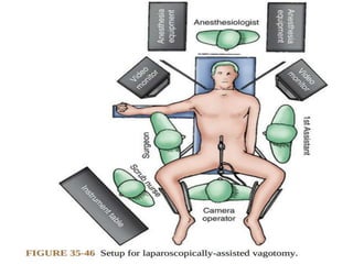
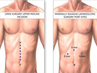
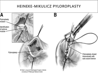
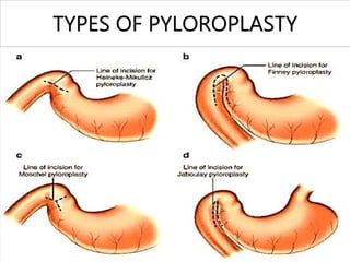
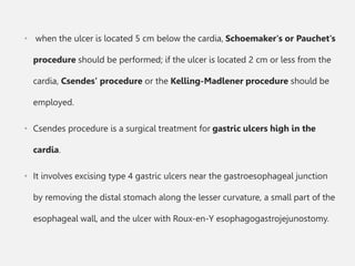
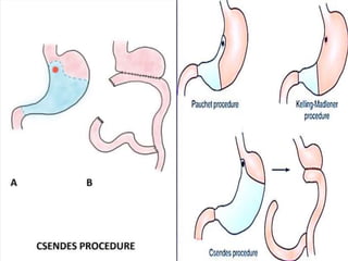
GASTRIC PERFORATION
Dr Reshma Chandrasekaran
DNB General Surgery Resident
UPCOMING SLIDES INCLUDE
• INTODUCTION
• BASIC ANATOMY OF STOMACH
• GASTRIC SECRETION PHYSIOLOGY
• APPROACH TO HVP CASE
• SURGICAL MANAGEMENT OF GASTRIC PERFORATION
• COMPLICATIONS
• CONCLUSION
INTRODUCTION
• Perforation of the stomach is a full-thickness injury of the wall of the
organ.
• The peritoneum completely covers the stomach and so perforation
of the wall creates a communication between the gastric lumen and
the peritoneal cavity.
• If the perforation occurs acutely, there is no time for an
inflammatory reaction to wall off the perforation, and the gastric
contents freely enter the general peritoneal cavity, causing chemical
peritonitis.
• Perforations occurring over a prolonged period may be contained
locally by the inflammatory reaction.
• Elective and emergency operations for benign gastric ulcer disease
has decreased over the decades.
• Annual incidence of peptic ulcer disease 0.1-3% (300,000 new
cases per year), ½ gastric ulcers
• Pharmacologic therapy for acid hypersecretion and H. pylori
treatment is the primary reason for reduction in surgical
intervention.
STOMACH
ANATOMY
ATTACHMENTS
RICH ARTERIAL SUPPLY
LYMPHATICS
VAGUS INNERVATION
GASTRIC SECRETION PHYSIOLOGY
ETIOLOGY OF GASTRIC PERFORATION
• MC : Secondary to peptic ulcer disease
• Other causes:
• Trauma:
• Malignancy:
• Interventional procedures:
• Intrinsic gastric pathology:
• Spontaneously in the newborn:
DISEASE PROGRESSION
FATE OF H.PYLORI INFECTION
• H. pylori, a group 1 carcinogen can lead to gastric adenocarcinoma
through a sequence of pathology starting from
• In patients with mucosa-associated lymphoid tissue (MALT) lymphoma, H.
pylori has been seen in more than 75% of cases.
• H. pylori testing is recommended in children having first-degree relatives
with gastric cancer.
Gastritis => atrophy => intestinal metaplasia => dysplasia => carcinoma.
ULCER OR PERFORATION SITE
PREDICTION WITH HISTORY
• Peptic ulceration is typically characterized by non-radiating epigastric
pain described as burning or stabbing.
• Referral of pain to the back may indicate posterior penetration of the
ulcer.
• H/O pain in relation to eating:
with duodenal ulcer pain relieved by eating,
gastric or marginal ulcer pain worsens with food intake.
ROLE OF UGIE
• About 10% of gastric ulcers are malignant or associated with malignancy,
so aggressive biopsy and brushings, as well as careful follow-up to
demonstrate healing, are mandatory.
• All gastric ulcers should undergo multiple biopsies, obtained from the
perimeter of the lesion.
• The addition of endoscopic brushings to multiple biopsies increases
diagnostic accuracy to approximately 95%.
SIGNS AND SYMPTOMS IN HVP
• Perforated Peptic ulcer-Sudden-onset, severe,
• Generalised abdominal pain-
• Tachycardia-
• Board-like rigidity-
• Distension-
• Obstipation-
• Fever(not initially)-
• Hypotension (later stage)
• POSTERIOR WALL PERFORATION USUALLY PRESENT LATER
THAN ANTERIOR WALL PERFORATION.
POSTERIOR WALL GASTRIC ULCERS PERFORATE AND THEY LEAK GASTRIC CONTENTS
INTO THE LESSER SAC, WHICH TENDS TO CONFINE THE PERITONITIS.
INVESTIGATIONS:
X-RAY ABDOMEN ERECT
FOOTBALL SIGN IN NEONATE WITH
GASTRIC PERFORATION
• 37-year-old woman with perforated gastric ulcer. Focal defect in lesser curvature of gastric
body is caused by deep ulcer (arrow) associated with surrounding mural thickening. Note
small air bubble (arrowhead) on anterior peritoneal surface of liver.
GASTRIC DECOMPRESSION
GASTRIC ASPIRATE
CURRENTINDICATIONFOR SURGICAL
INTERVENTION
1. Bleeding  Most Common Complication 
• 100 per 100.000 population
2. Perforation  11 per 100.000 population 
• highest rate of mortality
3.Obstruction  scarring of prepyloric and duodenal ulcers
4. Failed Medical therapy  PPIs
5. Risk of Malignancy  large gastric ulcers
ROLE OF SURGICAL MANAGEMENT
• The prognosis is improved if treatment is provided within 6 hours of
perforation.
• Delay in treatment beyond 12 hours  an increase in both morbidity and
mortality.
• A prospective study of patients  perforations >48 hours, pre-operative
shock, and concurrent medical illness were associated with an increase in
mortality.
• Emergency surgery for a perforated peptic ulcer has a 6–30% risk of
mortality.
“ Do not stitch the perforation but
plug it with viable omentum and
patch a perforation ulcer if you can,
if you cannot, then you must resect”
( Mosche Schein)
MODIFIEDJOHNSONCLASSIFICATION
SURGICAL MANAGEMENT
• Primary repair: The defect is primarily closed with suture, this is
appropriate for most traumatic perforations.
• Cellan-jones repair: The defect is simply plugged with a well-
vascularized omental pedicle and sutured.
• Graham patch repair: The ulcer is closed with omental cut patch
(no vascularity).
IN BOTH THESE METHODS PERFORTAION SITES ARE NOT CLOSED
WITH PRIMARY REPAIR.
• Karanjia technique: modified Cellan-Jones: omental pedicle is secured to the
tip of a NGT passed through the ULCER site.
NGT is withdrawn for 5-6 cms before the omentum is secured to healthy
serosa
• Modified Graham patch repair/ omentoplasty: primary closure of the defect
and then application of the omental tongue which is secured with same
suture thread.
• Wedge resection: The perforated area may be resected from healthy tissue,
particularly if it is on the greater curvature and distant from the
gastroesophageal junction or the pylorus
THOROUGH PERITONEAL TOILET:
• Irrigation with warm NS or antibiotics.
• One of the most Important parts of surgery 
6-10 liters even up to 30 litres of warm saline
are recommended.
CONTAMINATED PERITONEAL CAVITY
RE-LEAKFOLLOWINGOMENTOPLASTY
Expected in following Patients:
1.Age>60 years
2. Pulse rate >110/minute
3. Blood pressure <90 mmhg
4. Hb < 10 g/dl
5. Serum albumin < 2.5 g/dl
6.Total lymphocyte count < 1800 cells/mm3
7. Size of perforation> 0.5 cm
DRAINAGENEEDEDOR NOT?
• Still controversial.
• 80% no need.
• DrainWill not reduce the incidence of
intraabdominal fluid collections or abcesses
(Schein.M)
• 10% can become infected and intestinal
obstruction
ACID HYPERSECRETION SITES
10mm
5 mm
5 mm
OPEN SURGERY UPPER MIDLINE
INCISION
MINIMALLY INVASIVE LAPAROSCOPIC
SURGERY PORT SITES
HEINEKE-MIKULICZ PYLOROPLASTY
TYPES OF PYLOROPLASTY
The Heineke−Mikulicz pyloroplasty consists of a longitudinal
incision of the pyloric sphincter extending into the antrum and the
duodenum.
The incision is closed transversely, eliminating sphincteric closure and
increasing the lumen of the pyloric channel.
ROLE OF VAGOTOMY AND GASTRIC
DRAINAGE PROCEDURES
• Vagotomy and pyloroplasty has an 10–15% ulcer recurrence rate.
• Vagotomy with antrectomy : ulcer recurrence rate is very low.
• when the ulcer is located 5 cm below the cardia, Schoemaker's or Pauchet's
procedure should be performed; if the ulcer is located 2 cm or less from the
cardia, Csendes' procedure or the Kelling-Madlener procedure should be
employed.
• Csendes procedure is a surgical treatment for gastric ulcers high in the
cardia.
• It involves excising type 4 gastric ulcers near the gastroesophageal junction
by removing the distal stomach along the lesser curvature, a small part of the
esophageal wall, and the ulcer with Roux-en-Y esophagogastrojejunostomy.
OPTIONS FOR RECONSTRUCTION:
• Billroth I: Gastroduodenostomy, anastomosis between
the gastric remnant and the duodenum
• Billroth II: Gastrojejunostomy, side to side anastomosis
between gastric remnant and loop of jejunum with the
closure of duodenal stump
• Roux-en-Y gastrojejunostomy: The creation of jejuno-
jejunostomy forming y shaped figure of the small
bowel.
SURGICALRECONSTRUCTION
BEST PROCEDURE WHEN CONSIDERING REVISION SURGERY
SUTURING TECHNIQUES
Common Suture techniques:
1. Continuous over-and-over
suture
2. Lembert's suture
3. Connell suture
4. Cushing suture
Double layer anastomosis
• Posterior outer lembert's
sutures
• Posterior inner → over-and-
over continuous sutures
• Anterior inner connell's
sutures
• Anterior outer → lembert's
sutures
Lambert suture
Connell stitch
CIRCULAR ENDO GI STAPLER USAGE
Intestinal anastomosis is a common surgery
• Anastomotic healing is similar to other tissue healing
• Hand sewn anastomosis is not inferior to stapler
• Anastomosis must be tension free, with good blood supply and
minimal fecal contamination
Two main benefits of stapler anastomosis:
• Takes less time
• No objective variations – easy to use by all surgeons
SURGICALRECONSTRUCTION
• Both Billroth reconstruction  lead to bile reflux  5-35%
• Toavoid that  Roux-en-Y reconstruction (Roux1897)
• Roux-en-Yreconstruction  plaqued with a Roux stasis syndrome
• Braun variaton Billroth (1893)  lower incidence of Bile reflux 
some authors recommend this as standard reconstruction.
COMPLICATIONOF ULCER OPERATIONS
1. Early Satiety
2.postvagotomy syndrome  30%
3. DumpingSyndrome  20%
4.Alkaline Reflux gastritis  10%
5.Afferent and Efferent loop syndrome 
Mechanical obstruction of the limb kinking, anastomosis
narrowing, or adhesion
6. Roux stasis syndrome
7. RecurrentUlceration
8.Anastomotic leak
POST SURGICAL FOLLOW-UP
• UGIE WITH HP TEST
• TO TREAT RESISTANT VARIETY OF H.PYLORI.
THANKYOU

GASTRIC PERFORATION: A BRIEF SURGICAL MANAGEMNT

  • 1.
    GASTRIC PERFORATION Dr ReshmaChandrasekaran DNB General Surgery Resident
  • 2.
    UPCOMING SLIDES INCLUDE •INTODUCTION • BASIC ANATOMY OF STOMACH • GASTRIC SECRETION PHYSIOLOGY • APPROACH TO HVP CASE • SURGICAL MANAGEMENT OF GASTRIC PERFORATION • COMPLICATIONS • CONCLUSION
  • 3.
    INTRODUCTION • Perforation ofthe stomach is a full-thickness injury of the wall of the organ. • The peritoneum completely covers the stomach and so perforation of the wall creates a communication between the gastric lumen and the peritoneal cavity. • If the perforation occurs acutely, there is no time for an inflammatory reaction to wall off the perforation, and the gastric contents freely enter the general peritoneal cavity, causing chemical peritonitis. • Perforations occurring over a prolonged period may be contained locally by the inflammatory reaction.
  • 4.
    • Elective andemergency operations for benign gastric ulcer disease has decreased over the decades. • Annual incidence of peptic ulcer disease 0.1-3% (300,000 new cases per year), ½ gastric ulcers • Pharmacologic therapy for acid hypersecretion and H. pylori treatment is the primary reason for reduction in surgical intervention.
  • 5.
  • 6.
  • 7.
  • 8.
  • 10.
  • 11.
  • 12.
    ETIOLOGY OF GASTRICPERFORATION • MC : Secondary to peptic ulcer disease • Other causes: • Trauma: • Malignancy: • Interventional procedures: • Intrinsic gastric pathology: • Spontaneously in the newborn:
  • 14.
  • 15.
    FATE OF H.PYLORIINFECTION • H. pylori, a group 1 carcinogen can lead to gastric adenocarcinoma through a sequence of pathology starting from • In patients with mucosa-associated lymphoid tissue (MALT) lymphoma, H. pylori has been seen in more than 75% of cases. • H. pylori testing is recommended in children having first-degree relatives with gastric cancer. Gastritis => atrophy => intestinal metaplasia => dysplasia => carcinoma.
  • 16.
    ULCER OR PERFORATIONSITE PREDICTION WITH HISTORY • Peptic ulceration is typically characterized by non-radiating epigastric pain described as burning or stabbing. • Referral of pain to the back may indicate posterior penetration of the ulcer. • H/O pain in relation to eating: with duodenal ulcer pain relieved by eating, gastric or marginal ulcer pain worsens with food intake.
  • 17.
    ROLE OF UGIE •About 10% of gastric ulcers are malignant or associated with malignancy, so aggressive biopsy and brushings, as well as careful follow-up to demonstrate healing, are mandatory. • All gastric ulcers should undergo multiple biopsies, obtained from the perimeter of the lesion. • The addition of endoscopic brushings to multiple biopsies increases diagnostic accuracy to approximately 95%.
  • 18.
    SIGNS AND SYMPTOMSIN HVP • Perforated Peptic ulcer-Sudden-onset, severe, • Generalised abdominal pain- • Tachycardia- • Board-like rigidity- • Distension- • Obstipation- • Fever(not initially)- • Hypotension (later stage) • POSTERIOR WALL PERFORATION USUALLY PRESENT LATER THAN ANTERIOR WALL PERFORATION.
  • 19.
    POSTERIOR WALL GASTRICULCERS PERFORATE AND THEY LEAK GASTRIC CONTENTS INTO THE LESSER SAC, WHICH TENDS TO CONFINE THE PERITONITIS.
  • 21.
  • 22.
    FOOTBALL SIGN INNEONATE WITH GASTRIC PERFORATION
  • 23.
    • 37-year-old womanwith perforated gastric ulcer. Focal defect in lesser curvature of gastric body is caused by deep ulcer (arrow) associated with surrounding mural thickening. Note small air bubble (arrowhead) on anterior peritoneal surface of liver.
  • 24.
  • 25.
  • 27.
    CURRENTINDICATIONFOR SURGICAL INTERVENTION 1. Bleeding Most Common Complication  • 100 per 100.000 population 2. Perforation  11 per 100.000 population  • highest rate of mortality 3.Obstruction  scarring of prepyloric and duodenal ulcers 4. Failed Medical therapy  PPIs 5. Risk of Malignancy  large gastric ulcers
  • 28.
    ROLE OF SURGICALMANAGEMENT • The prognosis is improved if treatment is provided within 6 hours of perforation. • Delay in treatment beyond 12 hours  an increase in both morbidity and mortality. • A prospective study of patients  perforations >48 hours, pre-operative shock, and concurrent medical illness were associated with an increase in mortality. • Emergency surgery for a perforated peptic ulcer has a 6–30% risk of mortality.
  • 29.
    “ Do notstitch the perforation but plug it with viable omentum and patch a perforation ulcer if you can, if you cannot, then you must resect” ( Mosche Schein)
  • 30.
  • 32.
    SURGICAL MANAGEMENT • Primaryrepair: The defect is primarily closed with suture, this is appropriate for most traumatic perforations. • Cellan-jones repair: The defect is simply plugged with a well- vascularized omental pedicle and sutured. • Graham patch repair: The ulcer is closed with omental cut patch (no vascularity). IN BOTH THESE METHODS PERFORTAION SITES ARE NOT CLOSED WITH PRIMARY REPAIR.
  • 33.
    • Karanjia technique:modified Cellan-Jones: omental pedicle is secured to the tip of a NGT passed through the ULCER site. NGT is withdrawn for 5-6 cms before the omentum is secured to healthy serosa • Modified Graham patch repair/ omentoplasty: primary closure of the defect and then application of the omental tongue which is secured with same suture thread. • Wedge resection: The perforated area may be resected from healthy tissue, particularly if it is on the greater curvature and distant from the gastroesophageal junction or the pylorus
  • 36.
    THOROUGH PERITONEAL TOILET: •Irrigation with warm NS or antibiotics. • One of the most Important parts of surgery  6-10 liters even up to 30 litres of warm saline are recommended. CONTAMINATED PERITONEAL CAVITY
  • 37.
    RE-LEAKFOLLOWINGOMENTOPLASTY Expected in followingPatients: 1.Age>60 years 2. Pulse rate >110/minute 3. Blood pressure <90 mmhg 4. Hb < 10 g/dl 5. Serum albumin < 2.5 g/dl 6.Total lymphocyte count < 1800 cells/mm3 7. Size of perforation> 0.5 cm
  • 38.
    DRAINAGENEEDEDOR NOT? • Stillcontroversial. • 80% no need. • DrainWill not reduce the incidence of intraabdominal fluid collections or abcesses (Schein.M) • 10% can become infected and intestinal obstruction
  • 39.
  • 44.
    10mm 5 mm 5 mm OPENSURGERY UPPER MIDLINE INCISION MINIMALLY INVASIVE LAPAROSCOPIC SURGERY PORT SITES
  • 45.
  • 46.
  • 47.
    The Heineke−Mikulicz pyloroplastyconsists of a longitudinal incision of the pyloric sphincter extending into the antrum and the duodenum. The incision is closed transversely, eliminating sphincteric closure and increasing the lumen of the pyloric channel.
  • 49.
    ROLE OF VAGOTOMYAND GASTRIC DRAINAGE PROCEDURES • Vagotomy and pyloroplasty has an 10–15% ulcer recurrence rate. • Vagotomy with antrectomy : ulcer recurrence rate is very low.
  • 51.
    • when theulcer is located 5 cm below the cardia, Schoemaker's or Pauchet's procedure should be performed; if the ulcer is located 2 cm or less from the cardia, Csendes' procedure or the Kelling-Madlener procedure should be employed. • Csendes procedure is a surgical treatment for gastric ulcers high in the cardia. • It involves excising type 4 gastric ulcers near the gastroesophageal junction by removing the distal stomach along the lesser curvature, a small part of the esophageal wall, and the ulcer with Roux-en-Y esophagogastrojejunostomy.
  • 55.
    OPTIONS FOR RECONSTRUCTION: •Billroth I: Gastroduodenostomy, anastomosis between the gastric remnant and the duodenum • Billroth II: Gastrojejunostomy, side to side anastomosis between gastric remnant and loop of jejunum with the closure of duodenal stump • Roux-en-Y gastrojejunostomy: The creation of jejuno- jejunostomy forming y shaped figure of the small bowel.
  • 56.
  • 58.
    BEST PROCEDURE WHENCONSIDERING REVISION SURGERY
  • 59.
    SUTURING TECHNIQUES Common Suturetechniques: 1. Continuous over-and-over suture 2. Lembert's suture 3. Connell suture 4. Cushing suture Double layer anastomosis • Posterior outer lembert's sutures • Posterior inner → over-and- over continuous sutures • Anterior inner connell's sutures • Anterior outer → lembert's sutures
  • 60.
  • 62.
    CIRCULAR ENDO GISTAPLER USAGE
  • 63.
    Intestinal anastomosis isa common surgery • Anastomotic healing is similar to other tissue healing • Hand sewn anastomosis is not inferior to stapler • Anastomosis must be tension free, with good blood supply and minimal fecal contamination Two main benefits of stapler anastomosis: • Takes less time • No objective variations – easy to use by all surgeons
  • 64.
    SURGICALRECONSTRUCTION • Both Billrothreconstruction  lead to bile reflux  5-35% • Toavoid that  Roux-en-Y reconstruction (Roux1897) • Roux-en-Yreconstruction  plaqued with a Roux stasis syndrome • Braun variaton Billroth (1893)  lower incidence of Bile reflux  some authors recommend this as standard reconstruction.
  • 66.
    COMPLICATIONOF ULCER OPERATIONS 1.Early Satiety 2.postvagotomy syndrome  30% 3. DumpingSyndrome  20% 4.Alkaline Reflux gastritis  10% 5.Afferent and Efferent loop syndrome  Mechanical obstruction of the limb kinking, anastomosis narrowing, or adhesion 6. Roux stasis syndrome 7. RecurrentUlceration 8.Anastomotic leak
  • 67.
    POST SURGICAL FOLLOW-UP •UGIE WITH HP TEST • TO TREAT RESISTANT VARIETY OF H.PYLORI.
  • 68.