Downloaded 80 times
![Cardiac death and reinfarction after 1 year in the Thrombus Aspiration during
Percutaneous coronary intervention in Acute myocardial infarction Study
(TAPAS): a 1-year follow-up study
Findings
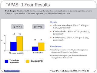
Cardiac death at 1 year was 3·6% (19 of 535 patients) in the
thrombus aspiration group and 6·7% (36 of 536) in the
conventional PCI group (hazard ratio [HR] 1·93; 95% CI 1·11—
3·37; p=0·020). 1-year cardiac death or non-fatal reinfarction
occurred in 5·6% (30 of 535) of patients in the thrombus
aspiration group and 9·9% (53 of 536) of patients in the
conventional PCI group (HR 1·81; 95% CI 1·16—2·84;
p=0·009).
Interpretation
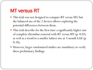
Compared with conventional PCI, thrombus aspiration before
stenting of the infarcted artery seems to improve the 1-year
clinical outcome after PCI for ST-elevation myocardial infarction.
Lancet 2008;371:1915–20.](https://image.slidesharecdn.com/journalmustelatrial-130111115735-phpapp02/85/Thromboectomy-trial-5-320.jpg)


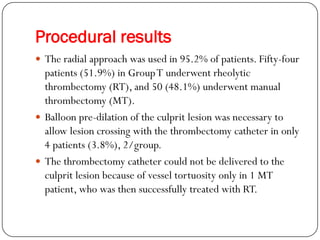
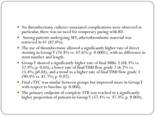
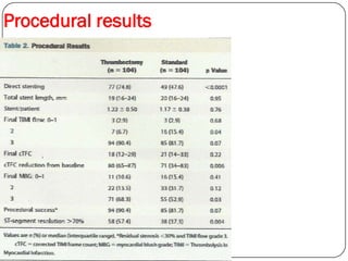
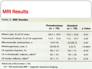
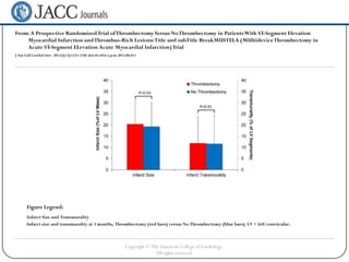
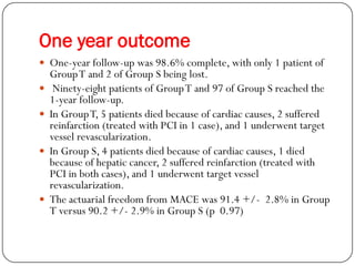


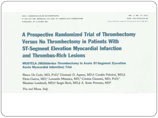
1) The study compared outcomes of STEMI patients undergoing primary PCI with thrombectomy (Group T) versus without thrombectomy (Group S). 2) MRI results at 3 months showed significantly smaller infarct size and less transmurality in Group T compared to Group S. 3) Procedural results favored Group T with higher rates of TIMI 3 flow and complete ST resolution. One-year outcomes also favored Group T with lower rates of MACE.


