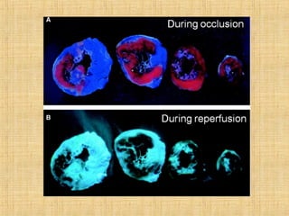
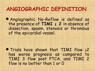
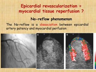
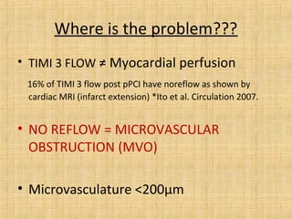
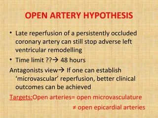
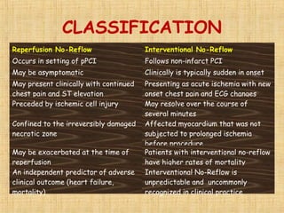
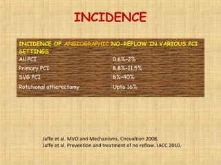
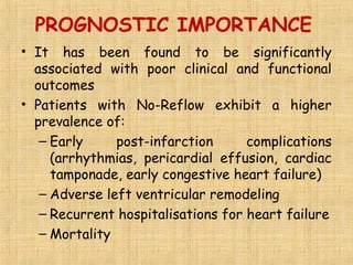
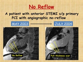
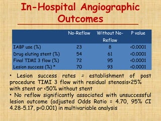
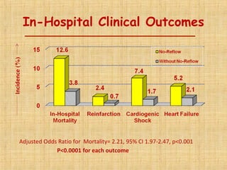
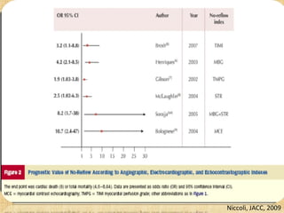
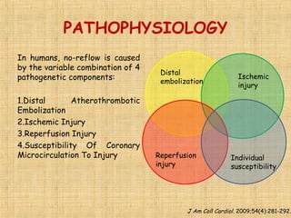
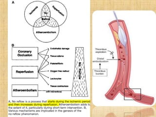
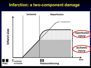
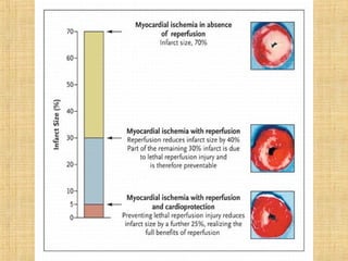
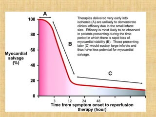
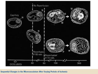
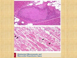
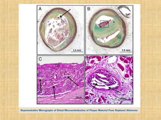
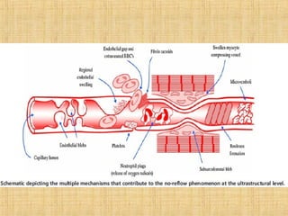
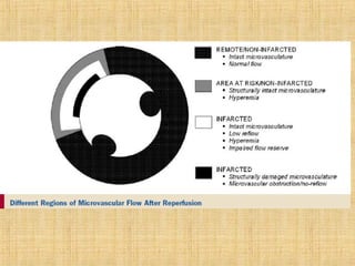
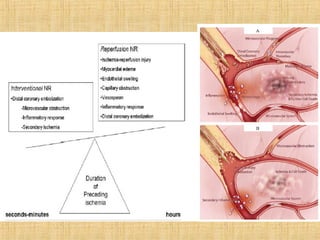
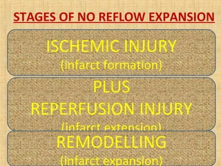
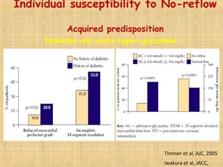
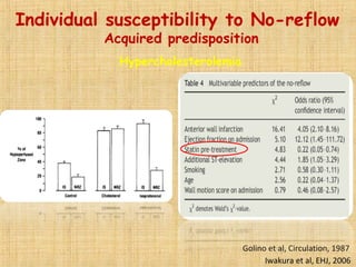
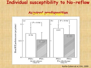
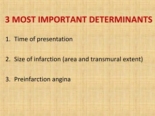
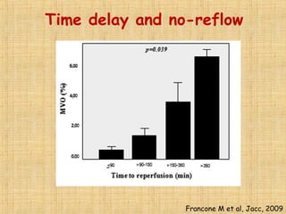
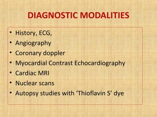
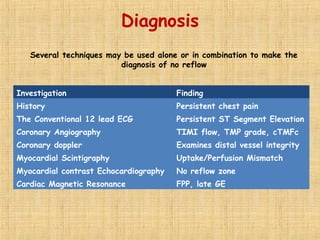
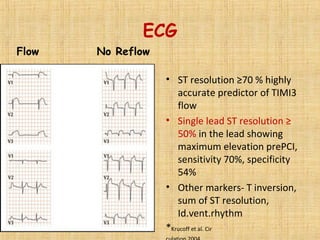
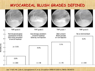
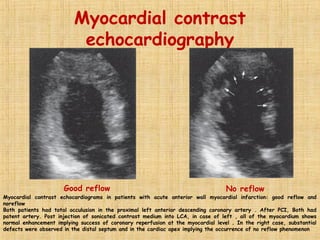
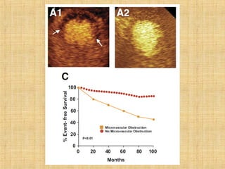
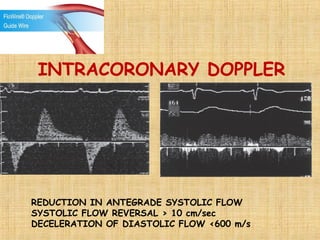
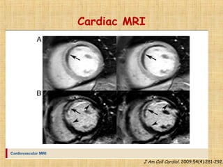
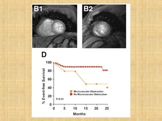
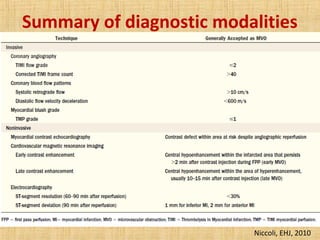
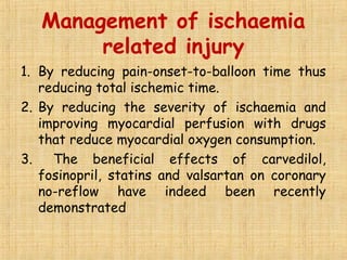
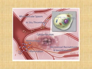
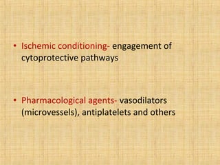
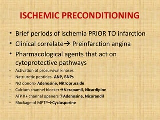
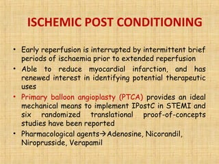
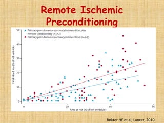
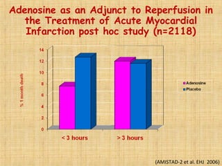
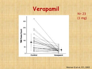
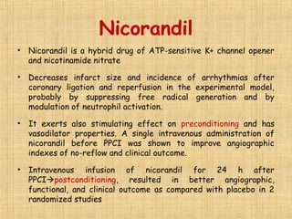
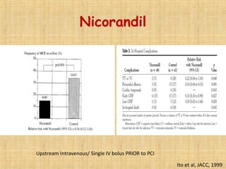
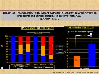
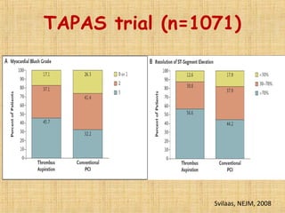
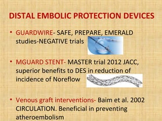
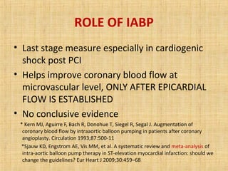
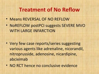
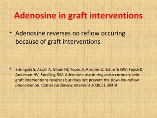
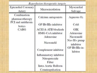
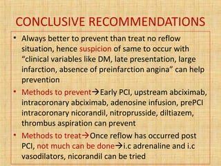
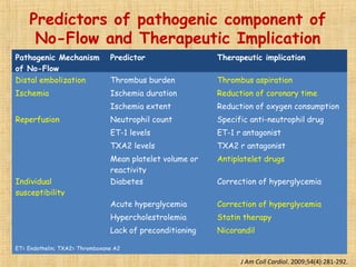
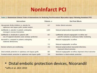
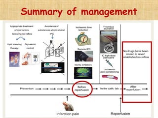
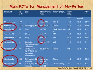
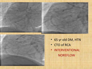
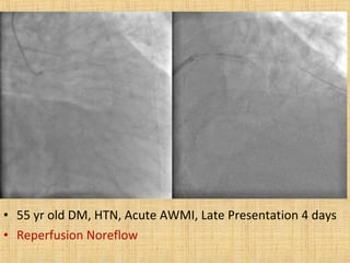
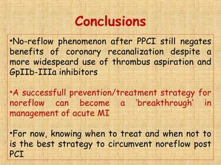
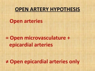
This document discusses the no-reflow phenomenon, which occurs when restoration of coronary artery patency after procedures like primary percutaneous coronary intervention (PCI) does not translate to improved tissue perfusion. No-reflow occurs in 30% of patients after reperfusion for acute myocardial infarction and is associated with worse outcomes. It is caused by microvascular obstruction from distal embolization, ischemic injury, reperfusion injury, and individual patient susceptibility. Methods to diagnose no-reflow include angiography, coronary Doppler, cardiac MRI, and myocardial contrast echocardiography. Prevention strategies target reducing ischemic time, microvascular spasm, and distal embolization through early reperfusion, pharmacological agents, and ischemic conditioning techniques.
Clinical and Angiographic Predictors of No-Reflow Phenomenon after Percutaneous Coronary Intervention in ST-Segment Elevation Myocardial Infarction Patients; Mansoura Experience
Various coronary physiological measurements can be made in the cardiac catheterization laboratory using sensor-tipped guidewires; they include the measurement of poststenotic absolute coronary flow reserve, the relative coronary flow reserve, and the pressure-derived fractional flow reserve of the myocardium. Ambiguity regarding abnormal microcirculation has been reduced or eliminated with measurements of relative coronary flow reserve and fractional flow reserve. The role of microvascular flow impairment can be separately determined with coronary flow velocity reserve measurements. In addition to lesion assessment before and after intervention, emerging applications of coronary physiology include the determination of physiological responses to new pharmacological agents, such as glycoprotein IIb/IIIa blockers, in patients with acute myocardial infarction. Measurements of coronary physiology in the catheterization laboratory provide objective data that complement angiography for clinical decision-making
How to deal with CALCIFIED CORONARY ARTERY LESIONS .Coronary artery calcification (CAC) is highly prevalent in patients with coronary heart disease (CHD) and is associated with major adverse cardiovascular events. There are two recognized type of CAC—intimal and medial calcification, and each of them have specific risk factors. Several theories about the mechanism of vascular calcification have been put forward, and we currently believe that vascular calcification is an active, regulated process. CAC can usually be found in patients with severe CHD, and this asymptomatic phenomenon make early diagnosis of CAC important. Coronary computed tomographic angiography is the main noninvasive tool to detect calcified lesions. Measurement of coronary artery calcification by scoring is a reasonable metric for cardiovascular risk assessment in asymptomatic adults at intermediate risk. To date, effective medical treatment of CAC has not been identified. Several strategies of percutaneous coronary intervention have been applied to CHD patients with CAC, but with unsatisfactory results. Prognosis of CAC is still a major problem of CHD patients. Thus, more details about the mechanisms of CAC need to be elucidated in order to improve the understanding and treatment of CAC.
SCAD is a rare, sometimes fatal, traumatic condition with approximately eighty percent of cases affecting women. The coronary artery can suddenly develop a tear, causing blood to flow between the layers which forces them apart, potentially causing a blockage of blood flow through the artery and a resulting heart attack. The condition may be related to female hormone levels, as it is often seen in post-partum women, or in women during or very near menstruation, but not always. It is not uncommon for SCAD to occur in people in good physical shape and with no known prior history of heart related illness. It is also not uncommon for SCAD to occur in people in their 20's, 30's, and 40's, as well as older.
Coronary CTO is characterized by heavy atherosclerotic plaque burden within the artery, resulting in complete (or nearly complete) occlusion of the vessel. Although the duration of the occlusion is difficult to determine on clinical grounds, a total occlusion must be present for at least 3 months to be considered a true CTO. Patients with CTO typically have collateralization of the distal vessel on coronary angiography, but these collaterals may not provide sufficient blood flow to the myocardial bed, resulting in ischemia and anginal symptoms. CTO is clinically distinct from acute coronary occlusion, which occurs in the setting of ST-segment–elevation myocardial infarction, or subacute coronary occlusion, discovered with delayed presentation after ST-segment–elevation myocardial infarction. Clinical features and treatment considerations of these entities differ considerably from CTO.
Among patients who have a clinical indication for coronary angiography, the incidence of CTO has been reported to be as high as 15% to 30%. Patients with CTO are referred for angiography because of anginal symptoms or significant ischemia on noninvasive ischemia testing. Patients who are symptomatic will have stable exertional angina resulting from a limitation of collateral vessel flow to meet myocardial oxygen demand with stress. Of patients referred for PCI in clinical trials of CTO PCI, only 10% to 15% of patients are asymptomatic. It is likewise uncommon for patients with CTO to present with an acute coronary syndrome caused by the CTO itself.
Each month, join us as we highlight and discuss hot topics ranging from the future of higher education to wearable technology, best productivity hacks and secrets to hiring top talent. Upload your SlideShares, and share your expertise with the world!
Not sure what to share on SlideShare?
SlideShares that inform, inspire and educate attract the most views. Beyond that, ideas for what you can upload are limitless. We’ve selected a few popular examples to get your creative juices flowing.
How to Make Awesome SlideShares: Tips & TricksSlideShare
Turbocharge your online presence with SlideShare. We provide the best tips and tricks for succeeding on SlideShare. Get ideas for what to upload, tips for designing your deck and more.
How to evaluate shock by echo. Echo is an essential tool in ICU and it´s the cornerstone in the management of shock nowadays, learning how to use this procedure must be mandatory
Physician should have a high suspicion to diagnose patient with pulmonary Embolism, this slides will give you precise Diagnosis, Investigation and guideline directed Treatment.
Does Preoperative Coronary Revascularization Improve Perioperative Cardiac Ou...Guilherme Barcellos
Draft que encontrei de apresentação em 201: Primeiro Encontro de Medicina Hospitalista da Argentina. Slides alguns já traduzidos, outros não - não encontrei versão final. De brasileiros no evento participaram eu, Lucas Zambon e Tiago Daltoé. Boas lembranças! Resgatei agora porque trata de evidência consolidada desde aquela época, e seguimos sobreutilizando o recurso. Ou algo novo que justifique?
A study to assess the effectiveness of structured teaching program on knowledge regarding care of patients after cardiac surgery among staff nurses at Shree Narayana, Hospital, Raipur, chhattisgarh.
Edward Fohrman | Neuroanesthesia for Cerebral Arteriovenous MalformationsEdward Fohrman
Edward Fohrman shares his presentation about Neuroanesthesia for Cerebral Arteriovenous Malformations.
Visit EdwardFohrman.com for more information about Edward and his industry insights.
Similar to No reflow phenomenon by dr. deepchandh (20)
Explore natural remedies for syphilis treatment in Singapore. Discover alternative therapies, herbal remedies, and lifestyle changes that may complement conventional treatments. Learn about holistic approaches to managing syphilis symptoms and supporting overall health.
These lecture slides, by Dr Sidra Arshad, offer a quick overview of physiological basis of a normal electrocardiogram.
Learning objectives:
1. Define an electrocardiogram (ECG) and electrocardiography
2. Describe how dipoles generated by the heart produce the waveforms of the ECG
3. Describe the components of a normal electrocardiogram of a typical bipolar leads (limb II)
4. Differentiate between intervals and segments
5. Enlist some common indications for obtaining an ECG
Study Resources:
1. Chapter 11, Guyton and Hall Textbook of Medical Physiology, 14th edition
2. Chapter 9, Human Physiology - From Cells to Systems, Lauralee Sherwood, 9th edition
3. Chapter 29, Ganong’s Review of Medical Physiology, 26th edition
4. Electrocardiogram, StatPearls - https://www.ncbi.nlm.nih.gov/books/NBK549803/
5. ECG in Medical Practice by ABM Abdullah, 4th edition
6. ECG Basics, http://www.nataliescasebook.com/tag/e-c-g-basics
MANAGEMENT OF ATRIOVENTRICULAR CONDUCTION BLOCK.pdfJim Jacob Roy
Cardiac conduction defects can occur due to various causes.
Atrioventricular conduction blocks ( AV blocks ) are classified into 3 types.
This document describes the acute management of AV block.
Pulmonary Thromboembolism - etilogy, types, medical- Surgical and nursing man...VarunMahajani
Disruption of blood supply to lung alveoli due to blockage of one or more pulmonary blood vessels is called as Pulmonary thromboembolism. In this presentation we will discuss its causes, types and its management in depth.
Flu Vaccine Alert in Bangalore Karnatakaaddon Scans
As flu season approaches, health officials in Bangalore, Karnataka, are urging residents to get their flu vaccinations. The seasonal flu, while common, can lead to severe health complications, particularly for vulnerable populations such as young children, the elderly, and those with underlying health conditions.
Dr. Vidisha Kumari, a leading epidemiologist in Bangalore, emphasizes the importance of getting vaccinated. "The flu vaccine is our best defense against the influenza virus. It not only protects individuals but also helps prevent the spread of the virus in our communities," he says.
This year, the flu season is expected to coincide with a potential increase in other respiratory illnesses. The Karnataka Health Department has launched an awareness campaign highlighting the significance of flu vaccinations. They have set up multiple vaccination centers across Bangalore, making it convenient for residents to receive their shots.
To encourage widespread vaccination, the government is also collaborating with local schools, workplaces, and community centers to facilitate vaccination drives. Special attention is being given to ensuring that the vaccine is accessible to all, including marginalized communities who may have limited access to healthcare.
Residents are reminded that the flu vaccine is safe and effective. Common side effects are mild and may include soreness at the injection site, mild fever, or muscle aches. These side effects are generally short-lived and far less severe than the flu itself.
Healthcare providers are also stressing the importance of continuing COVID-19 precautions. Wearing masks, practicing good hand hygiene, and maintaining social distancing are still crucial, especially in crowded places.
Protect yourself and your loved ones by getting vaccinated. Together, we can help keep Bangalore healthy and safe this flu season. For more information on vaccination centers and schedules, residents can visit the Karnataka Health Department’s official website or follow their social media pages.
Stay informed, stay safe, and get your flu shot today!
These simplified slides by Dr. Sidra Arshad present an overview of the non-respiratory functions of the respiratory tract.
Learning objectives:
1. Enlist the non-respiratory functions of the respiratory tract
2. Briefly explain how these functions are carried out
3. Discuss the significance of dead space
4. Differentiate between minute ventilation and alveolar ventilation
5. Describe the cough and sneeze reflexes
Study Resources:
1. Chapter 39, Guyton and Hall Textbook of Medical Physiology, 14th edition
2. Chapter 34, Ganong’s Review of Medical Physiology, 26th edition
3. Chapter 17, Human Physiology by Lauralee Sherwood, 9th edition
4. Non-respiratory functions of the lungs https://academic.oup.com/bjaed/article/13/3/98/278874
Factory Supply Best Quality Pmk Oil CAS 28578–16–7 PMK Powder in Stockrebeccabio
Factory Supply Best Quality Pmk Oil CAS 28578–16–7 PMK Powder in Stock
Telegram: bmksupplier
signal: +85264872720
threema: TUD4A6YC
You can contact me on Telegram or Threema
Communicate promptly and reply
Free of customs clearance, Double Clearance 100% pass delivery to USA, Canada, Spain, Germany, Netherland, Poland, Italy, Sweden, UK, Czech Republic, Australia, Mexico, Russia, Ukraine, Kazakhstan.Door to door service
Hot Selling Organic intermediates
Couples presenting to the infertility clinic- Do they really have infertility...Sujoy Dasgupta
Dr Sujoy Dasgupta presented the study on "Couples presenting to the infertility clinic- Do they really have infertility? – The unexplored stories of non-consummation" in the 13th Congress of the Asia Pacific Initiative on Reproduction (ASPIRE 2024) at Manila on 24 May, 2024.
ARTIFICIAL INTELLIGENCE IN HEALTHCARE.pdfAnujkumaranit
Artificial intelligence (AI) refers to the simulation of human intelligence processes by machines, especially computer systems. It encompasses tasks such as learning, reasoning, problem-solving, perception, and language understanding. AI technologies are revolutionizing various fields, from healthcare to finance, by enabling machines to perform tasks that typically require human intelligence.
Title: Sense of Taste
Presenter: Dr. Faiza, Assistant Professor of Physiology
Qualifications:
MBBS (Best Graduate, AIMC Lahore)
FCPS Physiology
ICMT, CHPE, DHPE (STMU)
MPH (GC University, Faisalabad)
MBA (Virtual University of Pakistan)
Learning Objectives:
Describe the structure and function of taste buds.
Describe the relationship between the taste threshold and taste index of common substances.
Explain the chemical basis and signal transduction of taste perception for each type of primary taste sensation.
Recognize different abnormalities of taste perception and their causes.
Key Topics:
Significance of Taste Sensation:
Differentiation between pleasant and harmful food
Influence on behavior
Selection of food based on metabolic needs
Receptors of Taste:
Taste buds on the tongue
Influence of sense of smell, texture of food, and pain stimulation (e.g., by pepper)
Primary and Secondary Taste Sensations:
Primary taste sensations: Sweet, Sour, Salty, Bitter, Umami
Chemical basis and signal transduction mechanisms for each taste
Taste Threshold and Index:
Taste threshold values for Sweet (sucrose), Salty (NaCl), Sour (HCl), and Bitter (Quinine)
Taste index relationship: Inversely proportional to taste threshold
Taste Blindness:
Inability to taste certain substances, particularly thiourea compounds
Example: Phenylthiocarbamide
Structure and Function of Taste Buds:
Composition: Epithelial cells, Sustentacular/Supporting cells, Taste cells, Basal cells
Features: Taste pores, Taste hairs/microvilli, and Taste nerve fibers
Location of Taste Buds:
Found in papillae of the tongue (Fungiform, Circumvallate, Foliate)
Also present on the palate, tonsillar pillars, epiglottis, and proximal esophagus
Mechanism of Taste Stimulation:
Interaction of taste substances with receptors on microvilli
Signal transduction pathways for Umami, Sweet, Bitter, Sour, and Salty tastes
Taste Sensitivity and Adaptation:
Decrease in sensitivity with age
Rapid adaptation of taste sensation
Role of Saliva in Taste:
Dissolution of tastants to reach receptors
Washing away the stimulus
Taste Preferences and Aversions:
Mechanisms behind taste preference and aversion
Influence of receptors and neural pathways
Impact of Sensory Nerve Damage:
Degeneration of taste buds if the sensory nerve fiber is cut
Abnormalities of Taste Detection:
Conditions: Ageusia, Hypogeusia, Dysgeusia (parageusia)
Causes: Nerve damage, neurological disorders, infections, poor oral hygiene, adverse drug effects, deficiencies, aging, tobacco use, altered neurotransmitter levels
Neurotransmitters and Taste Threshold:
Effects of serotonin (5-HT) and norepinephrine (NE) on taste sensitivity
Supertasters:
25% of the population with heightened sensitivity to taste, especially bitterness
Increased number of fungiform papillae
Report Back from SGO 2024: What’s the Latest in Cervical Cancer?bkling
Are you curious about what’s new in cervical cancer research or unsure what the findings mean? Join Dr. Emily Ko, a gynecologic oncologist at Penn Medicine, to learn about the latest updates from the Society of Gynecologic Oncology (SGO) 2024 Annual Meeting on Women’s Cancer. Dr. Ko will discuss what the research presented at the conference means for you and answer your questions about the new developments.
3. INTRODUCTION
• Main goal of any therapeutic intervention is
restoration of patency of the epicardial
coronary artery
• But restoration of this patency does not
translate into improved tissue perfusion
• And there comes the dreaded phenomenon
during primary PCI, with poor clinical outcome
known as “NO REFLOW PHENOMENON”
4. Historical perspective
The first clinical observation of coronary no-
reflow was reported by Schofer et al. in 1985,
first coined by Ames et al in 1970s in Brain
Ischemia
Innumerable experimental models in animals have
been studied to understand the pathogenesis of
no reflow
Likewise, many drugs have been found effective
in animal models of no reflow BUT WITH LITTLE
SUCCESS IN HUMANS
5.
6. DEFINITION
• The phenomenon of no-reflow is defined as
‘Inadequate myocardial perfusion through a given
segment of the coronary circulation without
angiographic evidence of mechanical vessel
obstruction’
• No-reflow has been documented in ≥ 30% of patients
after thrombolysis or mechanical intervention for
acute myocardial infarction in studies with
MRI/myocardial contrast echo
7. ANGIOGRAPHIC DEFINITION
Angiographic No-Reflow is defined as
the presence of TIMI ≤ 2 in absence of
dissection, spasm, stenosis or thrombus
of the epicardial vessel.
Trials have shown that TIMI flow ≤2
has worse prognosis as compared to
TIMI 3 flow post PTCA, and TIMI 2
flow is no better than 1 or 0
9. Where is the problem???
• TIMI 3 FLOW ≠ Myocardial perfusion
16% of TIMI 3 flow post pPCI have noreflow as shown by
cardiac MRI (infarct extension) *Ito et al. Circulation 2007.
• NO REFLOW = MICROVASCULAR
OBSTRUCTION (MVO)
• Microvasculature <200µm
10. OPEN ARTERY HYPOTHESIS
• Late reperfusion of a persistently occluded
coronary artery can still stop adverse left
ventricular remodelling
• Time limit ?? 48 hours
Antagonists view If one can establish
‘microvascular’ reperfusion, better clinical
outcomes can be achieved
Targets:Open arteries= open microvasculature
≠ open epicardial arteries
11. %age of optimal reperfusion, CADILLAC TRIAL
100 patients with STEMI
treated by PPCI
93 patients with TIMI 3
49 patients with TIMI 3
and MBG 2 or 3
35 patients with TIMI 3
and MBG 2 or 3 and
STR>70 %
1 pt with TIMI 0-1
6 pts with TIMI 2
44 pts with MBG
0/1
14 pts with STR
< 70%
Evaluation of
post procedural
TIMI flow
Evaluation of
post procedural
MBG
Evaluation of post
procedural STR>
70%
12. CLASSIFICATION OF NO-REFLOW
MYOCARDIAL INFARCTION REPERFUSION NO-REFOW
Definition no-reflow in the setting of pharmacological and/or
mechanical revascularization for acute myocardial
infarction
INTERVENTIONAL NO REFLOW
Definition no-reflow during percutaneous coronary interventions
especially rotational atherectomy, vein graft
interventions
13. CLASSIFICATION
Reperfusion No-Reflow Interventional No-Reflow
Occurs in setting of pPCI Follows non-infarct PCI
May be asymptomatic Clinically is typically sudden in onset
May present clinically with continued
chest pain and ST elevation
Presenting as acute ischemia with new
onset chest pain and ECG changes
Preceded by ischemic cell injury May resolve over the course of
several minutes
Confined to the irreversibly damaged
necrotic zone
Affected myocardium that was not
subjected to prolonged ischemia
before procedure
May be exacerbated at the time of
reperfusion
Patients with interventional no-reflow
have higher rates of mortality
An independent predictor of adverse
clinical outcome (heart failure,
mortality)
Interventional No-Reflow is
unpredictable and uncommonly
recognized in clinical practice
14. TYPES OF NO REFLOW
Sustained
• Result of anatomical
irreversible changes of
coronary
microcirculation
• Undergo ADVERSE LV
remodeling
Reversible
• Result of functional &
thus reversible changes
of microcirculation
• Maintain their left
ventricle volumes
unchanged over time
15.
16. INCIDENCE
INCIDENCE OF ANGIOGRAPHIC NO-REFLOW IN VARIOUS PCI
SETTINGS
All PCI 0.6%–2%
Primary PCI 8.8%–11.5%
SVG PCI 8%–40%
Rotational atherectomy Upto 16%
Jaffe et al. MVO and Mechanisms. Circualtion 2008.
Jaffe et al. Prevention and treatment of no reflow. JACC 2010.
17. PROGNOSTIC IMPORTANCE
• It has been found to be significantly
associated with poor clinical and functional
outcomes
• Patients with No-Reflow exhibit a higher
prevalence of:
– Early post-infarction complications
(arrhythmias, pericardial effusion, cardiac
tamponade, early congestive heart failure)
– Adverse left ventricular remodeling
– Recurrent hospitalisations for heart failure
– Mortality
18. No Reflow
A patient with anterior STEMI s/p primary
PCI with angiographic no-reflow
MAY 2003 JULY 2004
Full-thickness scarNo Reflow
19. No reflow occurs frequently
during PCI in STEMI and is
associated with reduced survival
20. In-Hospital Angiographic
Outcomes
No-Reflow Without No-
Reflow
P value
IABP use (%) 23 8 <0.0001
Drug eluting stent (%) 54 61 <0.0001
Final TIMI 3 flow (%) 72 95 <0.0001
Lesion success (%) * 70 93 <0.0001
• Lesion success rates = establishment of post
procedure TIMI 3 flow with residual stenosis<25%
with stent or <50% without stent
• No reflow significantly associated with unsuccessful
lesion outcome (adjusted Odds Ratio = 4.70, 95% CI
4.28-5.17, p<0.001) in multivariable analysis
24. PATHOPHYSIOLOGY
In humans, no-reflow is caused
by the variable combination of 4
pathogenetic components:
1.Distal Atherothrombotic
Embolization
2.Ischemic Injury
3.Reperfusion Injury
4.Susceptibility Of Coronary
Microcirculation To Injury
Distal
embolization
Ischemic
injury
Individual
susceptibility
Reperfusion
injury
J Am Coll Cardiol. 2009;54(4):281-292.
25. DURING MI
• Metabolism shifts to anerobic glycolysis
• Less ATP generated compared to the usual
fatty acid metabolism
• Less functioning of ATP dependent channels
• Cell swelling, increased cell Ca+, increased
intracellular lactate, decreased pH
• Ischemic injury sets in
26. STAGES OF NO REFLOW EXPANSION
• MYOCYTE CHANGES- subendocardium, swollen cells,
tissue edema
• VASCULAR CHANGES- intraluminal protrusions, cell
swelling, extrinsic compression due to tissue edema
• REPERFUSION INJURY- microembolisation of thrombi,
cholesterol crystals, oxygen free radicalsaggravate
myocyte & vascular changes, microvascular spasm,
microvascular pluggingloss of cell integrity, cell break,
extravasation
• SCAR- irreversible noreflowcontracture
ISCHEMIC INJURY
(infarct formation)
PLUS
REPERFUSION INJURY
(infarct extension)
REMODELLING
(infarct expansion)
27.
28. Reperfusion injury
• Braunwald described reperfusion as a
“Double edged sword”
• Death of viable myocardium
* Braunwald E, Kloner RA. Myocardial
reperfusion: a double-edged sword! J Clin
Invest 1985;76:1713–9.
49. Diagnosis
Investigation Finding
History Persistent chest pain
The Conventional 12 lead ECG Persistent ST Segment Elevation
Coronary Angiography TIMI flow, TMP grade, cTMFc
Coronary doppler Examines distal vessel integrity
Myocardial Scintigraphy Uptake/Perfusion Mismatch
Myocardial contrast Echocardiography No reflow zone
Cardiac Magnetic Resonance FPP, late GE
Several techniques may be used alone or in combination to make the
diagnosis of no reflow
50. ECG
• ST resolution ≥70 % highly
accurate predictor of TIMI3
flow
• Single lead ST resolution ≥
50% in the lead showing
maximum elevation prePCI,
sensitivity 70%, specificity
54%
• Other markers- T inversion,
sum of ST resolution,
Id.vent.rhythm
*Krucoff et al. Cir
Flow No Reflow
51. ANGIOGRAPHIC NO REFLOW
• TIMI FLOW- semiquantitative, indirect,
subjective, TIMI 3 flow does not mean no
Noreflow
• Corrected TIMI frame count: LAD > 40
correlates with MRI estimation of noreflow
• TMP grade
52. MYOCARDIAL BLUSH GRADES DEFINED
van 't Hof AW, Liem A, Suryapranata H, et al. Circulation 1998;97:2302-6. PMID: 9639373.
53. Myocardial contrast
echocardiography
Good reflow No reflow
Myocardial contrast echocardiograms in patients with acute anterior wall myocardial infarction: good reflow and
noreflow
Both patients had total occulusion in the proximal left anterior descending coronary artery . After PCI, Both had
patent artery. Post injection of sonicated contrast medium into LCA, in case of left , all of the myocardium shows
normal enhancement implying success of coronary reperfusion at the myocardial level . In the right case, substantial
defects were observed in the distal septum and in the cardiac apex implying the occurrence of no reflow phenomenon
54.
55. REDUCTION IN ANTEGRADE SYSTOLIC FLOW
SYSTOLIC FLOW REVERSAL > 10 cm/sec
DECELERATION OF DIASTOLIC FLOW <600 m/s
INTRACORONARY DOPPLER
62. Management of ischaemia
related injury
1. By reducing pain-onset-to-balloon time thus
reducing total ischemic time.
2. By reducing the severity of ischaemia and
improving myocardial perfusion with drugs
that reduce myocardial oxygen consumption.
3. The beneficial effects of carvedilol,
fosinopril, statins and valsartan on coronary
no-reflow have indeed been recently
demonstrated
63. 3 TARGETS FOR INTERVENTION
• Microvascular spasm
• Distal embolisation-platelet rich thrombi
• Engagement of cytoprotective pathways
64.
65. • Ischemic conditioning- engagement of
cytoprotective pathways
• Pharmacological agents- vasodilators
(microvessels), antiplatelets and others
66. ISCHEMIC CONDITIONING
(role in cardioprotection and prevention of noreflow)
• PRE CONDITIONING
• POST CONDITIONING
• REMOTE CONDITIONING
67. ISCHEMIC PRECONDITIONING
• Brief periods of ischemia PRIOR TO infarction
• Clinical correlate Preinfarction angina
• Pharmacological agents that act on
cytoprotective pathways
- Activation of prosurvival kinases
- Natriuretic peptides- ANP, BNPs
- NO donors- Adenosine, Nitroprusside
- Calcium channel blockerVerapamil, Nicardipine
- ATP K+ channel openersAdenosine, Nicorandil
- Blockage of MPTPCyclosporine
68. ISCHEMIC POST CONDITIONING
• Early reperfusion is interrupted by intermittent brief
periods of ischaemia prior to extended reperfusion
• Able to reduce myocardial infarction, and has
renewed interest in identifying potential therapeutic
uses
• Primary balloon angioplasty (PTCA) provides an ideal
mechanical means to implement IPostC in STEMI and
six randomized translational proof-of-concepts
studies have been reported
• Pharmacological agentsAdenosine, Nicorandil,
Niroprusside, Verapamil
71. Abciximab
• Platelet inhibition - reduce downstream embolization
and local generation of thrombus, and
• Reduce release of vasoactive and chemotactic
mediators from platelets
72. ABCIXIMAB
• RELAX-AMI study 2007 Upstream beneficial
in reducing infarct size, no reflow incidence
• Thiele et al (CIRCULATION
2008)Intracoronary administration prior to
PTCA beneficial
• CADILLAC 2002 No superior benefit to
placebo in absence of thienopyridine loading
74. Tirofiban
• ONTIME 2 STUDY 2008 Infusion of tirofiban
upstream beneficial
• Reduces no reflow incidence, infarct size on
follow up
75. Role of abciximab in
saphenous vein graft ??
• For patients with saphenous vein graft disease,
microvascular protection with glycoprotein IIb/IIIa
antagonists may not occur.
• EPIC and EPILOG trials failed to demonstrate any
clinical benefit with the active drug treatment with
an 18·6% incidence of death, myocardial infarction
and urgent revascularization at 30 days compared to
16·3% for placebo.
• They hypothesized that distal embolization of
atheromatous plaque from the vein graft wall is less
sensitive to the antiplatelet effect of abciximab.
77. Adenosine
• Adenosine is an endogenous nucleoside mainly produced by the
degradation of adenosine triphosphate, which antagonizes
platelets and neutrophils, reduces calcium overload and oxygen-
free radicals, and induces vasodilation.
• Interestingly, in a small randomized trial, intracoronary
administration of 4 mg of adenosine before complete vessel re-
opening resulted in a lower rate of no-reflow when compared
with the control arm.
• Of note, a large trial of a lower dose of adenosine (120 µg) after
thrombus aspiration did not result in better STR when compared
with placebo, thus suggesting that appropriate doses may be
relevant.
78. AMISTAD II InfarctAMISTAD II Infarct
SizeSize
57% reduction in median infarct size with 70 μg/kg/min x 3hrs,
relative to placebo
p=0.122
26%
23%
11%
10%
20%
30%
40%
Placebo 50 μg 70 μg
Median LV Infarct Size (%)
p=0.028
0%
79. Adenosine as an Adjunct to Reperfusion in
the Treatment of Acute Myocardial
Infarction post hoc study (n=2118)
(AMISTAD-2 et al. EHJ 2006)
80. Adenosine Dosage
• 1ml=2 mg1:51:10 dilution 40 μg bolus
(upto 4 doses)
• 70 μg/kg/mt x 3 hrs studied in AMISTAD II
81. Nitroprusside
• Nitroprusside is a nitric oxide donor that does not
depend on intracellular metabolism to derive nitric
oxide UNLIKE NITROGLYCERINE
• Nitroglycerine DOES NOT act on microvasculature
• Potent vasodilator properties as well as antiplatelet
effects
• Clinical setting conflicting evidence
83. Verapamil
• Verapamil is a calcium-channel blocker that has been
utilized for the prevention of no-reflow.
• In a small randomized study, 40 patients with first
STEMI, intracoronary verapamil as compared with
placebo was associated with better microvascular
function as assessed by MCE.
• Accordingly, intracoronary verapamil has been
successfully used to reverse no-reflow after PPCI
85. NICARDIPINE
• More potent then diltiazem/ verapamil
• 200µg intracoronary bolus
*Fuji et al. Journal of Invasive cardiology 2000
86. Nicorandil
• Nicorandil is a hybrid drug of ATP-sensitive K+ channel opener
and nicotinamide nitrate
• Decreases infarct size and incidence of arrhythmias after
coronary ligation and reperfusion in the experimental model,
probably by suppressing free radical generation and by
modulation of neutrophil activation.
• It exerts also stimulating effect on preconditioning and has
vasodilator properties. A single intravenous administration of
nicorandil before PPCI was shown to improve angiographic
indexes of no-reflow and clinical outcome.
• Intravenous infusion of nicorandil for 24 h after
PPCIpostconditioning, resulted in better angiographic,
functional, and clinical outcome as compared with placebo in 2
randomized studies
88. SUGGESTED INTRACORONARY DRUG ADMINISTRATION
REGIMENS FOR PREVENTION/TREATMENT OF NO-REFLOW
Verapamil Boluses of 100–200 µg up to four doses upto
1000µg
Nicardipine 200µg bolus intracoronary
Adenosine Boluses of 24 µg up to four doses or
70µg/kg/mt infusion for 3 hours
Sodium
nitroprusside
Boluses of 100 µg up to total of 1,000 µg
Nitroglycerin Boluses of 100–200 µg up to four doses
Nicorandil Bolus of 2 mg intracoronary
89. Other drugs…
• Atrial natriuretic peptide has been tested recently in a large-
scale randomized trial- J-WIND trial (n=227) demonstrated
that atrial natriuretic peptide treatment was associated with a
reduction of 14.7% in infarct size, an increase in the 6 to 12
months of LV ejection fraction by 5%, and an improved
myocardial perfusion
• Cyclosporine, which blocks the m-PTP, has been recently shown
to reduce infarct size by 20% when administered intravenously
in patients undergoing pPCI. Finally, ischemic pre-conditioning
might also reduce infarct size by blockade of m-PTP (Piot et al,
NEJM 2008)
• Intracoronary papaverine/ Leucocyte antibodies / ET
antagonists/ C1 esterase inhibitors / Fucoidin-p selectin
inhibitor/ erythropoeitin
91. Intracoronary thrombolysis
• Only 1 small RCT showing reduction in infarct
size
• Not recommended for clinical practice
• However the study throws light on the role of
microvascular thombi in pathogenesis of no
reflow
* Sezer et al. NEJM 2007. Role of intracoronary
STK after pPCI
94. THROMBUS ASPIRATION
• Conflict of evidence
• DEAR MI, REMEDIA, TAPAS vs TASTE, TOTAL
trials
• For now, recommended for large thrombus
burden
95. Impact of Thrombectomy with EXPort catheter in Infarct Related Artery on
procedural and clinical outcome in patients with AMI
(EXPIRA Trial).
(G.Sardella et al J. Am. Coll. Cardiol 2009;53;309-315 )
TGCG
TG
CG
97. DISTAL EMBOLIC PROTECTION DEVICES
• GUARDWIRE- SAFE, PREPARE, EMERALD
studies-NEGATIVE trials
• MGUARD STENT- MASTER trial 2012 JACC,
superior benefits to DES in reduction of
incidence of Noreflow
• Venous graft interventions- Baim et al. 2002
CIRCULATION. Beneficial in preventing
atheroembolism
98. DIRECT STENTING
• Hypothesis avoiding predilation can reduce
microembolisation and hence has lesser
infarct sizes than those with predilation and
stenting
• Thrombus aspiration + Direct stenting is a
preferred strategy by many to reduce
incidence of noreflow
*Morice et al. JACC 2002. Direct stenting vs Conventional PCI
99. ROLE OF IABP
• Last stage measure especially in cardiogenic
shock post PCI
• Helps improve coronary blood flow at
microvascular level, ONLY AFTER EPICARDIAL
FLOW IS ESTABLISHED
• No conclusive evidence
* Kern MJ, Aguirre F, Bach R, Donohue T, Siegel R, Segal J. Augmentation of
coronary blood flow by intraaortic balloon pumping in patients after coronary
angioplasty. Circulation 1993;87:500-11
*Sjauw KD, Engstrom AE, Vis MM, et al. A systematic review and meta-analysis of
intra-aortic balloon pump therapy in ST-elevation myocardial infarction: should we
change the guidelines? Eur Heart J 2009;30:459–68
101. Treatment of No Reflow
• Means REVERSAL OF NO REFLOW
• NoREFLOW postPCI suggests SEVERE MVO
WITH LARGE INFARCTION
• Very few case reports/series suggesting
various agents like adrenaline, nicorandil,
nitroprusside, adenosine, nicardipine,
abciximab
• NO RCT hence no conclusive evidence
103. Adenosine in graft interventions
• Adenosine reverses no reflow occuring
because of graft interventions
* Sdringola S, Assali A, Ghani M, Yepes A, Rosales O, Schroth GW, Fujise K,
Anderson HV, Smalling RW. Adenosine use during aorto coronary vein
graft interventions reverses but does not prevent the slow- No-reflow
phenomenon. Cathet cardiovasc Intervent 2000;51:394-9
104.
105. CONCLUSIVE RECOMMENDATIONS
• Always better to prevent than treat no reflow
situation, hence suspicion of same to occur with
“clinical variables like DM, late presentation, large
infarction, absence of preinfarction angina” can help
prevention
• Methods to preventEarly PCI, upstream abciximab,
intracoronary abciximab, adenosine infusion, prePCI
intracoronary nicorandil, nitroprusside, diltiazem,
thrombus aspiration can prevent
• Methods to treatOnce reflow has occurred post
PCI, not much can be donei.c adrenaline and i.c
vasodilators, nicorandil can be tried
109. Management of individual susceptibility
to microcirculatory injury
• The DIGAMI (Diabetes Mellitus Insulin-Glucose Infusion in
Acute Myocardial Infarction) study demonstrated that
periprocedural reduction of blood glucose was associated with a
reduction of infarct size
• Iwakura et al. have demonstrated that chronic statin therapy in
patients with or without hypercholesterolemia is associated with
lower prevalence of no-reflow and better functional recovery
• Avoidance of substances potentially blocking pre-conditioning
like Glibenclamide (Glyburide) and high doses of alcohol block
ATP K+ channels
110. Rotational atherectomy
• The following preventive technical measures have
been suggested:
1. a low burr to artery ratio (0·6–9·8) followed by
conventional PTCA (conservative rotational
atherectomy) and/or
2. a low rotational speed (140 000 rounds per minute).
3. Cocktail saline flush with verapamil (10μg/mL),
nitroglycerin (4μg/mL), and heparin (20U/mL) for
intracoronary perfusion, under pressure, in the
lateral sheath of the rotablator
112. Main RCTs for Management of No-Reflow
Treatment No.
of Pt
Dose Administration
Timing
Primary End pt. Event
Rate
NNT
T/T Control
Thrombectomy 1071 - During PCI MBG 0–1 17.1 26.3 10.7
Adenosine IV 2118 50/70 μg/kg/min Pre-post PCI Clinical 16.3 17.9 59.0
Adenosine IC 54 4 mg Pre-PCI TIMI flow grade
3
0.0 30.0 3.4
Adenosine IC 51 60 mg Post-PCI STR 67.0 91.0 4.1
Nitroprusside IC 98 60 μg During PCI STR 48.3 48.8 1200
Nicorandil IV 81 4mg bolus+
6mg/infusion+ora
l nicorandil
Pre-post PCI MCE 15.0 33.0 5.2
Nicorandil IV+IC 92 0.5 mg IC +4
mg IV bolus and
continuous
infusion of 6
mg/h
Pre-post PCI Clinical 9.6 33.3 4.2
Abciximab IV 2082 0.25 mg/kg +12
h infusion
Pre-during-
post PCI
Clinical 10.2 20.0 10.0
Abciximab IV 90 0.25 mg/kg +12
h infusion
Pre-during-
post PCI
LV Remodelling 7.0 30.0 4.3
J Am Coll Cardiol. 2009;54(4):281-292
113. Future Perspectives
The understanding of the prevailing pathogenetic mechanisms of
No-Reflow in the individual patients is probably important in the
selection of the most appropriate therapeutic approach.
New drugs such as ET/1 and TxA2 antagonists and the
combination of old drugs should be tested in large controlled
randomized trials in patients at high risk of reperfusion injury.
Optimal and prompt risk factor control and induction of
preconditioning represent additional therapeutic options, that,
should be tested in large controlled randomized trials.
115. • 65 yr old DM, HTN
• CTO of RCA
• INTERVENTIONAL
NOREFLOW
116. • 55 yr old DM, HTN, Acute AWMI, Late Presentation 4 days
• Reperfusion Noreflow
117. • 60 yr old HTN, Acute AWMI, Presentation 10 hrs.
• Successful TIMI 3 flow
118. Conclusions
•No-reflow phenomenon after PPCI still negates
benefits of coronary recanalization despite a
more widespeard use of thrombus aspiration and
GpIIb-IIIa inhibitors
•A successfull prevention/treatment strategy for
noreflow can become a ‘breakthrough’ in
management of acute MI
•For now, knowing when to treat and when not to
is the best strategy to circumvent noreflow post
PCI
119. OPEN ARTERY HYPOTHESIS
Open arteries
= Open microvasculature +
epicardial arteries
≠ Open epicardial arteries only
Because total coronary artery occlusion was found in the
early hours of transmural myocardial infarction, most of
our research interest and treatment strategies focus on epicardial
coronary arteries.1 Little attention, however, is paid to the
coronary microvasculature.
Animal model….reperfusion failing to reduce the infarct size…..
Thifalvin S dye in B.
Supravital staining in A.
Not a problem of epicardial coronary arteries….
Estimate of the number of patients (pts) receiving optimal reperfusion according to Thrombolysis In Myocardial Infarction (TIMI) flow grade, myocardial blush grade (MBG), and ST-segment resolution (STR) of 100 patients without cardiogenic shock treated by primary percutaneous coronary intervention (PPCI). *Estimation derived from 20 randomized trials comparing standard percutaneous coronary intervention with thrombectomy or distal protection (75). **Estimation derived from core laboratory analysis of the CADILLAC (Controlled Abciximab and Device Investigation to Lower Late Angioplasty Complications) trial (8). STEMI ST-segment elevation myocardial infarction.
Galiuto et al. (14), with sequential measurements of myocardial perfusion by myocardial contrast echocardiography (MCE), have recently shown that in humans no-reflow detected 24 h after successful PCI spontaneously improves over time in approximately 50% of patients. Thus, no-reflow can be categorized as sustained and reversible. Sustained no-reflow is probably the result of anatomical irreversible changes of coronary microcirculation, whereas reversible no-reflow is the result of functional and, thus reversible, changes of microcirculation. Interestingly, whereas patients with sustained no-reflow undergo unfavorable left ventricle (LV) remodeling, patients with reversible no-reflow maintain their LV volumes unchanged over time (14). Similar findings were shown by Hoffman et al. (15) by analyzing changes of myocardial blush grade (MBG) over time. In this study also the evolution of MBG was a potent predictor of LV remodeling.
Case series….
Although, Rotational Atherectomy has highest incidence of no reflow it has most favourable reaction to pharmacological therapy with restoration of normal TIMI flow in 63% of cases.
In fact the Hazards ratio…..
Extension is new necrosis,,,expansion is disprprorptionate thinning and dialtation of the infarct zone
V- vesicles---p-prteusions, r nd w –rbc and wbc causing capillary plugging
Obstructed cappilary lumen with thrombus….arrows show contraction band necrosis with red and white blood cell infiltraion
a. Site of plaque rupture….b. distal to site floating thombus c. lesion site showing thrombus and cholesterol clefts……d/ micro vessels distal to the occlusion site showing cobstructed capillary lumen with cholesterol clefts, rbc and wbc….
Mechanisms different in different types of no reflow…
Extension is new necrosis,,,expansion is disprprorptionate thinning and dialtation of the infarct zone
Krucoff MW, Johanson P, Baeza R, Crater SW, Dellborg M. Clinical
utility of serial and continuous ST-segment recovery assessment in
patients with acute ST-elevation myocardial infarction: assessing the
dynamics of epicardial and myocardial reperfusion. Circulation 2004;
110:e533–9.
*van &apos;t Hof AW, Liem A, Suryapranata H, et al; Zwolle Myocardial Infarction Study Group. Angiographic assessment of myocardial reperfusion in patients treated with primary angioplasty for acute myocardial infarction: myocardial blush grade. Circulation 1998;97:2302-6. PMID: 9639373.
A1 no relow…a2 normal
Early and late gadolinium enhancement images….B MVO within the area of infarct (gadolinium enahced
)
At 3 levels…
Targets for intervention
Recruitment of cytoporotective pathways…
Upper limb occlusion…..
Previous studies in backgroung of aspirin therapy……recent studies in background of potent antiplatelt therpy with theinopyridines…
Multicentre trial involving around 2000 patients…
can
Only indiacted when pharmacological measures measures of increasing cardiac output have been tried….
Summary of RCT…state of the art article JACC 2010….Jafe etal…
Clinical:Occurrence of in-hospital heart failure, repeat hospital stay for heart failure, or 6-month death. †Composite incidence of reperfusion arrhythmias, chest pain, no-reflow/slow flow. ‡Death, recurrent acute
myocardial infarction, target vessel revascularization, major stroke.
IC intracoronary; IV intravenous; LV left ventricular; MBG myocardial blush grade; MCE myocardial contrast echocardiography; NNT number needed to treat; PCI percutaneous coronary
intervention; STR ST-segment elevation resolution; TIMI Thrombolysis In Myocardial Infarction.