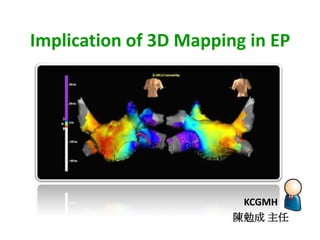
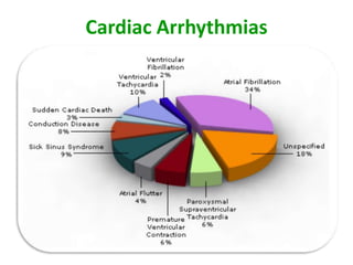
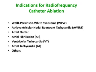
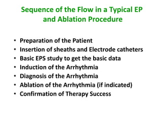
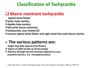
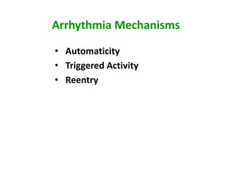
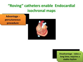
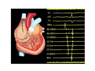
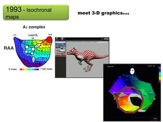
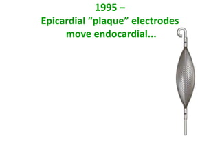
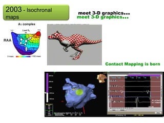
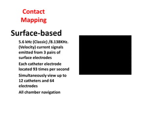
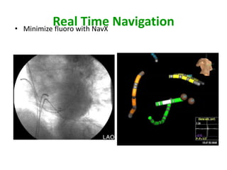
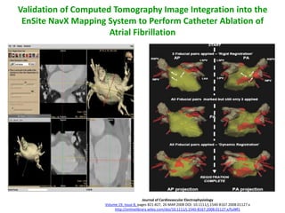
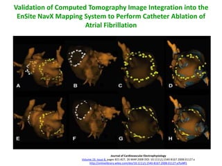
This document discusses the implications of 3D mapping in electrophysiology procedures. It provides an overview of common arrhythmias treated with catheter ablation such as WPW syndrome, AVNRT, atrial flutter, and atrial fibrillation. It describes the typical sequence of an EP study and ablation procedure. It also discusses classification of tachycardias as focal or macroreentrant, and different reentry patterns. The document highlights the development of 3D mapping technologies including contact and non-contact mapping systems, and their ability to create 3D geometry and electroanatomic maps with integration of CT/MRI images. It reviews studies validating the reduction of fluoroscopy time with 3D mapping approaches.
There are many interventional cardiac procedure those need a trans septal puncture of the interatrial septum. This presentation clearly elaborates everything you need to know about the TSP.
Our concepts of heart disease are based on the enormous reservoir of physiologic and anatomic knowledge derived from the past 70 years' of experience in the cardiac catheterization laboratory.
As Andre Cournand remarked in his Nobel lecture of December 11, 1956, the cardiac catheter was the key in the lock.
By turning this key, Cournand and his colleagues led us into a new era in the understanding of normal and disordered cardiac function in huma
rotablation is procedure used in complex pci with heavily calcified lesion for adequate expansion of stent.if used in indicated case and well aware of contraindication is necessary for achieving good results.
Speckle tracking echocardiography (STE) is an echocardiographic imaging technique that analyzes the motion of tissues in the heart by using the naturally occurring speckle pattern in the myocardium or blood when imaged by ultrasound.
Electrical mapping of the heart is a medical procedure that is use to diagnose Arrhythmias in patients. This is done by using sensitive catheter to map the electrical activity in the chambers of the heart.
To begin an electrical mapping procedure, a thin tube called a catheter sheath is inserted into a small incision in the arm or upper thigh. This process is usually visualized using x-rays and a special dye that helps reveal the arteries (called angiography). This catheter is carefully guided through the blood vessels until it is inside the heart.
New Directions in Targeted Therapeutic Approaches for Older Adults With Mantl...i3 Health
i3 Health is pleased to make the speaker slides from this activity available for use as a non-accredited self-study or teaching resource.
This slide deck presented by Dr. Kami Maddocks, Professor-Clinical in the Division of Hematology and
Associate Division Director for Ambulatory Operations
The Ohio State University Comprehensive Cancer Center, will provide insight into new directions in targeted therapeutic approaches for older adults with mantle cell lymphoma.
STATEMENT OF NEED
Mantle cell lymphoma (MCL) is a rare, aggressive B-cell non-Hodgkin lymphoma (NHL) accounting for 5% to 7% of all lymphomas. Its prognosis ranges from indolent disease that does not require treatment for years to very aggressive disease, which is associated with poor survival (Silkenstedt et al, 2021). Typically, MCL is diagnosed at advanced stage and in older patients who cannot tolerate intensive therapy (NCCN, 2022). Although recent advances have slightly increased remission rates, recurrence and relapse remain very common, leading to a median overall survival between 3 and 6 years (LLS, 2021). Though there are several effective options, progress is still needed towards establishing an accepted frontline approach for MCL (Castellino et al, 2022). Treatment selection and management of MCL are complicated by the heterogeneity of prognosis, advanced age and comorbidities of patients, and lack of an established standard approach for treatment, making it vital that clinicians be familiar with the latest research and advances in this area. In this activity chaired by Michael Wang, MD, Professor in the Department of Lymphoma & Myeloma at MD Anderson Cancer Center, expert faculty will discuss prognostic factors informing treatment, the promising results of recent trials in new therapeutic approaches, and the implications of treatment resistance in therapeutic selection for MCL.
Target Audience
Hematology/oncology fellows, attending faculty, and other health care professionals involved in the treatment of patients with mantle cell lymphoma (MCL).
Learning Objectives
1.) Identify clinical and biological prognostic factors that can guide treatment decision making for older adults with MCL
2.) Evaluate emerging data on targeted therapeutic approaches for treatment-naive and relapsed/refractory MCL and their applicability to older adults
3.) Assess mechanisms of resistance to targeted therapies for MCL and their implications for treatment selection
Lung Cancer: Artificial Intelligence, Synergetics, Complex System Analysis, S...Oleg Kshivets
RESULTS: Overall life span (LS) was 2252.1±1742.5 days and cumulative 5-year survival (5YS) reached 73.2%, 10 years – 64.8%, 20 years – 42.5%. 513 LCP lived more than 5 years (LS=3124.6±1525.6 days), 148 LCP – more than 10 years (LS=5054.4±1504.1 days).199 LCP died because of LC (LS=562.7±374.5 days). 5YS of LCP after bi/lobectomies was significantly superior in comparison with LCP after pneumonectomies (78.1% vs.63.7%, P=0.00001 by log-rank test). AT significantly improved 5YS (66.3% vs. 34.8%) (P=0.00000 by log-rank test) only for LCP with N1-2. Cox modeling displayed that 5YS of LCP significantly depended on: phase transition (PT) early-invasive LC in terms of synergetics, PT N0—N12, cell ratio factors (ratio between cancer cells- CC and blood cells subpopulations), G1-3, histology, glucose, AT, blood cell circuit, prothrombin index, heparin tolerance, recalcification time (P=0.000-0.038). Neural networks, genetic algorithm selection and bootstrap simulation revealed relationships between 5YS and PT early-invasive LC (rank=1), PT N0—N12 (rank=2), thrombocytes/CC (3), erythrocytes/CC (4), eosinophils/CC (5), healthy cells/CC (6), lymphocytes/CC (7), segmented neutrophils/CC (8), stick neutrophils/CC (9), monocytes/CC (10); leucocytes/CC (11). Correct prediction of 5YS was 100% by neural networks computing (area under ROC curve=1.0; error=0.0).
CONCLUSIONS: 5YS of LCP after radical procedures significantly depended on: 1) PT early-invasive cancer; 2) PT N0--N12; 3) cell ratio factors; 4) blood cell circuit; 5) biochemical factors; 6) hemostasis system; 7) AT; 8) LC characteristics; 9) LC cell dynamics; 10) surgery type: lobectomy/pneumonectomy; 11) anthropometric data. Optimal diagnosis and treatment strategies for LC are: 1) screening and early detection of LC; 2) availability of experienced thoracic surgeons because of complexity of radical procedures; 3) aggressive en block surgery and adequate lymph node dissection for completeness; 4) precise prediction; 5) adjuvant chemoimmunoradiotherapy for LCP with unfavorable prognosis.
Title: Sense of Smell
Presenter: Dr. Faiza, Assistant Professor of Physiology
Qualifications:
MBBS (Best Graduate, AIMC Lahore)
FCPS Physiology
ICMT, CHPE, DHPE (STMU)
MPH (GC University, Faisalabad)
MBA (Virtual University of Pakistan)
Learning Objectives:
Describe the primary categories of smells and the concept of odor blindness.
Explain the structure and location of the olfactory membrane and mucosa, including the types and roles of cells involved in olfaction.
Describe the pathway and mechanisms of olfactory signal transmission from the olfactory receptors to the brain.
Illustrate the biochemical cascade triggered by odorant binding to olfactory receptors, including the role of G-proteins and second messengers in generating an action potential.
Identify different types of olfactory disorders such as anosmia, hyposmia, hyperosmia, and dysosmia, including their potential causes.
Key Topics:
Olfactory Genes:
3% of the human genome accounts for olfactory genes.
400 genes for odorant receptors.
Olfactory Membrane:
Located in the superior part of the nasal cavity.
Medially: Folds downward along the superior septum.
Laterally: Folds over the superior turbinate and upper surface of the middle turbinate.
Total surface area: 5-10 square centimeters.
Olfactory Mucosa:
Olfactory Cells: Bipolar nerve cells derived from the CNS (100 million), with 4-25 olfactory cilia per cell.
Sustentacular Cells: Produce mucus and maintain ionic and molecular environment.
Basal Cells: Replace worn-out olfactory cells with an average lifespan of 1-2 months.
Bowman’s Gland: Secretes mucus.
Stimulation of Olfactory Cells:
Odorant dissolves in mucus and attaches to receptors on olfactory cilia.
Involves a cascade effect through G-proteins and second messengers, leading to depolarization and action potential generation in the olfactory nerve.
Quality of a Good Odorant:
Small (3-20 Carbon atoms), volatile, water-soluble, and lipid-soluble.
Facilitated by odorant-binding proteins in mucus.
Membrane Potential and Action Potential:
Resting membrane potential: -55mV.
Action potential frequency in the olfactory nerve increases with odorant strength.
Adaptation Towards the Sense of Smell:
Rapid adaptation within the first second, with further slow adaptation.
Psychological adaptation greater than receptor adaptation, involving feedback inhibition from the central nervous system.
Primary Sensations of Smell:
Camphoraceous, Musky, Floral, Pepperminty, Ethereal, Pungent, Putrid.
Odor Detection Threshold:
Examples: Hydrogen sulfide (0.0005 ppm), Methyl-mercaptan (0.002 ppm).
Some toxic substances are odorless at lethal concentrations.
Characteristics of Smell:
Odor blindness for single substances due to lack of appropriate receptor protein.
Behavioral and emotional influences of smell.
Transmission of Olfactory Signals:
From olfactory cells to glomeruli in the olfactory bulb, involving lateral inhibition.
Primitive, less old, and new olfactory systems with different path
TEST BANK for Operations Management, 14th Edition by William J. Stevenson, Ve...kevinkariuki227
TEST BANK for Operations Management, 14th Edition by William J. Stevenson, Verified Chapters 1 - 19, Complete Newest Version.pdf
Prix Galien International 2024 Forum ProgramLevi Shapiro
June 20, 2024, Prix Galien International and Jerusalem Ethics Forum in ROME. Detailed agenda including panels:
- ADVANCES IN CARDIOLOGY: A NEW PARADIGM IS COMING
- WOMEN’S HEALTH: FERTILITY PRESERVATION
- WHAT’S NEW IN THE TREATMENT OF INFECTIOUS,
ONCOLOGICAL AND INFLAMMATORY SKIN DISEASES?
- ARTIFICIAL INTELLIGENCE AND ETHICS
- GENE THERAPY
- BEYOND BORDERS: GLOBAL INITIATIVES FOR DEMOCRATIZING LIFE SCIENCE TECHNOLOGIES AND PROMOTING ACCESS TO HEALTHCARE
- ETHICAL CHALLENGES IN LIFE SCIENCES
- Prix Galien International Awards Ceremony
These simplified slides by Dr. Sidra Arshad present an overview of the non-respiratory functions of the respiratory tract.
Learning objectives:
1. Enlist the non-respiratory functions of the respiratory tract
2. Briefly explain how these functions are carried out
3. Discuss the significance of dead space
4. Differentiate between minute ventilation and alveolar ventilation
5. Describe the cough and sneeze reflexes
Study Resources:
1. Chapter 39, Guyton and Hall Textbook of Medical Physiology, 14th edition
2. Chapter 34, Ganong’s Review of Medical Physiology, 26th edition
3. Chapter 17, Human Physiology by Lauralee Sherwood, 9th edition
4. Non-respiratory functions of the lungs https://academic.oup.com/bjaed/article/13/3/98/278874
Anti ulcer drugs and their Advance pharmacology ||
Anti-ulcer drugs are medications used to prevent and treat ulcers in the stomach and upper part of the small intestine (duodenal ulcers). These ulcers are often caused by an imbalance between stomach acid and the mucosal lining, which protects the stomach lining.
||Scope: Overview of various classes of anti-ulcer drugs, their mechanisms of action, indications, side effects, and clinical considerations.
Ethanol (CH3CH2OH), or beverage alcohol, is a two-carbon alcohol
that is rapidly distributed in the body and brain. Ethanol alters many
neurochemical systems and has rewarding and addictive properties. It
is the oldest recreational drug and likely contributes to more morbidity,
mortality, and public health costs than all illicit drugs combined. The
5th edition of the Diagnostic and Statistical Manual of Mental Disorders
(DSM-5) integrates alcohol abuse and alcohol dependence into a single
disorder called alcohol use disorder (AUD), with mild, moderate,
and severe subclassifications (American Psychiatric Association, 2013).
In the DSM-5, all types of substance abuse and dependence have been
combined into a single substance use disorder (SUD) on a continuum
from mild to severe. A diagnosis of AUD requires that at least two of
the 11 DSM-5 behaviors be present within a 12-month period (mild
AUD: 2–3 criteria; moderate AUD: 4–5 criteria; severe AUD: 6–11 criteria).
The four main behavioral effects of AUD are impaired control over
drinking, negative social consequences, risky use, and altered physiological
effects (tolerance, withdrawal). This chapter presents an overview
of the prevalence and harmful consequences of AUD in the U.S.,
the systemic nature of the disease, neurocircuitry and stages of AUD,
comorbidities, fetal alcohol spectrum disorders, genetic risk factors, and
pharmacotherapies for AUD.
Report Back from SGO 2024: What’s the Latest in Cervical Cancer?bkling
Are you curious about what’s new in cervical cancer research or unsure what the findings mean? Join Dr. Emily Ko, a gynecologic oncologist at Penn Medicine, to learn about the latest updates from the Society of Gynecologic Oncology (SGO) 2024 Annual Meeting on Women’s Cancer. Dr. Ko will discuss what the research presented at the conference means for you and answer your questions about the new developments.
Ozempic: Preoperative Management of Patients on GLP-1 Receptor Agonists Saeid Safari
Preoperative Management of Patients on GLP-1 Receptor Agonists like Ozempic and Semiglutide
ASA GUIDELINE
NYSORA Guideline
2 Case Reports of Gastric Ultrasound
4. Sequence of the Flow in a Typical EP
and Ablation Procedure
• Preparation of the Patient
• Insertion of sheaths and Electrode catheters
• Basic EPS study to get the basic data
• Induction of the Arrhythmia
• Diagnosis of the Arrhythmia
• Ablation of the Arrhythmia (if indicated)
• Confirmation of Therapy Success
5. Classification of Tachycardia
Tachycardia are classified as focal or
macro reentrant tachycardia
Focal tachycardia exhibit activation spreading
from a single focus either radically, circularly or
centrifugally without an electrical activation
spanning the tachycardia cycle length
(right atrial activation is considerably shorter
than the tachycardia cycle length–about 14%)
(Zipes DP, Jalife J. Cardiac Electrophysiology: From cell to bedside, 4th edition. 2004; pg. 500-501)
6. Classification of Tachycardia
Macro reentrant tachycardia
typical atrial flutter
lower loop reentry,
double loop reentry,
left atrial macro reentrant
tachycardia, scar-related AT,
reverse typical atrial flutter and right atrial free wall macro reentry
The various patterns are:
Single loop (like typical atrial flutter)
Figure of eight (made up of two loops)
Reentry through narrow channels adjacent to scar,
anatomic barriers (i.e. tricuspid annulus)
(Zipes DP, Jalife J. Cardiac Electrophysiology: From cell to bedside, 4th edition. 2004; pg. 500-501)
33. Double Potentials
• Double potentials are indicative of a line of block
• Lines of block are either fixed or functional
– Atriotomy sites and the Eustachian ridge are examples of fixed lines
of block
– Evidence exists that block in region of Cristal terminals during atrial
flutter is a form of functional conduction block
39. 2003 - Isochronal meet 3-D graphics...
maps meet 3-D graphics...
Contact Mapping is born
40. Contact
Mapping
Surface-based
5.6 kHz (Classic) /8.138KHz.
(Velocity) current signals
emitted from 3 pairs of
surface electrodes
Each catheter electrode
located 93 times per second
Simultaneously view up to
12 catheters and 64
electrodes
All chamber navigation
45. Validation of Computed Tomography Image Integration into the
EnSite NavX Mapping System to Perform Catheter Ablation of
Atrial Fibrillation
Journal of Cardiovascular Electrophysiology
Volume 19, Issue 8, pages 821-827, 26 MAR 2008 DOI: 10.1111/j.1540-8167.2008.01127.x
http://onlinelibrary.wiley.com/doi/10.1111/j.1540-8167.2008.01127.x/full#f1
46. Validation of Computed Tomography Image Integration into the
EnSite NavX Mapping System to Perform Catheter Ablation of
Atrial Fibrillation
Journal of Cardiovascular Electrophysiology
Volume 19, Issue 8, pages 821-827, 26 MAR 2008 DOI: 10.1111/j.1540-8167.2008.01127.x
http://onlinelibrary.wiley.com/doi/10.1111/j.1540-8167.2008.01127.x/full#f1
47. No fluoroscopic Three Dimensional Mapping
for Arrhythmia Ablation : Tool or Toy ?
Methods
We compared the results of 88 arrhythmia ablations (79
patients) using CARTO with 100 ablations (94 patients)
using the conventional technique.
The ablations were separated into four groups:
AV nodal reentrant tachycardia (AVNRT)
Atrial tachycardia/flutter
ventricular tachycardia (VT)
bypass tract tachycardia.
J Cardiovasc Electrohysio, Vol. 11, pp.239-243,March 2000
48. No fluoroscopic Three Dimensional Mapping
for Arrhythmia Ablation : Tool or Toy ?
Result
The ablation outcomes were excellent and comparable in all four types of the
arrhythmias between the two techniques.
Fluoroscopy time was shorter using the CARTO technique:
10 ± 7 versus 27 ± 15 minutes for AVNRT (P <0.01),
18 ± 17 versus 44 ± 23 minutes for atrial tachycardia and flutter (P < 0.01)
15 ± 12 versus 34 ± 31 minutes for VT (P < 0.05)
21 ± 14 versus 53 ± 32 minutes for by pass tract tachycardia (P < 0.01).
Conclusion
The electro anatomic three-dimensional mapping technique reduced fluoroscopy
time
J Cardiovasc Electrohysio, Vol. 11, pp.239-243,March 2000
The array catheter has a 9 French inner lumen, over-thewiredesign, and a 110 cm usable length.Other catheter features include:• Soft, compliant pigtail tip to reduce trauma. The tip ismade of polyurethane and has a radiopaque marker.• Balloon inflation port to inflate and deflate the arrayballoon.• Hemostasis valve with a sideport for flushing thecatheter during use.• Positive gripping mechanism (PGM) to grip thecentral catheter shaft.• 96-pin connector that connects to the BOB.• Data module that includes the number, status, andexact location of each array electrode. Electrode datais unique for each catheter and is established duringmanufacture. The array is functional with up to eightdisabled electrodes.
The EnSite System uses the chamber geometry to solve theinverse problem for Laplace’s equation at 64 locations onthe geometry surface. The process creates an isopotentialmap surface with more than 3000 virtual EGMs.• The Laplace equation states that if the voltages measuredon an outer boundary (endocardium) are known, voltageseverywhere inside the chamber can be calculated.− The potential field at any one electrode (pointshown) is influenced by electrical potentials fromthe entire endocardium, with the degree of influencediminishing as distance between the electrode andeach endocardial point increases.− The potential field created on the multielectrode array(MEA) surface depends on the shape of the MEA andendocardium as well as their locations relative to oneanother.• Using the known information about the MEA, chambergeometry, and position and distance of the MEA relativeto the endocardial surface, the EnSite System canconstruct endocardial EGMs (virtuals) from MEApotentials.
R and Z values identify the position of the multielectrodearray (MEA) relative to the located electrode on a roving EPcatheter – called the Active EnGuide. These values serve ascoordinates for optimizing the position of the MEA within achamber.• The R value is the distance in millimeters from the activeelectrode to the center of the MEA.• The Z value is the distance in millimeters of the activeelectrode above or below the MEA equator. The Zvalue is negative if the active electrode is distal the MEAequator and positive if it is proximal to the MEA equator.Note: The R and Z values are displayed in the lower portionof the map. The Active EnGuide is setup at the start of a newstudy (see CheckWizard).
The EnGuide catheter is a user-selected contact catheterused to collect location information, build the chambergeometry, and place anatomic and lesion markers. TheEnGuide locator displays the position of the EnGuidecatheter relative to the position of the E1 and E2 electrodes ofthe MEA.
Virtual EGMs display the direction and velocity ofcardiac activation and confirm the site of earliest activation(arrhythmia origin) in the cardiac chamber.• Virtuals are mathematically constructed from voltagemeasurements and plotted as amplitude (verticalaxis) vs. time (horizontal axis). When positioned onthe isopotential map, virtuals verify map color duringdepolarization and are useful in characterizing anarrhythmia.• The tracking virtual is a virtual EGM that automaticallytravels to the most negative location on the map in stepwith the time cursor. The tracking virtual is the red iconwithin the white area of the map. The computed trackingvirtual EGM is displayed in the lower righthand cornerof the map.
The EnSite System can be used with surface electrodepatches to navigate conventional EP catheters to criticalintracardiac sites, without using fluoroscopy. This techniqueis called contact mapping.Using the surface electrodes, the EnSite System:• Collects, records, and displays surface ECG andintracardiac EGM waveforms.• Locates and displays up to 64 EP catheter electrodes inthe cardiac chamber. The location information is usedto navigate EP catheters. It can also be used to createa chamber geometry and place anatomic labels, lesionmarkers, and EnGuide shadows.
Contact mapping works by creating an electrical fieldbetween opposing pairs of surface patch electrodes to locateand display an EP catheter in 3D space.
EnSiteNavX enables physicians to visualize and navigate a myriad of intracardiac catheters in any chamber of the heart. It also simultaneously displays more electrodes and catheters than any other 3-D mapping system currently available.EnSiteNavX is compatible with catheters from all manufacturers. The non-proprietary nature of our technology gives you freedom of choice for diagnostic and ablation catheters.
Respiration Compensation helps minimize the effects ofrespiration during contact mapping studies. It may facilitatemore consistent geometry creation and lesion marking andimproves navigation stability while placing lesions.During Respiration Compensation, the cardiac mappingsystem:• Instructs the clinician to leave all catheters in relativelystable positions.• Collects the x-y-z coordinates of all electrodes andimpedance values of all surface electrodes over a tensecondsampling period.• Identifies respiration through the surface electrodes as agradual rise in intrathoracic impedance and correlates thisrise with a motion artifact on each electrode.• Monitors surface electrodes for the impedance pattern.When respiration occurs, navigation on each electrode iscompensated gradually, in correlation with the degree ofthe impedance change.The cardiac mapping system accommodates changes inrespiration during sampling. Respiration Compensationcan be recalculated any time and is recommended afteradding electrodes, significantly moving the catheter, or whenrespiration motion is still visible.
Figure 3. The 3D lesions (actual lesion positions in 3D space) are shown in the upper panels and the surface lesions (projected onto the CT surface) are shown in the lower panels for a patient undergoing left atrial pulmonary vein (PV) circumferential ablation. Panels (A) and (E) show the lesion sets around both PV pairs, (B) and (F) around the left PVs, and (C) and (G) around the right PVs. Panels (D) and (H) show the same orientations as panels (C) and (G) but with the NavX geometry overlaid onto the CT (without any transparency). The close approximation of the NavX geometry and CT can be appreciated, as well as the relationship of the lesion rings to both. The circumferences of the 3D and surface lesion rings were calculated as described in the main text, and the values compared as a measure of accuracy of geometry/CT integration. A single catheter can be seen in the LA in panels (B) and (F).