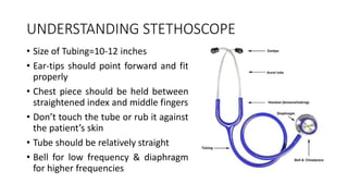
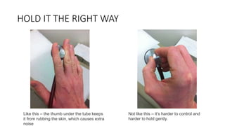
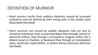
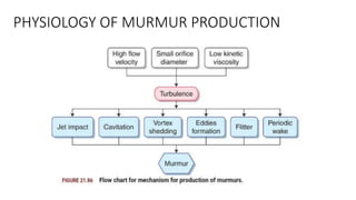
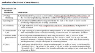
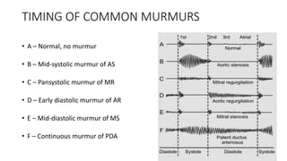
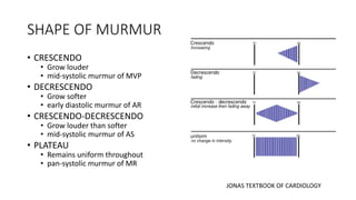
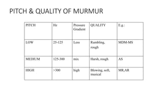
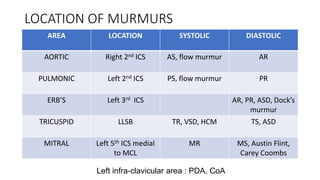
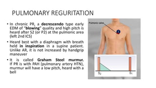
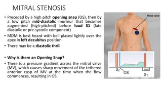
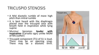
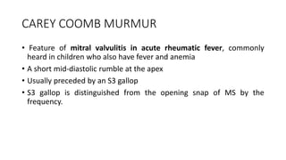
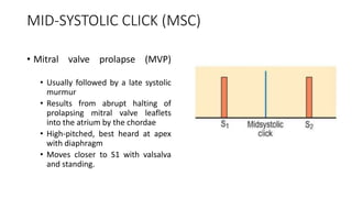
SlideShare a Scribd company logo
1 of 63
Cardiac Murmur &
Dynamic Auscultation
Dr Awadhesh Kr Sharma
Professor Cardiology,LPS Institute of Cardiology,Kanpur
UNDERSTANDING STETHOSCOPE
• Size of Tubing=10-12 inches
• Ear-tips should point forward and fit
properly
• Chest piece should be held between
straightened index and middle fingers
• Don’t touch the tube or rub it against
the patient’s skin
• Tube should be relatively straight
• Bell for low frequency & diaphragm
for higher frequencies
HOLD IT THE RIGHT WAY
Like this – the thumb under the tube keeps
it from rubbing the skin, which causes extra
noise
Not like this – it’s harder to control and
harder to hold gently.
DEFINITION OF MURMUR
• Heart murmur results from auditory vibrations caused by increased
turbulence and are defined by their timing with in the cardiac cycle
(Braunwald text book)
• Heart murmurs are caused by audible vibrations that are due to
increased turbulence from accelerated blood flow through normal or
abnormal orifices, flow through a narrowed or irregular orifice into a
dilated vessel or chamber, or backward flow through an incompetent
valve, ventricular septal defect, or patent ductus arteriosus (Harrison
text book)
PHYSIOLOGY OF MURMUR PRODUCTION
Description of a Murmur
 Timing in the cardiac cycle
 Site of murmur
 Shape of murmur
 Intensity/ Loudness
 Quality & Pitch
 Conduction/ Radiation
 Dynamic changes
TIMING OF MURMURS
• SYSTOLIC
• Early systolic i.e Acute severe MR, Acute TR
• Ejection(mid)-systolic i.e. AS, PS, HCMP ➜Late systolic i.e. MVP
• Pansystolic i.e. MR, TR, VSD
• DIASTOLIC
• Early diastolic i.e. AR, PR, Dock’s murmur
• Mid-diastolic i.e. MS, TS, Austin Flint, Carey-coombs, LA myxoma
• Late diastolic / pre-systolic i.e. MS
• CONTINUOUS
• PDA, severe COA, Severe AS with AR
TIMING OF COMMON MURMURS
• A – Normal, no murmur
• B – Mid-systolic murmur of AS
• C – Pansystolic murmur of MR
• D – Early diastolic murmur of AR
• E – Mid-diastolic murmur of MS
• F – Continuous murmur of PDA
SHAPE OF MURMUR
• CRESCENDO
• Grow louder
• mid-systolic murmur of MVP
• DECRESCENDO
• Grow softer
• early diastolic murmur of AR
• CRESCENDO-DECRESCENDO
• Grow louder than softer
• mid-systolic murmur of AS
• PLATEAU
• Remains uniform throughout
• pan-systolic murmur of MR
JONAS TEXTBOOK OF CARDIOLOGY
PITCH & QUALITY OF MURMUR
PITCH Hz Pressure
Gradient
QUALITY E.g.:
LOW 25-125 Less Rumbling,
rough
MDM-MS
MEDIUM 125-300 mix Harsh, rough AS
HIGH >300 high Blowing, soft,
musical
MR,AR
LOCATION OF MURMURS
AREA LOCATION SYSTOLIC DIASTOLIC
AORTIC Right 2nd ICS AS, flow murmur AR
PULMONIC Left 2nd ICS PS, flow murmur PR
ERB’S Left 3rd ICS AR, PR, ASD, Dock’s
murmur
TRICUSPID LLSB TR, VSD, HCM TS, ASD
MITRAL Left 5th ICS medial
to MCL
MR MS, Austin Flint,
Carey Coombs
Left infra-clavicular area : PDA, CoA
RADIATION OF MURMURS
Freeman and Levine Grading of Heart Murmur
Murmurs accentuated during
Inspiration
TS
TR (Carvallo's sign)
PR
Mild or moderate PS
Severe PS (no further
increase in gradient)
 No change of murmur on
deep inspiration in RV
failure and severe PAH
Expiration
MS
MR
AS
AR
VSD
Pericardial rub
VARIATION WITH RESPIRATION
VARIATION WITH MANEUVRES
• STANDING, VALSALVA
• Decreases venous return and ventricular filling
• Decreases intensity of all murmurs except MVP
• Since outflow obstruction is increased by decreasing preload, it increases the
intensity of HOCM murmur as well
• SQUATTING, LAYING DOWN and LEG RAISING
• Increases venous return and ventricular filling (preload), Hence, increases
cardiac output
• It increases the murmurs of AS, AR, MR and VSD
• It decreases the murmurs of HOCM and MVP
VARIATION WITH MANEUVRES
• HAND-GRIP and PHENYLEPHRINE
• Increases total peripheral resistance (afterload)
• Increases murmurs of AR, MR, VSD and MS
• Decreases murmurs of AS, MVP and HOCM
• INHALED AMYL NITRATE
• Vasodilation causes decreased TPR and afterload
• Increases murmurs of AS, MS, PS, MVP and HOCM
• Decreases murmurs of AR, MR and VSD
• SITTING UP and LEANING FORWARD
• Accentuates S2 and increases the murmur of AR
• LEFT LATERAL DECUBITUS POSITION
• Accentuates the murmur of MS
AORTIC STENOSIS
• An ejection click (EC) followed by a crescendo-
decrescendo ejection systolic murmur at the
aortic area. Heard best with breath held in
expiration in a leaning forward. Sometimes
radiates to the carotid arteries
• In mild AS, the diamond-shaped murmur has an
early peak whereas In severe AS, the murmur
has a late peak
• Gallavardin phenomenon?
It is the systolic murmur of fibrocalcific AS seen
with aging. It has a harsh and noisy quality in
aortic area but is musical over the apex area.
PULMONIC STENOSIS
• An ejection click (EC) followed by a
crescendo-decrescendo ejection
systolic murmur at the pulmonic area,
EC may be absent
• Murmur becomes louder with
inspiration unlike ESM of AS which
becomes softer
• May radiate towards the left clavicle
PS with or without VSD
• If IVS is intact, as obstruction
becomes more severe:
• The murmur lengthens
• Peak of murmur moves closer to
S2
• P2 becomes softer
• If VSD is present (e.g. tetralogy
of Fallot / TOF) as obstruction
becomes more severe:
• The murmur becomes shorter
• Peak becomes more closer to S1
• P2 is very soft or absent
TRICUSPID REGURGITATION
• A holosystolic murmur (HSM) at
LLSB (tricuspid area) that may
obscure S1
• Heard best with diaphragm with
breath held in inspiration
• Murmur becomes louder with
inspiration (Carvallo sign) unlike
murmur of MR
• Murmur may radiate upwards to
upper left sternal border
• Acute severe TR may present as an
early systolic murmur
MITRAL REGURGITATION
• A holosystolic murmur (HSM) at apex area that may
obscure S1
• Heard best with diaphragm
• Murmur may radiate into the precordium or axilla,
due to intra-atrial backflow jet
• Murmur becomes softer with inspiration unlike
murmur of TR (turns loud)
• Murmur of MR is ‘rumbling’ as opposed to musical
quality of fibro-calcific AS
VARIANTS OF MR
• Acute severe MR may present as
an early systolic murmur as left
atrium has a normal size with
limited distensibility.
• Papillary muscle dysfunction
(PMD) seen in acute MI may
present as an late systolic
murmur without a preceding
click. Also called ischemic MR
MVP
• A mid-systolic click (MSC) followed by a mid-
to-late systolic murmur at the apex area.
• It has a “whooping” musical quality.
• Maneuvers that preload (standing, Valsalva)
or afterload (amyl nitrate):
• earlier onset of click (closer to S1)
• longer murmur duration
• decreased murmur intensity
• Maneuvers that preload (leg elevation,
squatting) or afterload (hand grip) :
• Delayed onset of click (closer to S2),
• Shorten murmur duration
• Increase murmur intensity.
HOCM
• A crescendo-decrescendo systolic
murmur at the left sternal border
• Best heard with diaphragm in
standing position.
• Like in AS, there is LVOT*
obstruction but at a sub-valvular
level, due to ASH**
• Aortic valve is not involved so there
is no ejection click, as heard with AS
VSD
• A pansystolic murmur (PSM) at the left sternal
border, with a harsh blowing character, becomes
louder with expiration
• There may be an associated systolic thrill, louder
the murmur, smaller the defect
Roger’s murmur – the loud PSM of small VSD
• Murmur of supra-cristal type of VSD is heard in
the pulmonic area
• If pulmonary hypertension is present,
there will be a loud P2 and a right ventricular
heave. But if there is an associated severe
pulmonic stenosis (TOF), P2 will be soft/absent
ASD
• A ejection systolic murmur (ESM)
without a preceding click is heard at the
upper sternal border. It is due to the
excessive flow across the pulmonary
valve. It is soft and heard best with a
bell. Louder the murmur, smaller the
defect
• Wide fixed splitting of S2 at pulmonic
area is the most important feature
• A diastolic murmur may be heard at the
LLSB due to increased flow across
tricuspid valve
AORTIC REGURGITATION
• In chronic AR, a decrescendo type EDM of
“blowing” quality and high pitch is heard after S2
(or A2) at the left 3rd ICS- Erb’s area
• It is heard best with a diaphragm with breath held
in expiration in a patient leaning forward
• If the AR is due to aortic root dilatation, diastolic
murmur will radiate into the aortic area
• Any maneuver that increases the afterload will
increase the murmur of AR, as it will tend to
favour backflow into the ventricle. i.e. handgrip
Aortic regurgitation cont…
• There can also be a mid-systolic murmur with AR,
due to the increased flow across the aortic valve
during systole
• In AR, a third murmur may be heard, called Austin-
Flint murmur, which is a soft mid-to-late diastolic
murmur of low pitch and “rumbling” quality. It is
heard at the apex, best with a bell during expiration
• It is thought to be due to functional mitral valve
stenosis, as backflow jet from aorta presses on the
anterior mitral leaflet to slightly occlude the blood
flow from LA into LV during diastole
• Rarely, an early diastolic murmur, called Cole-Cecil
murmur, may be heard in axilla
PULMONARY REGURITATION
• In chronic PR, a decrescendo type early
EDM of “blowing” quality and high pitch is
heard after S2 (or P2) at the pulmonic area
(left 2nd ICS)
• Heard best with a diaphragm with breath
held in inspiration in a supine patient.
Unlike AR, it is not increased by handgrip
maneuver
• It is called Graham Steel murmur.
If PR is with PAH (pulmonary artery HTN),
murmur will have a low pitch, heard with a
bell
MITRAL STENOSIS
• Preceded by a high pitch opening snap (OS), then by
a low pitch mid-diastolic murmur that becomes
augmented (high-pitched) before loud S1 (late
diastolic or pre-systolic component)
• MDM is best heard with bell placed lightly over the
apex in left decubitus position
• There may be a diastolic thrill
• Why is there an Opening Snap?
• There is a pressure gradient across the mitral valve
(MV), which causes sharp movement of the tethered
anterior cusp of MV at the time when the flow
commences, resulting in OS.
AUSTIN FLINT VS MDM OF MS
TRICUSPID STENOSIS
• A Mid diastolic rumble of more high
pitch than mitral rumble
• It is best heard with the diaphragm
placed over the tricuspid area with
breath held in inspiration while lying
supine
• Murmur becomes louder with
inspiration (Carvallo sign) unlike MDM
of MS (softer)
• Tricuspid component (T1) of S1 is loud.
There may be an opening snap.
There may be a diastolic thrill.
CAREY COOMB MURMUR
• Feature of mitral valvulitis in acute rheumatic fever, commonly
heard in children who also have fever and anemia
• A short mid-diastolic rumble at the apex
• Usually preceded by an S3 gallop
• S3 gallop is distinguished from the opening snap of MS by the
frequency.
CABOT-LOCKE MURMUR
• Feature of severe anemia
• An early diastolic murmur, resembling the murmur of AR, can be
heard at the Erb’s area
• Unlike murmur of AR, it does not have a decrescendo
• It is an innocent murmur that resolves with treatment of anemia
• It is due to the increased flow from aorta to coronary arteries that
arise from the aortic root
DOCK’S MURMUR
• Feature of severe stenosis of left anterior descending coronary artery
(LAD)
• A crescendo-decrescendo type diastolic murmur with late
accentuation
• In a sharply localized area: 4 cm left of sternal border in left 4th ICS
• It is detected only if the patient is sitting upright
RYTAND MURMUR
• Feature of chronic complete heart block
• A mid-to-late diastolic murmur at the apex
• It is thought that murmur is due to increased flow across AV valves
due to a slow heart rate
• It may also be due to antegrade flow across the closing mitral valve
(when LA is contracting but LV also contracts before it is full)
KEY HODGKIN MURMUR
• Feature of syphilitic aortitis
• An early diastolic murmur, resembling the murmur of AR, but heard
only at the aortic area instead of Erb’s area
• It has a “raspy” quality (saw cutting through wood)
• It is due to the retroversion of aortic valve leaflets during diastole due
to inflammation
“Innocent murmur”
• “functional murmur or flow murmur”
• Doesn’t radiate
• Underlying cardiovascular system is entirely normal and murmur is
audible at rest
• Results from normal turbulence occurring during ejection of blood
from heart
• Typical innocent flow murmur arises in pulmonary artery
INNOCENT MURMUR CHARACTERSTICS
Physiologic murmurs
• Caused by transient increase in blood volume and/or velocity of
ejection
• Common causes-Anemia, anxiety, fever, thyrotoxicosis, pregnancy
• Murmur will no longer audible or become faint (grade 1-2/6)
innocent murmur after correction of increase blood flow or
augmented cardiac contractility
EJECTION CLICK (EC)
Results from sudden opening of a stenotic valve
• AORTIC EJECTION CLICK
High-pitched, best heard at the aortic area, but widely audible.
Occurs earlier as severity of stenosis increases ➜Followed by a
systolic murmur and not seen in calcific AS as the cusps are rigid
• PULMONARY EJECTION CLICK
High-pitched, best heard at the pulmonic area with a diaphragm,
softens with inspiration. ➜Coincides with a wave of JVP.
MID-SYSTOLIC CLICK (MSC)
• Mitral valve prolapse (MVP)
• Usually followed by a late systolic
murmur
• Results from abrupt halting of
prolapsing mitral valve leaflets
into the atrium by the chordae
• High-pitched, best heard at apex
with diaphragm
• Moves closer to S1 with valsalva
and standing.
OPENING SNAP (OS)
• Mitral stenosis (MS), rarely in
tricuspid stenosis
• Occur early in diastole after S2 but
before S3
• May be followed by apical diastolic
rumble in MS
• Results from sudden opening of a
stenotic valve
• High-pitched, best heard at apex by
diaphragm in left decubitus position
“Softer and closer to S2 in severe MS”
PERICARDIAL KNOCK (PK)
• Constrictive pericarditis
• Occur early in diastole, after S2
but before S3
• Sharper, higher pitch than S3 and
OS
• Heard widely but best at apex with
diaphragm
• Knock coincides with rapid Y
descent of JVP
• JVP increases with inspiration
(Kussmaul’s sign)
TUMOR PLOP
• Myxoma or vegetation in left
atrium
• Causing ventricular in-flow
obstruction
• Occurs in early diastole, after S2
• Low pitch, heard best at apex with
bell
• A diastolic murmur may also be
appreciated
PERICARDIAL FRICTION RUB
• A coarse scratching or leathery sound (acute viral pericarditis
and Dressler’s syndrome 24-72 hours after MI)
• Best heard over the bare area of heart with diaphragm with the
breath held in expiration. Vary in intensity over time, and with
position of patient (louder when leaning forward and over)
• No change with respiration. In contrast, pleural rub disappears
when breath is held
• 3 components : presystolic rub during atrial filling
ventricular systolic rub (loudest)
ventricular diastolic rub (after a2p2)
HAMMAN’S CRUNCH
• A series of precordial crackles that correlate with the heartbeat and
not with respiration
• “Crunching” sounds are produced by the heart beating against air-
filled tissues.
• A finding heard in pneumo-pericardium:
• Following heart surgery
• Vigorous resuscitation
• Iatrogenic i.e. pericardiocentesis, bronchoscopy
• Also in pneumo-mediastinum, spontaneous mediastinal emphysema,
Boerhaave syndrome
Physiological maneuver
1. Valsalva
2. Muller
3. Passive leg elevation
4. Sudden standing from
squatting or lying down
5. Sudden squatting
6. Isometric exercise
7. Respiration
Dynamic Auscultation
Physiological maneuver
1. Valsalva
2. Muller
3. Passive leg elevation
4. Sudden standing from
squatting or lying down
5. Sudden squatting
6. Isometric exercise
Pharmacological
• Amyl Nitrate inhalation
• Methoxamine/phenylephrine
Physiological maneuver
1. Valsalva
2. Muller
3. Passive leg elevation
4. Sudden standing from
squatting or lying down
5. Sudden squatting
6. Isometric exercise
7. Respiration
Pharmacological
• Amyl Nitrate inhalation
• Methoxamine/phenylephrine
Other postural changes
• Left lateral- MS
• Sitting up and leaning forward- AR
• Stretching of neck- venous hum
1.Valsalva maneuver
•Exhale forcefully in to a mercury manometer to generate
pressure of 40 mm Hg for 20 sec
•Deep inspiration followed forced expiration against a closed
glottis for 20 seconds
Stages
1.Onset of straining
2.Continued straining (Strain phase)
3.End of expiration (Release phase)
4.Recovery (5-10 sec after end of
expiration) (Overshoot phase)
Stages
1.Onset of straining
2.Continued straining
3.End of expiration
4.Recovery (5-10 sec after end of
expiration)
VALSALVA PHASE I
• increased intrathoracic pressure-> pulmonary
circulation is squeezed, so blood is pushed to left
heart and there is increase in BP and LV output
Stages
1.Onset of straining
2.Continued straining
3.End of expiration
4.Recovery(5-10 sec after end of expir)
VALSALVA PHASE I
• increased intrathoracic pressure-> pulmonary
circulation is squeezed, so blood is pushed to left
heart and there is increase in BP and LV output
VALSALVA PHASE II
• d/t increase intrathoracic pressure, systemic
venous return decrease
• Filling of right and then left side chamber reduced
• Stroke volume reduced
• Mean arterial and pulse pressures falls
• Reflex tachycardia
Stages
1.Onset of straining
2.Continued straining
3.End of expiration
4.Recovery(5-10 sec after end of expir)
VALSALVA PHASE I
• increased intrathoracic pressure-> pulmonary
circulation is squeezed, so blood is pushed to left
heart and there is increase in BP and LV output
VALSALVA PHASE II
• d/t increase intrathoracic pressure, systemic
venous return decrease
• Filling of right and then left side reduced
• Stroke volume reduced
• Mean arterial and pulse pressures falls
• Reflex tachycardia
PHASE III VALSALVA RELEASE
• As the pressure on chest is released, allowing
pulmonary vessels and aorta to re-expand causing
a further initial slight fall in SV due to decrease LA
return and increase aortic volume
• Venous blood can once more enter chest and
heart, CO begins to increase
Stages
1.Onset of straining
2.Continued straining
3.End of expiration
4.Recovery(5-10 sec after end of expir)
VALSALVA PHASE I
• increased intrathoracic pressure-> pulmonary
circulation is squeezed, so blood is pushed to left
heart and there is increase in BP and LV output
VALSALVA PHASE II
• d/t inc intrathoracic pressure, systemic venous
return decrease
• Filling of right and then left side reduced
• Stroke volume reduced
• Mean arterial and pulse pressures falls
• Reflex tachycardia
PHASE III VALSALVA (RELEASE PHASE)
• As the pressure on chest is released, allowing
pulmonary vessels and aorta to re-expand causing
a further initial slight fall in SV due to decr LA
return and incr aortic volume
• Venous blood can once more enter chest and
heart, CO begins to incr
PHASE IV (OVERSHOOT PHASE)
• Blood pressure over shoots
• increased Vagal stimulation in carotids
• heart rate decrease (bradycardia)
Importance of Valsalva
• The Valsalva maneuver is helpful in differentiating right-
sided systolic murmurs from those originating from the left
heart
• Most useful in identifying the systolic murmurs of HOCM
and MVP
• Upon release of the Valsalva maneuver, murmurs from the
right heart generally return to baseline intensity within 2-3
cardiac cycles whereas left sided murmurs generally takes
5-10 cycles
• Not useful in heart failure, ASD and MS
2.Muller’s maneuver
• Nares held closed, patient has to suck forcibly into a
manometer to generate a negative pressure of 40-50 mm Hg
for 10 seconds and see for changes in intensity of murmur at
end of 10 sec
• Nares pinched, mouth firmly sealed, and asked patient to
inspire for 10 sec
• Historically used to accentuate right sided murmur
• Objective studies has found not useful
• Infrequently performed
Dynamic auscultation helpful in
• AS vs HOCM : Squatting (A/v)/ Post VPCS
Valsalva/Standing (V/A)
• AS x MR : Handgrip (V/A)
Phenyl ephrine (V/A)
Post PVC (A/V)
Amyl nitrate (A/V)
• MS X Austin Flint murmur: Amyl nitrate(A/v)
• PR X AR : Squatting (_/A)
Sustained handgrip (-/A)
• PS X Small VSD : Amyl nitrate (A/V)
Respiration
Thanku

More Related Content

Similar to Cardiac Murmur & Dynamic Auscultation.pptx

Heart Sounds and Murmurs
Heart Sounds and MurmursHeart Sounds and Murmurs
Heart Sounds and Murmursshahzaibashraf7
 
MURMUR Cardio vascular system ready.pptx
MURMUR Cardio vascular system ready.pptxMURMUR Cardio vascular system ready.pptx
MURMUR Cardio vascular system ready.pptxAnujaJacob5
 
Continuous Murmurs
Continuous MurmursContinuous Murmurs
Continuous MurmursSujay Iyer
 
Systolic murmur
Systolic murmurSystolic murmur
Systolic murmurSujay Iyer
 
MULTI VALVULAR HEART DISEASE clinical presentation
MULTI VALVULAR HEART DISEASE clinical presentation MULTI VALVULAR HEART DISEASE clinical presentation
MULTI VALVULAR HEART DISEASE clinical presentation Kurian Joseph
 
[Int. med] heart murmurs from SIMS Lahore
[Int. med] heart murmurs from SIMS Lahore[Int. med] heart murmurs from SIMS Lahore
[Int. med] heart murmurs from SIMS LahoreMuhammad Ahmad
 
Heart Sounds And Murmurs
Heart Sounds And MurmursHeart Sounds And Murmurs
Heart Sounds And MurmursSherry Knowles
 
murmur-1.pptx
murmur-1.pptxmurmur-1.pptx
murmur-1.pptxHibaP5
 
Normal and abnormal Heart sounds (Murmurs).pptx
Normal and abnormal Heart sounds (Murmurs).pptxNormal and abnormal Heart sounds (Murmurs).pptx
Normal and abnormal Heart sounds (Murmurs).pptxDr. Irtaza Rehman
 
ADDED HEART SOUNDS- DR ANISH- 10-6-14.ppsx
ADDED HEART SOUNDS- DR ANISH- 10-6-14.ppsxADDED HEART SOUNDS- DR ANISH- 10-6-14.ppsx
ADDED HEART SOUNDS- DR ANISH- 10-6-14.ppsxAleenaRahim2
 
Percussion and Auscultation of CARDIOVASCULAR system.
Percussion and Auscultation of CARDIOVASCULAR system.Percussion and Auscultation of CARDIOVASCULAR system.
Percussion and Auscultation of CARDIOVASCULAR system.Manoz Marwin
 
Cardiology 1.6. Heart Sounds and Murmurs - by Dr. Farjad Ikram
Cardiology 1.6. Heart Sounds and Murmurs - by Dr. Farjad IkramCardiology 1.6. Heart Sounds and Murmurs - by Dr. Farjad Ikram
Cardiology 1.6. Heart Sounds and Murmurs - by Dr. Farjad IkramFarjad Ikram
 
Auscultation.pptx
Auscultation.pptxAuscultation.pptx
Auscultation.pptxAmrutha0013
 
Cvs examination nov 2020
Cvs examination nov 2020Cvs examination nov 2020
Cvs examination nov 2020rajasthan govt
 
Cns- percussion & auscultation
Cns- percussion & auscultation Cns- percussion & auscultation
Cns- percussion & auscultation Krishna Patel
 

Similar to Cardiac Murmur & Dynamic Auscultation.pptx (20)

Heart Sounds and Murmurs
Heart Sounds and MurmursHeart Sounds and Murmurs
Heart Sounds and Murmurs
 
MURMUR Cardio vascular system ready.pptx
MURMUR Cardio vascular system ready.pptxMURMUR Cardio vascular system ready.pptx
MURMUR Cardio vascular system ready.pptx
 
HEART MURMURS
HEART MURMURSHEART MURMURS
HEART MURMURS
 
Approach to Heart Murmurs
Approach to Heart MurmursApproach to Heart Murmurs
Approach to Heart Murmurs
 
Continuous Murmurs
Continuous MurmursContinuous Murmurs
Continuous Murmurs
 
Systolic murmur
Systolic murmurSystolic murmur
Systolic murmur
 
MULTI VALVULAR HEART DISEASE clinical presentation
MULTI VALVULAR HEART DISEASE clinical presentation MULTI VALVULAR HEART DISEASE clinical presentation
MULTI VALVULAR HEART DISEASE clinical presentation
 
[Int. med] heart murmurs from SIMS Lahore
[Int. med] heart murmurs from SIMS Lahore[Int. med] heart murmurs from SIMS Lahore
[Int. med] heart murmurs from SIMS Lahore
 
Heart Sounds And Murmurs
Heart Sounds And MurmursHeart Sounds And Murmurs
Heart Sounds And Murmurs
 
Ms mr
Ms mrMs mr
Ms mr
 
murmur-1.pptx
murmur-1.pptxmurmur-1.pptx
murmur-1.pptx
 
Normal and abnormal Heart sounds (Murmurs).pptx
Normal and abnormal Heart sounds (Murmurs).pptxNormal and abnormal Heart sounds (Murmurs).pptx
Normal and abnormal Heart sounds (Murmurs).pptx
 
ADDED HEART SOUNDS- DR ANISH- 10-6-14.ppsx
ADDED HEART SOUNDS- DR ANISH- 10-6-14.ppsxADDED HEART SOUNDS- DR ANISH- 10-6-14.ppsx
ADDED HEART SOUNDS- DR ANISH- 10-6-14.ppsx
 
Cvs examination
Cvs examinationCvs examination
Cvs examination
 
Percussion and Auscultation of CARDIOVASCULAR system.
Percussion and Auscultation of CARDIOVASCULAR system.Percussion and Auscultation of CARDIOVASCULAR system.
Percussion and Auscultation of CARDIOVASCULAR system.
 
Cardiology 1.6. Heart Sounds and Murmurs - by Dr. Farjad Ikram
Cardiology 1.6. Heart Sounds and Murmurs - by Dr. Farjad IkramCardiology 1.6. Heart Sounds and Murmurs - by Dr. Farjad Ikram
Cardiology 1.6. Heart Sounds and Murmurs - by Dr. Farjad Ikram
 
Diastolic murmurs
Diastolic murmursDiastolic murmurs
Diastolic murmurs
 
Auscultation.pptx
Auscultation.pptxAuscultation.pptx
Auscultation.pptx
 
Cvs examination nov 2020
Cvs examination nov 2020Cvs examination nov 2020
Cvs examination nov 2020
 
Cns- percussion & auscultation
Cns- percussion & auscultation Cns- percussion & auscultation
Cns- percussion & auscultation
 

More from LPS Institute of Cardiology Kanpur UP India

More from LPS Institute of Cardiology Kanpur UP India (20)

Pragmatic Use of Rosuvastatin for CVD Prevention
Pragmatic Use of Rosuvastatin for CVD PreventionPragmatic Use of Rosuvastatin for CVD Prevention
Pragmatic Use of Rosuvastatin for CVD Prevention
 
HEART FAILURE TREATMENT RECENT ADVANCES 2024
HEART FAILURE TREATMENT RECENT ADVANCES 2024HEART FAILURE TREATMENT RECENT ADVANCES 2024
HEART FAILURE TREATMENT RECENT ADVANCES 2024
 
CT angiography.pptx
CT angiography.pptxCT angiography.pptx
CT angiography.pptx
 
CT angiography Vs Invasive CAG.pptx
CT angiography Vs Invasive CAG.pptxCT angiography Vs Invasive CAG.pptx
CT angiography Vs Invasive CAG.pptx
 
Primary care management in Acute Coronary Syndrome
Primary care management in Acute Coronary SyndromePrimary care management in Acute Coronary Syndrome
Primary care management in Acute Coronary Syndrome
 
Heart sounds,murmurs & Dynamic auscultation.pptx
Heart sounds,murmurs & Dynamic auscultation.pptxHeart sounds,murmurs & Dynamic auscultation.pptx
Heart sounds,murmurs & Dynamic auscultation.pptx
 
acute rheumatic fever .pptx
acute rheumatic fever .pptxacute rheumatic fever .pptx
acute rheumatic fever .pptx
 
ffr.pptx
ffr.pptxffr.pptx
ffr.pptx
 
Mechanical Circulatory Support.pptx
Mechanical Circulatory Support.pptxMechanical Circulatory Support.pptx
Mechanical Circulatory Support.pptx
 
PACEMAKER BASIC AND TIMING CYCLE .pptx
PACEMAKER BASIC AND TIMING CYCLE .pptxPACEMAKER BASIC AND TIMING CYCLE .pptx
PACEMAKER BASIC AND TIMING CYCLE .pptx
 
Vitamin D and heart disease
Vitamin D and heart diseaseVitamin D and heart disease
Vitamin D and heart disease
 
CALCIFIED CORONARY ARTERY LESIONS
CALCIFIED CORONARY ARTERY LESIONSCALCIFIED CORONARY ARTERY LESIONS
CALCIFIED CORONARY ARTERY LESIONS
 
Azelnidipine.pptx
Azelnidipine.pptxAzelnidipine.pptx
Azelnidipine.pptx
 
DAPT & Statin Fixed dose combination.pptx
DAPT & Statin Fixed dose combination.pptxDAPT & Statin Fixed dose combination.pptx
DAPT & Statin Fixed dose combination.pptx
 
TRANSESOPHAGEAL ECHOCARDIOGRAPHY.pptx
TRANSESOPHAGEAL ECHOCARDIOGRAPHY.pptxTRANSESOPHAGEAL ECHOCARDIOGRAPHY.pptx
TRANSESOPHAGEAL ECHOCARDIOGRAPHY.pptx
 
Advances in treatment of Pulmonary Arterial Hypertension
Advances in treatment of Pulmonary Arterial HypertensionAdvances in treatment of Pulmonary Arterial Hypertension
Advances in treatment of Pulmonary Arterial Hypertension
 
pulmonary hypertension.pptx
pulmonary hypertension.pptxpulmonary hypertension.pptx
pulmonary hypertension.pptx
 
CALCIUM CHANNEL BLOCKERS AND CARDIOVASCULAR SAFETY.pptx
CALCIUM CHANNEL BLOCKERS AND CARDIOVASCULAR SAFETY.pptxCALCIUM CHANNEL BLOCKERS AND CARDIOVASCULAR SAFETY.pptx
CALCIUM CHANNEL BLOCKERS AND CARDIOVASCULAR SAFETY.pptx
 
Dapagliflozin
DapagliflozinDapagliflozin
Dapagliflozin
 
Ecg in emergency room
Ecg in emergency roomEcg in emergency room
Ecg in emergency room
 

Recently uploaded

Meghan Sutherland In Media Res Media Component
Meghan Sutherland In Media Res Media ComponentMeghan Sutherland In Media Res Media Component
Meghan Sutherland In Media Res Media ComponentInMediaRes1
 
Blooming Together_ Growing a Community Garden Worksheet.docx
Blooming Together_ Growing a Community Garden Worksheet.docxBlooming Together_ Growing a Community Garden Worksheet.docx
Blooming Together_ Growing a Community Garden Worksheet.docxUnboundStockton
 
Pharmacognosy Flower 3. Compositae 2023.pdf
Pharmacognosy Flower 3. Compositae 2023.pdfPharmacognosy Flower 3. Compositae 2023.pdf
Pharmacognosy Flower 3. Compositae 2023.pdfMahmoud M. Sallam
 
Capitol Tech U Doctoral Presentation - April 2024.pptx
Capitol Tech U Doctoral Presentation - April 2024.pptxCapitol Tech U Doctoral Presentation - April 2024.pptx
Capitol Tech U Doctoral Presentation - April 2024.pptxCapitolTechU
 
ENGLISH 7_Q4_LESSON 2_ Employing a Variety of Strategies for Effective Interp...
ENGLISH 7_Q4_LESSON 2_ Employing a Variety of Strategies for Effective Interp...ENGLISH 7_Q4_LESSON 2_ Employing a Variety of Strategies for Effective Interp...
ENGLISH 7_Q4_LESSON 2_ Employing a Variety of Strategies for Effective Interp...JhezDiaz1
 
Organic Name Reactions for the students and aspirants of Chemistry12th.pptx
Organic Name Reactions  for the students and aspirants of Chemistry12th.pptxOrganic Name Reactions  for the students and aspirants of Chemistry12th.pptx
Organic Name Reactions for the students and aspirants of Chemistry12th.pptxVS Mahajan Coaching Centre
 
MARGINALIZATION (Different learners in Marginalized Group
MARGINALIZATION (Different learners in Marginalized GroupMARGINALIZATION (Different learners in Marginalized Group
MARGINALIZATION (Different learners in Marginalized GroupJonathanParaisoCruz
 
ECONOMIC CONTEXT - LONG FORM TV DRAMA - PPT
ECONOMIC CONTEXT - LONG FORM TV DRAMA - PPTECONOMIC CONTEXT - LONG FORM TV DRAMA - PPT
ECONOMIC CONTEXT - LONG FORM TV DRAMA - PPTiammrhaywood
 
Like-prefer-love -hate+verb+ing & silent letters & citizenship text.pdf
Like-prefer-love -hate+verb+ing & silent letters & citizenship text.pdfLike-prefer-love -hate+verb+ing & silent letters & citizenship text.pdf
Like-prefer-love -hate+verb+ing & silent letters & citizenship text.pdfMr Bounab Samir
 
DATA STRUCTURE AND ALGORITHM for beginners
DATA STRUCTURE AND ALGORITHM for beginnersDATA STRUCTURE AND ALGORITHM for beginners
DATA STRUCTURE AND ALGORITHM for beginnersSabitha Banu
 
Alper Gobel In Media Res Media Component
Alper Gobel In Media Res Media ComponentAlper Gobel In Media Res Media Component
Alper Gobel In Media Res Media ComponentInMediaRes1
 
Computed Fields and api Depends in the Odoo 17
Computed Fields and api Depends in the Odoo 17Computed Fields and api Depends in the Odoo 17
Computed Fields and api Depends in the Odoo 17Celine George
 
ECONOMIC CONTEXT - PAPER 1 Q3: NEWSPAPERS.pptx
ECONOMIC CONTEXT - PAPER 1 Q3: NEWSPAPERS.pptxECONOMIC CONTEXT - PAPER 1 Q3: NEWSPAPERS.pptx
ECONOMIC CONTEXT - PAPER 1 Q3: NEWSPAPERS.pptxiammrhaywood
 
EPANDING THE CONTENT OF AN OUTLINE using notes.pptx
EPANDING THE CONTENT OF AN OUTLINE using notes.pptxEPANDING THE CONTENT OF AN OUTLINE using notes.pptx
EPANDING THE CONTENT OF AN OUTLINE using notes.pptxRaymartEstabillo3
 
CELL CYCLE Division Science 8 quarter IV.pptx
CELL CYCLE Division Science 8 quarter IV.pptxCELL CYCLE Division Science 8 quarter IV.pptx
CELL CYCLE Division Science 8 quarter IV.pptxJiesonDelaCerna
 
How to Configure Email Server in Odoo 17
How to Configure Email Server in Odoo 17How to Configure Email Server in Odoo 17
How to Configure Email Server in Odoo 17Celine George
 
“Oh GOSH! Reflecting on Hackteria's Collaborative Practices in a Global Do-It...
“Oh GOSH! Reflecting on Hackteria's Collaborative Practices in a Global Do-It...“Oh GOSH! Reflecting on Hackteria's Collaborative Practices in a Global Do-It...
“Oh GOSH! Reflecting on Hackteria's Collaborative Practices in a Global Do-It...Marc Dusseiller Dusjagr
 
Introduction to ArtificiaI Intelligence in Higher Education
Introduction to ArtificiaI Intelligence in Higher EducationIntroduction to ArtificiaI Intelligence in Higher Education
Introduction to ArtificiaI Intelligence in Higher Educationpboyjonauth
 
AmericanHighSchoolsprezentacijaoskolama.
AmericanHighSchoolsprezentacijaoskolama.AmericanHighSchoolsprezentacijaoskolama.
AmericanHighSchoolsprezentacijaoskolama.arsicmarija21
 

Recently uploaded (20)

Meghan Sutherland In Media Res Media Component
Meghan Sutherland In Media Res Media ComponentMeghan Sutherland In Media Res Media Component
Meghan Sutherland In Media Res Media Component
 
Blooming Together_ Growing a Community Garden Worksheet.docx
Blooming Together_ Growing a Community Garden Worksheet.docxBlooming Together_ Growing a Community Garden Worksheet.docx
Blooming Together_ Growing a Community Garden Worksheet.docx
 
Pharmacognosy Flower 3. Compositae 2023.pdf
Pharmacognosy Flower 3. Compositae 2023.pdfPharmacognosy Flower 3. Compositae 2023.pdf
Pharmacognosy Flower 3. Compositae 2023.pdf
 
Capitol Tech U Doctoral Presentation - April 2024.pptx
Capitol Tech U Doctoral Presentation - April 2024.pptxCapitol Tech U Doctoral Presentation - April 2024.pptx
Capitol Tech U Doctoral Presentation - April 2024.pptx
 
ENGLISH 7_Q4_LESSON 2_ Employing a Variety of Strategies for Effective Interp...
ENGLISH 7_Q4_LESSON 2_ Employing a Variety of Strategies for Effective Interp...ENGLISH 7_Q4_LESSON 2_ Employing a Variety of Strategies for Effective Interp...
ENGLISH 7_Q4_LESSON 2_ Employing a Variety of Strategies for Effective Interp...
 
Organic Name Reactions for the students and aspirants of Chemistry12th.pptx
Organic Name Reactions  for the students and aspirants of Chemistry12th.pptxOrganic Name Reactions  for the students and aspirants of Chemistry12th.pptx
Organic Name Reactions for the students and aspirants of Chemistry12th.pptx
 
MARGINALIZATION (Different learners in Marginalized Group
MARGINALIZATION (Different learners in Marginalized GroupMARGINALIZATION (Different learners in Marginalized Group
MARGINALIZATION (Different learners in Marginalized Group
 
ECONOMIC CONTEXT - LONG FORM TV DRAMA - PPT
ECONOMIC CONTEXT - LONG FORM TV DRAMA - PPTECONOMIC CONTEXT - LONG FORM TV DRAMA - PPT
ECONOMIC CONTEXT - LONG FORM TV DRAMA - PPT
 
Like-prefer-love -hate+verb+ing & silent letters & citizenship text.pdf
Like-prefer-love -hate+verb+ing & silent letters & citizenship text.pdfLike-prefer-love -hate+verb+ing & silent letters & citizenship text.pdf
Like-prefer-love -hate+verb+ing & silent letters & citizenship text.pdf
 
DATA STRUCTURE AND ALGORITHM for beginners
DATA STRUCTURE AND ALGORITHM for beginnersDATA STRUCTURE AND ALGORITHM for beginners
DATA STRUCTURE AND ALGORITHM for beginners
 
Alper Gobel In Media Res Media Component
Alper Gobel In Media Res Media ComponentAlper Gobel In Media Res Media Component
Alper Gobel In Media Res Media Component
 
Computed Fields and api Depends in the Odoo 17
Computed Fields and api Depends in the Odoo 17Computed Fields and api Depends in the Odoo 17
Computed Fields and api Depends in the Odoo 17
 
ECONOMIC CONTEXT - PAPER 1 Q3: NEWSPAPERS.pptx
ECONOMIC CONTEXT - PAPER 1 Q3: NEWSPAPERS.pptxECONOMIC CONTEXT - PAPER 1 Q3: NEWSPAPERS.pptx
ECONOMIC CONTEXT - PAPER 1 Q3: NEWSPAPERS.pptx
 
EPANDING THE CONTENT OF AN OUTLINE using notes.pptx
EPANDING THE CONTENT OF AN OUTLINE using notes.pptxEPANDING THE CONTENT OF AN OUTLINE using notes.pptx
EPANDING THE CONTENT OF AN OUTLINE using notes.pptx
 
ESSENTIAL of (CS/IT/IS) class 06 (database)
ESSENTIAL of (CS/IT/IS) class 06 (database)ESSENTIAL of (CS/IT/IS) class 06 (database)
ESSENTIAL of (CS/IT/IS) class 06 (database)
 
CELL CYCLE Division Science 8 quarter IV.pptx
CELL CYCLE Division Science 8 quarter IV.pptxCELL CYCLE Division Science 8 quarter IV.pptx
CELL CYCLE Division Science 8 quarter IV.pptx
 
How to Configure Email Server in Odoo 17
How to Configure Email Server in Odoo 17How to Configure Email Server in Odoo 17
How to Configure Email Server in Odoo 17
 
“Oh GOSH! Reflecting on Hackteria's Collaborative Practices in a Global Do-It...
“Oh GOSH! Reflecting on Hackteria's Collaborative Practices in a Global Do-It...“Oh GOSH! Reflecting on Hackteria's Collaborative Practices in a Global Do-It...
“Oh GOSH! Reflecting on Hackteria's Collaborative Practices in a Global Do-It...
 
Introduction to ArtificiaI Intelligence in Higher Education
Introduction to ArtificiaI Intelligence in Higher EducationIntroduction to ArtificiaI Intelligence in Higher Education
Introduction to ArtificiaI Intelligence in Higher Education
 
AmericanHighSchoolsprezentacijaoskolama.
AmericanHighSchoolsprezentacijaoskolama.AmericanHighSchoolsprezentacijaoskolama.
AmericanHighSchoolsprezentacijaoskolama.
 

Cardiac Murmur & Dynamic Auscultation.pptx

  • 1. Cardiac Murmur & Dynamic Auscultation Dr Awadhesh Kr Sharma Professor Cardiology,LPS Institute of Cardiology,Kanpur
  • 2. UNDERSTANDING STETHOSCOPE • Size of Tubing=10-12 inches • Ear-tips should point forward and fit properly • Chest piece should be held between straightened index and middle fingers • Don’t touch the tube or rub it against the patient’s skin • Tube should be relatively straight • Bell for low frequency & diaphragm for higher frequencies
  • 3. HOLD IT THE RIGHT WAY Like this – the thumb under the tube keeps it from rubbing the skin, which causes extra noise Not like this – it’s harder to control and harder to hold gently.
  • 4. DEFINITION OF MURMUR • Heart murmur results from auditory vibrations caused by increased turbulence and are defined by their timing with in the cardiac cycle (Braunwald text book) • Heart murmurs are caused by audible vibrations that are due to increased turbulence from accelerated blood flow through normal or abnormal orifices, flow through a narrowed or irregular orifice into a dilated vessel or chamber, or backward flow through an incompetent valve, ventricular septal defect, or patent ductus arteriosus (Harrison text book)
  • 5. PHYSIOLOGY OF MURMUR PRODUCTION
  • 6.
  • 7. Description of a Murmur  Timing in the cardiac cycle  Site of murmur  Shape of murmur  Intensity/ Loudness  Quality & Pitch  Conduction/ Radiation  Dynamic changes
  • 8. TIMING OF MURMURS • SYSTOLIC • Early systolic i.e Acute severe MR, Acute TR • Ejection(mid)-systolic i.e. AS, PS, HCMP ➜Late systolic i.e. MVP • Pansystolic i.e. MR, TR, VSD • DIASTOLIC • Early diastolic i.e. AR, PR, Dock’s murmur • Mid-diastolic i.e. MS, TS, Austin Flint, Carey-coombs, LA myxoma • Late diastolic / pre-systolic i.e. MS • CONTINUOUS • PDA, severe COA, Severe AS with AR
  • 9. TIMING OF COMMON MURMURS • A – Normal, no murmur • B – Mid-systolic murmur of AS • C – Pansystolic murmur of MR • D – Early diastolic murmur of AR • E – Mid-diastolic murmur of MS • F – Continuous murmur of PDA
  • 10. SHAPE OF MURMUR • CRESCENDO • Grow louder • mid-systolic murmur of MVP • DECRESCENDO • Grow softer • early diastolic murmur of AR • CRESCENDO-DECRESCENDO • Grow louder than softer • mid-systolic murmur of AS • PLATEAU • Remains uniform throughout • pan-systolic murmur of MR JONAS TEXTBOOK OF CARDIOLOGY
  • 11. PITCH & QUALITY OF MURMUR PITCH Hz Pressure Gradient QUALITY E.g.: LOW 25-125 Less Rumbling, rough MDM-MS MEDIUM 125-300 mix Harsh, rough AS HIGH >300 high Blowing, soft, musical MR,AR
  • 12. LOCATION OF MURMURS AREA LOCATION SYSTOLIC DIASTOLIC AORTIC Right 2nd ICS AS, flow murmur AR PULMONIC Left 2nd ICS PS, flow murmur PR ERB’S Left 3rd ICS AR, PR, ASD, Dock’s murmur TRICUSPID LLSB TR, VSD, HCM TS, ASD MITRAL Left 5th ICS medial to MCL MR MS, Austin Flint, Carey Coombs Left infra-clavicular area : PDA, CoA
  • 14. Freeman and Levine Grading of Heart Murmur
  • 15. Murmurs accentuated during Inspiration TS TR (Carvallo's sign) PR Mild or moderate PS Severe PS (no further increase in gradient)  No change of murmur on deep inspiration in RV failure and severe PAH Expiration MS MR AS AR VSD Pericardial rub VARIATION WITH RESPIRATION
  • 16. VARIATION WITH MANEUVRES • STANDING, VALSALVA • Decreases venous return and ventricular filling • Decreases intensity of all murmurs except MVP • Since outflow obstruction is increased by decreasing preload, it increases the intensity of HOCM murmur as well • SQUATTING, LAYING DOWN and LEG RAISING • Increases venous return and ventricular filling (preload), Hence, increases cardiac output • It increases the murmurs of AS, AR, MR and VSD • It decreases the murmurs of HOCM and MVP
  • 17. VARIATION WITH MANEUVRES • HAND-GRIP and PHENYLEPHRINE • Increases total peripheral resistance (afterload) • Increases murmurs of AR, MR, VSD and MS • Decreases murmurs of AS, MVP and HOCM • INHALED AMYL NITRATE • Vasodilation causes decreased TPR and afterload • Increases murmurs of AS, MS, PS, MVP and HOCM • Decreases murmurs of AR, MR and VSD • SITTING UP and LEANING FORWARD • Accentuates S2 and increases the murmur of AR • LEFT LATERAL DECUBITUS POSITION • Accentuates the murmur of MS
  • 18.
  • 19. AORTIC STENOSIS • An ejection click (EC) followed by a crescendo- decrescendo ejection systolic murmur at the aortic area. Heard best with breath held in expiration in a leaning forward. Sometimes radiates to the carotid arteries • In mild AS, the diamond-shaped murmur has an early peak whereas In severe AS, the murmur has a late peak • Gallavardin phenomenon? It is the systolic murmur of fibrocalcific AS seen with aging. It has a harsh and noisy quality in aortic area but is musical over the apex area.
  • 20. PULMONIC STENOSIS • An ejection click (EC) followed by a crescendo-decrescendo ejection systolic murmur at the pulmonic area, EC may be absent • Murmur becomes louder with inspiration unlike ESM of AS which becomes softer • May radiate towards the left clavicle
  • 21. PS with or without VSD • If IVS is intact, as obstruction becomes more severe: • The murmur lengthens • Peak of murmur moves closer to S2 • P2 becomes softer • If VSD is present (e.g. tetralogy of Fallot / TOF) as obstruction becomes more severe: • The murmur becomes shorter • Peak becomes more closer to S1 • P2 is very soft or absent
  • 22. TRICUSPID REGURGITATION • A holosystolic murmur (HSM) at LLSB (tricuspid area) that may obscure S1 • Heard best with diaphragm with breath held in inspiration • Murmur becomes louder with inspiration (Carvallo sign) unlike murmur of MR • Murmur may radiate upwards to upper left sternal border • Acute severe TR may present as an early systolic murmur
  • 23. MITRAL REGURGITATION • A holosystolic murmur (HSM) at apex area that may obscure S1 • Heard best with diaphragm • Murmur may radiate into the precordium or axilla, due to intra-atrial backflow jet • Murmur becomes softer with inspiration unlike murmur of TR (turns loud) • Murmur of MR is ‘rumbling’ as opposed to musical quality of fibro-calcific AS
  • 24. VARIANTS OF MR • Acute severe MR may present as an early systolic murmur as left atrium has a normal size with limited distensibility. • Papillary muscle dysfunction (PMD) seen in acute MI may present as an late systolic murmur without a preceding click. Also called ischemic MR
  • 25. MVP • A mid-systolic click (MSC) followed by a mid- to-late systolic murmur at the apex area. • It has a “whooping” musical quality. • Maneuvers that preload (standing, Valsalva) or afterload (amyl nitrate): • earlier onset of click (closer to S1) • longer murmur duration • decreased murmur intensity • Maneuvers that preload (leg elevation, squatting) or afterload (hand grip) : • Delayed onset of click (closer to S2), • Shorten murmur duration • Increase murmur intensity.
  • 26. HOCM • A crescendo-decrescendo systolic murmur at the left sternal border • Best heard with diaphragm in standing position. • Like in AS, there is LVOT* obstruction but at a sub-valvular level, due to ASH** • Aortic valve is not involved so there is no ejection click, as heard with AS
  • 27. VSD • A pansystolic murmur (PSM) at the left sternal border, with a harsh blowing character, becomes louder with expiration • There may be an associated systolic thrill, louder the murmur, smaller the defect Roger’s murmur – the loud PSM of small VSD • Murmur of supra-cristal type of VSD is heard in the pulmonic area • If pulmonary hypertension is present, there will be a loud P2 and a right ventricular heave. But if there is an associated severe pulmonic stenosis (TOF), P2 will be soft/absent
  • 28. ASD • A ejection systolic murmur (ESM) without a preceding click is heard at the upper sternal border. It is due to the excessive flow across the pulmonary valve. It is soft and heard best with a bell. Louder the murmur, smaller the defect • Wide fixed splitting of S2 at pulmonic area is the most important feature • A diastolic murmur may be heard at the LLSB due to increased flow across tricuspid valve
  • 29. AORTIC REGURGITATION • In chronic AR, a decrescendo type EDM of “blowing” quality and high pitch is heard after S2 (or A2) at the left 3rd ICS- Erb’s area • It is heard best with a diaphragm with breath held in expiration in a patient leaning forward • If the AR is due to aortic root dilatation, diastolic murmur will radiate into the aortic area • Any maneuver that increases the afterload will increase the murmur of AR, as it will tend to favour backflow into the ventricle. i.e. handgrip
  • 30. Aortic regurgitation cont… • There can also be a mid-systolic murmur with AR, due to the increased flow across the aortic valve during systole • In AR, a third murmur may be heard, called Austin- Flint murmur, which is a soft mid-to-late diastolic murmur of low pitch and “rumbling” quality. It is heard at the apex, best with a bell during expiration • It is thought to be due to functional mitral valve stenosis, as backflow jet from aorta presses on the anterior mitral leaflet to slightly occlude the blood flow from LA into LV during diastole • Rarely, an early diastolic murmur, called Cole-Cecil murmur, may be heard in axilla
  • 31. PULMONARY REGURITATION • In chronic PR, a decrescendo type early EDM of “blowing” quality and high pitch is heard after S2 (or P2) at the pulmonic area (left 2nd ICS) • Heard best with a diaphragm with breath held in inspiration in a supine patient. Unlike AR, it is not increased by handgrip maneuver • It is called Graham Steel murmur. If PR is with PAH (pulmonary artery HTN), murmur will have a low pitch, heard with a bell
  • 32. MITRAL STENOSIS • Preceded by a high pitch opening snap (OS), then by a low pitch mid-diastolic murmur that becomes augmented (high-pitched) before loud S1 (late diastolic or pre-systolic component) • MDM is best heard with bell placed lightly over the apex in left decubitus position • There may be a diastolic thrill • Why is there an Opening Snap? • There is a pressure gradient across the mitral valve (MV), which causes sharp movement of the tethered anterior cusp of MV at the time when the flow commences, resulting in OS.
  • 33. AUSTIN FLINT VS MDM OF MS
  • 34. TRICUSPID STENOSIS • A Mid diastolic rumble of more high pitch than mitral rumble • It is best heard with the diaphragm placed over the tricuspid area with breath held in inspiration while lying supine • Murmur becomes louder with inspiration (Carvallo sign) unlike MDM of MS (softer) • Tricuspid component (T1) of S1 is loud. There may be an opening snap. There may be a diastolic thrill.
  • 35. CAREY COOMB MURMUR • Feature of mitral valvulitis in acute rheumatic fever, commonly heard in children who also have fever and anemia • A short mid-diastolic rumble at the apex • Usually preceded by an S3 gallop • S3 gallop is distinguished from the opening snap of MS by the frequency.
  • 36. CABOT-LOCKE MURMUR • Feature of severe anemia • An early diastolic murmur, resembling the murmur of AR, can be heard at the Erb’s area • Unlike murmur of AR, it does not have a decrescendo • It is an innocent murmur that resolves with treatment of anemia • It is due to the increased flow from aorta to coronary arteries that arise from the aortic root
  • 37. DOCK’S MURMUR • Feature of severe stenosis of left anterior descending coronary artery (LAD) • A crescendo-decrescendo type diastolic murmur with late accentuation • In a sharply localized area: 4 cm left of sternal border in left 4th ICS • It is detected only if the patient is sitting upright
  • 38. RYTAND MURMUR • Feature of chronic complete heart block • A mid-to-late diastolic murmur at the apex • It is thought that murmur is due to increased flow across AV valves due to a slow heart rate • It may also be due to antegrade flow across the closing mitral valve (when LA is contracting but LV also contracts before it is full)
  • 39. KEY HODGKIN MURMUR • Feature of syphilitic aortitis • An early diastolic murmur, resembling the murmur of AR, but heard only at the aortic area instead of Erb’s area • It has a “raspy” quality (saw cutting through wood) • It is due to the retroversion of aortic valve leaflets during diastole due to inflammation
  • 40. “Innocent murmur” • “functional murmur or flow murmur” • Doesn’t radiate • Underlying cardiovascular system is entirely normal and murmur is audible at rest • Results from normal turbulence occurring during ejection of blood from heart • Typical innocent flow murmur arises in pulmonary artery
  • 42. Physiologic murmurs • Caused by transient increase in blood volume and/or velocity of ejection • Common causes-Anemia, anxiety, fever, thyrotoxicosis, pregnancy • Murmur will no longer audible or become faint (grade 1-2/6) innocent murmur after correction of increase blood flow or augmented cardiac contractility
  • 43. EJECTION CLICK (EC) Results from sudden opening of a stenotic valve • AORTIC EJECTION CLICK High-pitched, best heard at the aortic area, but widely audible. Occurs earlier as severity of stenosis increases ➜Followed by a systolic murmur and not seen in calcific AS as the cusps are rigid • PULMONARY EJECTION CLICK High-pitched, best heard at the pulmonic area with a diaphragm, softens with inspiration. ➜Coincides with a wave of JVP.
  • 44. MID-SYSTOLIC CLICK (MSC) • Mitral valve prolapse (MVP) • Usually followed by a late systolic murmur • Results from abrupt halting of prolapsing mitral valve leaflets into the atrium by the chordae • High-pitched, best heard at apex with diaphragm • Moves closer to S1 with valsalva and standing.
  • 45. OPENING SNAP (OS) • Mitral stenosis (MS), rarely in tricuspid stenosis • Occur early in diastole after S2 but before S3 • May be followed by apical diastolic rumble in MS • Results from sudden opening of a stenotic valve • High-pitched, best heard at apex by diaphragm in left decubitus position “Softer and closer to S2 in severe MS”
  • 46. PERICARDIAL KNOCK (PK) • Constrictive pericarditis • Occur early in diastole, after S2 but before S3 • Sharper, higher pitch than S3 and OS • Heard widely but best at apex with diaphragm • Knock coincides with rapid Y descent of JVP • JVP increases with inspiration (Kussmaul’s sign)
  • 47. TUMOR PLOP • Myxoma or vegetation in left atrium • Causing ventricular in-flow obstruction • Occurs in early diastole, after S2 • Low pitch, heard best at apex with bell • A diastolic murmur may also be appreciated
  • 48. PERICARDIAL FRICTION RUB • A coarse scratching or leathery sound (acute viral pericarditis and Dressler’s syndrome 24-72 hours after MI) • Best heard over the bare area of heart with diaphragm with the breath held in expiration. Vary in intensity over time, and with position of patient (louder when leaning forward and over) • No change with respiration. In contrast, pleural rub disappears when breath is held • 3 components : presystolic rub during atrial filling ventricular systolic rub (loudest) ventricular diastolic rub (after a2p2)
  • 49. HAMMAN’S CRUNCH • A series of precordial crackles that correlate with the heartbeat and not with respiration • “Crunching” sounds are produced by the heart beating against air- filled tissues. • A finding heard in pneumo-pericardium: • Following heart surgery • Vigorous resuscitation • Iatrogenic i.e. pericardiocentesis, bronchoscopy • Also in pneumo-mediastinum, spontaneous mediastinal emphysema, Boerhaave syndrome
  • 50. Physiological maneuver 1. Valsalva 2. Muller 3. Passive leg elevation 4. Sudden standing from squatting or lying down 5. Sudden squatting 6. Isometric exercise 7. Respiration Dynamic Auscultation
  • 51. Physiological maneuver 1. Valsalva 2. Muller 3. Passive leg elevation 4. Sudden standing from squatting or lying down 5. Sudden squatting 6. Isometric exercise Pharmacological • Amyl Nitrate inhalation • Methoxamine/phenylephrine
  • 52. Physiological maneuver 1. Valsalva 2. Muller 3. Passive leg elevation 4. Sudden standing from squatting or lying down 5. Sudden squatting 6. Isometric exercise 7. Respiration Pharmacological • Amyl Nitrate inhalation • Methoxamine/phenylephrine Other postural changes • Left lateral- MS • Sitting up and leaning forward- AR • Stretching of neck- venous hum
  • 53. 1.Valsalva maneuver •Exhale forcefully in to a mercury manometer to generate pressure of 40 mm Hg for 20 sec •Deep inspiration followed forced expiration against a closed glottis for 20 seconds
  • 54. Stages 1.Onset of straining 2.Continued straining (Strain phase) 3.End of expiration (Release phase) 4.Recovery (5-10 sec after end of expiration) (Overshoot phase)
  • 55. Stages 1.Onset of straining 2.Continued straining 3.End of expiration 4.Recovery (5-10 sec after end of expiration) VALSALVA PHASE I • increased intrathoracic pressure-> pulmonary circulation is squeezed, so blood is pushed to left heart and there is increase in BP and LV output
  • 56. Stages 1.Onset of straining 2.Continued straining 3.End of expiration 4.Recovery(5-10 sec after end of expir) VALSALVA PHASE I • increased intrathoracic pressure-> pulmonary circulation is squeezed, so blood is pushed to left heart and there is increase in BP and LV output VALSALVA PHASE II • d/t increase intrathoracic pressure, systemic venous return decrease • Filling of right and then left side chamber reduced • Stroke volume reduced • Mean arterial and pulse pressures falls • Reflex tachycardia
  • 57. Stages 1.Onset of straining 2.Continued straining 3.End of expiration 4.Recovery(5-10 sec after end of expir) VALSALVA PHASE I • increased intrathoracic pressure-> pulmonary circulation is squeezed, so blood is pushed to left heart and there is increase in BP and LV output VALSALVA PHASE II • d/t increase intrathoracic pressure, systemic venous return decrease • Filling of right and then left side reduced • Stroke volume reduced • Mean arterial and pulse pressures falls • Reflex tachycardia PHASE III VALSALVA RELEASE • As the pressure on chest is released, allowing pulmonary vessels and aorta to re-expand causing a further initial slight fall in SV due to decrease LA return and increase aortic volume • Venous blood can once more enter chest and heart, CO begins to increase
  • 58. Stages 1.Onset of straining 2.Continued straining 3.End of expiration 4.Recovery(5-10 sec after end of expir) VALSALVA PHASE I • increased intrathoracic pressure-> pulmonary circulation is squeezed, so blood is pushed to left heart and there is increase in BP and LV output VALSALVA PHASE II • d/t inc intrathoracic pressure, systemic venous return decrease • Filling of right and then left side reduced • Stroke volume reduced • Mean arterial and pulse pressures falls • Reflex tachycardia PHASE III VALSALVA (RELEASE PHASE) • As the pressure on chest is released, allowing pulmonary vessels and aorta to re-expand causing a further initial slight fall in SV due to decr LA return and incr aortic volume • Venous blood can once more enter chest and heart, CO begins to incr PHASE IV (OVERSHOOT PHASE) • Blood pressure over shoots • increased Vagal stimulation in carotids • heart rate decrease (bradycardia)
  • 59. Importance of Valsalva • The Valsalva maneuver is helpful in differentiating right- sided systolic murmurs from those originating from the left heart • Most useful in identifying the systolic murmurs of HOCM and MVP • Upon release of the Valsalva maneuver, murmurs from the right heart generally return to baseline intensity within 2-3 cardiac cycles whereas left sided murmurs generally takes 5-10 cycles • Not useful in heart failure, ASD and MS
  • 60. 2.Muller’s maneuver • Nares held closed, patient has to suck forcibly into a manometer to generate a negative pressure of 40-50 mm Hg for 10 seconds and see for changes in intensity of murmur at end of 10 sec • Nares pinched, mouth firmly sealed, and asked patient to inspire for 10 sec
  • 61. • Historically used to accentuate right sided murmur • Objective studies has found not useful • Infrequently performed
  • 62. Dynamic auscultation helpful in • AS vs HOCM : Squatting (A/v)/ Post VPCS Valsalva/Standing (V/A) • AS x MR : Handgrip (V/A) Phenyl ephrine (V/A) Post PVC (A/V) Amyl nitrate (A/V) • MS X Austin Flint murmur: Amyl nitrate(A/v) • PR X AR : Squatting (_/A) Sustained handgrip (-/A) • PS X Small VSD : Amyl nitrate (A/V) Respiration