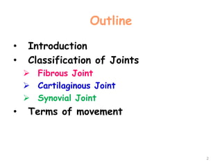
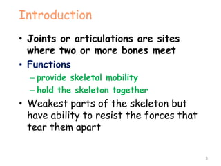
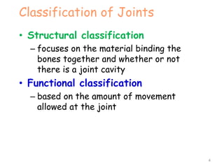
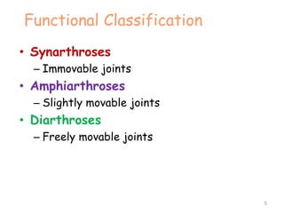
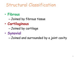
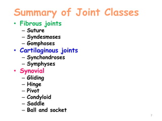
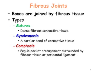
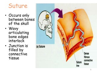
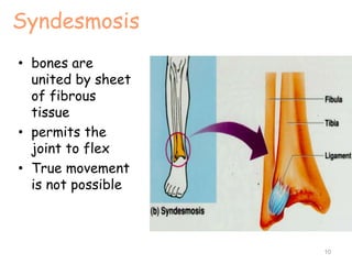
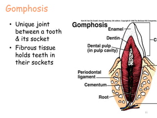
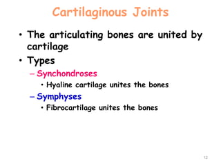
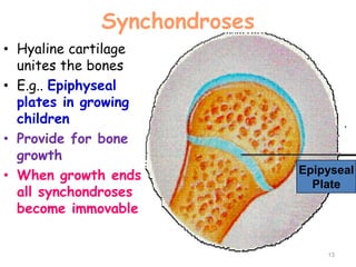
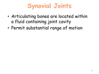
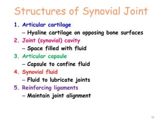
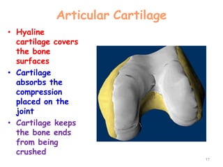
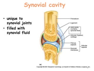
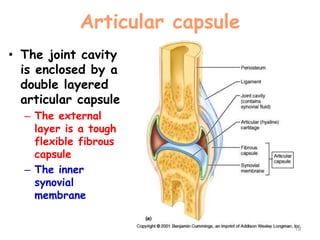
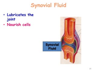
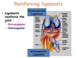
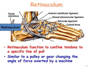
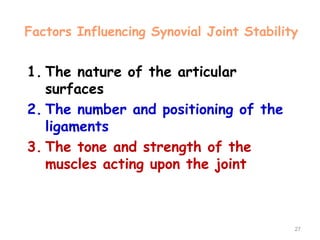
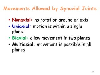
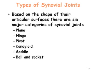
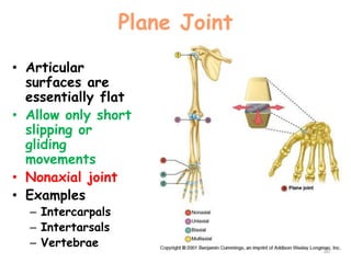
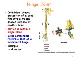
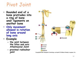
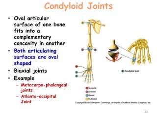
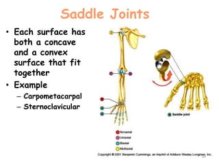
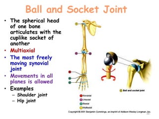
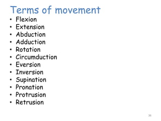
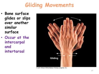
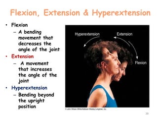
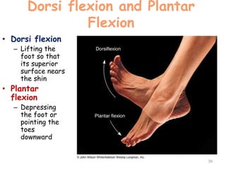
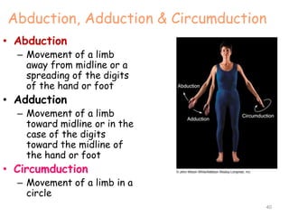
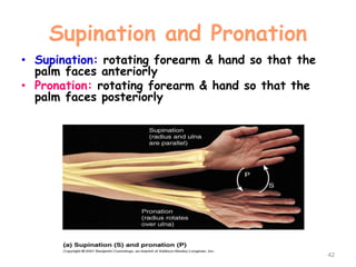
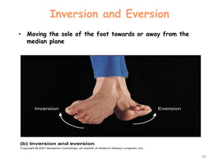
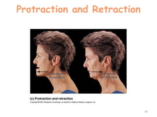
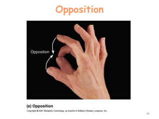
Arthrology
1
Dr. Abdulrahman ( MD )
Outline
• Introduction
• Classification of Joints
 Fibrous Joint
 Cartilaginous Joint
 Synovial Joint
• Terms of movement
2
Introduction
• Joints or articulations are sites
where two or more bones meet
• Functions
– provide skeletal mobility
– hold the skeleton together
• Weakest parts of the skeleton but
have ability to resist the forces that
tear them apart
3
Classification of Joints
• Structural classification
– focuses on the material binding the
bones together and whether or not
there is a joint cavity
• Functional classification
– based on the amount of movement
allowed at the joint
4
Functional Classification
• Synarthroses
– Immovable joints
• Amphiarthroses
– Slightly movable joints
• Diarthroses
– Freely movable joints
5
Structural Classification
• Fibrous
– Joined by fibrous tissue
• Cartilaginous
– Joined by cartilage
• Synovial
– Joined and surrounded by a joint cavity
6
Summary of Joint Classes
• Fibrous joints
– Suture
– Syndesmoses
– Gomphoses
• Cartilaginous joints
– Synchondroses
– Symphyses
• Synovial
– Gliding
– Hinge
– Pivot
– Condyloid
– Saddle
– Ball and socket
7
Fibrous Joints
• Bones are joined by fibrous tissue
• Types
– Sutures
• Dense fibrous connective tissue
– Syndesmosis
• A cord or band of connective tissue
– Gomphosis
• Peg-in-socket arrangement surrounded by
fibrous tissue or peridontal ligament
8
Suture
• Occurs only
between bones
of the skull
• Wavy
articulating
bone edges
interlock
• Junction is
filled by
connective
tissue
9
Syndesmosis
• bones are
united by sheet
of fibrous
tissue
• permits the
joint to flex
• True movement
is not possible
10
Gomphosis
• Unique joint
between a tooth
& its socket
• Fibrous tissue
holds teeth in
their sockets
11
Cartilaginous Joints
• The articulating bones are united by
cartilage
• Types
– Synchondroses
• Hyaline cartilage unites the bones
– Symphyses
• Fibrocartilage unites the bones
12
Synchondroses
• Hyaline cartilage
unites the bones
• E.g.. Epiphyseal
plates in growing
children
• Provide for bone
growth
• When growth ends
all synchondroses
become immovable
13
Epipyseal
Plate
Symphyses
• Bone surfaces are
covered with
articular hyaline
cartilage which is
fused to a pad of
fibrocartilage
• Fibrocartilage is
resilient and acts
as a shock
absorber and
permits limited
movement Pubic Symphysis
14
Synovial Joints
• Articulating bones are located within
a fluid containing joint cavity
• Permit substantial range of motion
15
Structures of Synovial Joint
1. Articular cartilage
– Hyaline cartilage on opposing bone surfaces
2. Joint (synovial) cavity
– Space filled with fluid
3. Articular capsule
– Capsule to confine fluid
4. Synovial fluid
– Fluid to lubricate joints
5. Reinforcing ligaments
– Maintain joint alignment
16
Articular Cartilage
• Hyaline
cartilage covers
the bone
surfaces
• Cartilage
absorbs the
compression
placed on the
joint
• Cartilage keeps
the bone ends
from being
crushed
17
Synovial cavity
• unique to
synovial joints
• filled with
synovial fluid
18
Articular capsule
• The joint cavity
is enclosed by a
double layered
articular capsule
– The external
layer is a tough
flexible fibrous
capsule
– The inner
synovial
membrane
19
Synovial Fluid
• Lubricates the
joint
• Nourish cells
20
Synovial
Fluid
Reinforcing ligaments
• Ligaments
reinforce the
joint
– Extracapsular
– Intracapsular
21
Extracapsular
Ligament
Intracapsular
Ligament
Synovial Joints
• Certain synovial joints have additional
structural features
–Fatty pads cushion the knee and hip
joints
–Fibrocartilage articular discs
(menisci) separates articular
surfaces
• Articular discs improve the fit
between the articulating surface
22
Bursae and Tendon Sheaths
• Bursae and tendon sheaths are
closely associated with synovial
joints
• They are sacs of lubricant
• Function: reduce friction between
adjacent structures
23
Bursae
• Bursae are
flattened fibrous
sacs lined with
synovial
membrane and
containing a thin
film of synovial
fluid
• Common at sites
where ligaments,
skin, muscles or
tendons rub
against a bone
24
Tendon Sheaths
• An elongated bursa
that wraps
completely around a
tendon subjected to
friction
• Tendon slides within
this lubricated
sleeve
• Common at sites
where the tendon is
subject to friction
from other tendons
or bone features
25
Tendon
Sheath
Retinaculum
• Retinaculum function to confine tendons to
a specific line of pull
• Similar to a pulley or gear changing the
angle of force exerted by a machine
26
Retinaculum
Factors Influencing Synovial Joint Stability
1. The nature of the articular
surfaces
2. The number and positioning of the
ligaments
3. The tone and strength of the
muscles acting upon the joint
27
Movements Allowed by Synovial Joints
• Nonaxial: no rotation around an axis
• Uniaxial: motion is within a single
plane
• Biaxial: allow movement in two planes
• Multiaxial: movement is possible in all
planes
28
Types of Synovial Joints
• Based on the shape of their
articular surfaces there are six
major categories of synovial joints
– Plane
– Hinge
– Pivot
– Condyloid
– Saddle
– Ball and socket
29
Plane Joint
• Articular
surfaces are
essentially flat
• Allow only short
slipping or
gliding
movements
• Nonaxial joint
• Examples
– Intercarpals
– Intertarsals
– Vertebrae
30
Hinge Joint
• Cylindrical shaped
projection of a bone
fits into a trough
shaped surface of
another bone
• Motion is within a
single plane
• Joint components
resemble that of a
mechanical hinge
• Example
– elbow joint
31
Pivot Joint
• Rounded end of a
bone protrudes into
a ring of bone
and/ ligaments on
another bone
• Only movement
allowed is rotation
of bone around
long axis
• Example
– the joint between
the atlas and axis
Atlantoaxial Joint
– proximal radioulnar
joint
32
Condyloid Joints
• Oval articular
surface of one bone
fits into a
complementary
concavity in another
• Both articulating
surfaces are oval
shaped
• Biaxial joints
• Example
– Metacarpo-phalangeal
joints
– Atlanto-occipital
Joint
33
Saddle Joints
• Each surface has
both a concave
and a convex
surface that fit
together
• Example
– Carpometacarpal
– Sternoclavicular
34
Ball and Socket Joint
• The spherical head
of one bone
articulates with the
cuplike socket of
another
• Multiaxial
• The most freely
moving synovial
joint
• Movements in all
planes is allowed
• Examples
– Shoulder joint
– Hip joint
35
Terms of movement
• Flexion
• Extension
• Abduction
• Adduction
• Rotation
• Circumduction
• Eversion
• Inversion
• Supination
• Pronation
• Protrusion
• Retrusion
36
Gliding Movements
• Bone surface
glides or slips
over another
similar
surface
• Occur at the
intercarpal
and
intertarsal
37
Flexion, Extension & Hyperextension
• Flexion
– A bending
movement that
decreases the
angle of the joint
• Extension
– A movement
that increases
the angle of the
joint
• Hyperextension
– Bending beyond
the upright
position
38
Dorsi flexion and Plantar
Flexion
• Dorsi flexion
– Lifting the
foot so that
its superior
surface nears
the shin
• Plantar
flexion
– Depressing
the foot or
pointing the
toes
downward
39
Abduction, Adduction & Circumduction
• Abduction
– Movement of a limb
away from midline or a
spreading of the digits
of the hand or foot
• Adduction
– Movement of a limb
toward midline or in the
case of the digits
toward the midline of
the hand or foot
• Circumduction
– Movement of a limb in a
circle
40
Rotation
• Rotation is the
turning of a bone
around its own long
axis
– Only movement
possible between C1
& C2
– Common at the hip
and shoulder joints
41
Supination and Pronation
• Supination: rotating forearm & hand so that the
palm faces anteriorly
• Pronation: rotating forearm & hand so that the
palm faces posteriorly
42
Inversion and Eversion
• Moving the sole of the foot towards or away from the
median plane
43
Protraction and Retraction
44
Elevation and Depression
45
Opposition
46

arthrology

  • 1.
  • 2.
    Outline • Introduction • Classificationof Joints  Fibrous Joint  Cartilaginous Joint  Synovial Joint • Terms of movement 2
  • 3.
    Introduction • Joints orarticulations are sites where two or more bones meet • Functions – provide skeletal mobility – hold the skeleton together • Weakest parts of the skeleton but have ability to resist the forces that tear them apart 3
  • 4.
    Classification of Joints •Structural classification – focuses on the material binding the bones together and whether or not there is a joint cavity • Functional classification – based on the amount of movement allowed at the joint 4
  • 5.
    Functional Classification • Synarthroses –Immovable joints • Amphiarthroses – Slightly movable joints • Diarthroses – Freely movable joints 5
  • 6.
    Structural Classification • Fibrous –Joined by fibrous tissue • Cartilaginous – Joined by cartilage • Synovial – Joined and surrounded by a joint cavity 6
  • 7.
    Summary of JointClasses • Fibrous joints – Suture – Syndesmoses – Gomphoses • Cartilaginous joints – Synchondroses – Symphyses • Synovial – Gliding – Hinge – Pivot – Condyloid – Saddle – Ball and socket 7
  • 8.
    Fibrous Joints • Bonesare joined by fibrous tissue • Types – Sutures • Dense fibrous connective tissue – Syndesmosis • A cord or band of connective tissue – Gomphosis • Peg-in-socket arrangement surrounded by fibrous tissue or peridontal ligament 8
  • 9.
    Suture • Occurs only betweenbones of the skull • Wavy articulating bone edges interlock • Junction is filled by connective tissue 9
  • 10.
    Syndesmosis • bones are unitedby sheet of fibrous tissue • permits the joint to flex • True movement is not possible 10
  • 11.
    Gomphosis • Unique joint betweena tooth & its socket • Fibrous tissue holds teeth in their sockets 11
  • 12.
    Cartilaginous Joints • Thearticulating bones are united by cartilage • Types – Synchondroses • Hyaline cartilage unites the bones – Symphyses • Fibrocartilage unites the bones 12
  • 13.
    Synchondroses • Hyaline cartilage unitesthe bones • E.g.. Epiphyseal plates in growing children • Provide for bone growth • When growth ends all synchondroses become immovable 13 Epipyseal Plate
  • 14.
    Symphyses • Bone surfacesare covered with articular hyaline cartilage which is fused to a pad of fibrocartilage • Fibrocartilage is resilient and acts as a shock absorber and permits limited movement Pubic Symphysis 14
  • 15.
    Synovial Joints • Articulatingbones are located within a fluid containing joint cavity • Permit substantial range of motion 15
  • 16.
    Structures of SynovialJoint 1. Articular cartilage – Hyaline cartilage on opposing bone surfaces 2. Joint (synovial) cavity – Space filled with fluid 3. Articular capsule – Capsule to confine fluid 4. Synovial fluid – Fluid to lubricate joints 5. Reinforcing ligaments – Maintain joint alignment 16
  • 17.
    Articular Cartilage • Hyaline cartilagecovers the bone surfaces • Cartilage absorbs the compression placed on the joint • Cartilage keeps the bone ends from being crushed 17
  • 18.
    Synovial cavity • uniqueto synovial joints • filled with synovial fluid 18
  • 19.
    Articular capsule • Thejoint cavity is enclosed by a double layered articular capsule – The external layer is a tough flexible fibrous capsule – The inner synovial membrane 19
  • 20.
    Synovial Fluid • Lubricatesthe joint • Nourish cells 20 Synovial Fluid
  • 21.
    Reinforcing ligaments • Ligaments reinforcethe joint – Extracapsular – Intracapsular 21 Extracapsular Ligament Intracapsular Ligament
  • 22.
    Synovial Joints • Certainsynovial joints have additional structural features –Fatty pads cushion the knee and hip joints –Fibrocartilage articular discs (menisci) separates articular surfaces • Articular discs improve the fit between the articulating surface 22
  • 23.
    Bursae and TendonSheaths • Bursae and tendon sheaths are closely associated with synovial joints • They are sacs of lubricant • Function: reduce friction between adjacent structures 23
  • 24.
    Bursae • Bursae are flattenedfibrous sacs lined with synovial membrane and containing a thin film of synovial fluid • Common at sites where ligaments, skin, muscles or tendons rub against a bone 24
  • 25.
    Tendon Sheaths • Anelongated bursa that wraps completely around a tendon subjected to friction • Tendon slides within this lubricated sleeve • Common at sites where the tendon is subject to friction from other tendons or bone features 25 Tendon Sheath
  • 26.
    Retinaculum • Retinaculum functionto confine tendons to a specific line of pull • Similar to a pulley or gear changing the angle of force exerted by a machine 26 Retinaculum
  • 27.
    Factors Influencing SynovialJoint Stability 1. The nature of the articular surfaces 2. The number and positioning of the ligaments 3. The tone and strength of the muscles acting upon the joint 27
  • 28.
    Movements Allowed bySynovial Joints • Nonaxial: no rotation around an axis • Uniaxial: motion is within a single plane • Biaxial: allow movement in two planes • Multiaxial: movement is possible in all planes 28
  • 29.
    Types of SynovialJoints • Based on the shape of their articular surfaces there are six major categories of synovial joints – Plane – Hinge – Pivot – Condyloid – Saddle – Ball and socket 29
  • 30.
    Plane Joint • Articular surfacesare essentially flat • Allow only short slipping or gliding movements • Nonaxial joint • Examples – Intercarpals – Intertarsals – Vertebrae 30
  • 31.
    Hinge Joint • Cylindricalshaped projection of a bone fits into a trough shaped surface of another bone • Motion is within a single plane • Joint components resemble that of a mechanical hinge • Example – elbow joint 31
  • 32.
    Pivot Joint • Roundedend of a bone protrudes into a ring of bone and/ ligaments on another bone • Only movement allowed is rotation of bone around long axis • Example – the joint between the atlas and axis Atlantoaxial Joint – proximal radioulnar joint 32
  • 33.
    Condyloid Joints • Ovalarticular surface of one bone fits into a complementary concavity in another • Both articulating surfaces are oval shaped • Biaxial joints • Example – Metacarpo-phalangeal joints – Atlanto-occipital Joint 33
  • 34.
    Saddle Joints • Eachsurface has both a concave and a convex surface that fit together • Example – Carpometacarpal – Sternoclavicular 34
  • 35.
    Ball and SocketJoint • The spherical head of one bone articulates with the cuplike socket of another • Multiaxial • The most freely moving synovial joint • Movements in all planes is allowed • Examples – Shoulder joint – Hip joint 35
  • 36.
    Terms of movement •Flexion • Extension • Abduction • Adduction • Rotation • Circumduction • Eversion • Inversion • Supination • Pronation • Protrusion • Retrusion 36
  • 37.
    Gliding Movements • Bonesurface glides or slips over another similar surface • Occur at the intercarpal and intertarsal 37
  • 38.
    Flexion, Extension &Hyperextension • Flexion – A bending movement that decreases the angle of the joint • Extension – A movement that increases the angle of the joint • Hyperextension – Bending beyond the upright position 38
  • 39.
    Dorsi flexion andPlantar Flexion • Dorsi flexion – Lifting the foot so that its superior surface nears the shin • Plantar flexion – Depressing the foot or pointing the toes downward 39
  • 40.
    Abduction, Adduction &Circumduction • Abduction – Movement of a limb away from midline or a spreading of the digits of the hand or foot • Adduction – Movement of a limb toward midline or in the case of the digits toward the midline of the hand or foot • Circumduction – Movement of a limb in a circle 40
  • 41.
    Rotation • Rotation isthe turning of a bone around its own long axis – Only movement possible between C1 & C2 – Common at the hip and shoulder joints 41
  • 42.
    Supination and Pronation •Supination: rotating forearm & hand so that the palm faces anteriorly • Pronation: rotating forearm & hand so that the palm faces posteriorly 42
  • 43.
    Inversion and Eversion •Moving the sole of the foot towards or away from the median plane 43
  • 44.
  • 45.
  • 46.