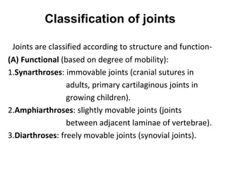
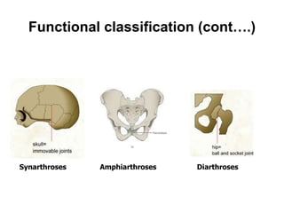
This document discusses the classification and movement of joints in the human body. It begins by defining joints as junctions between bones that allow for movement. Joints are classified based on their structure and degree of mobility. There are three main types of joints: synarthroses which are immovable, amphiarthroses which allow slight movement, and diarthroses which are freely movable. Diarthroses include synovial joints, which have a fluid-filled cavity and include ball-and-socket shoulders and hinge knee joints. Synovial joints are further classified based on their shape and plane of movement, and can be uniaxial, biaxial, or multiaxial. The
Classification and Applied Aspects of JointsMathew Joseph
The structural classification divides joints into fibrous, cartilaginous, and synovial joints depending on the material composing the joint and the presence or absence of a cavity in the joint. The functional classification divides joints into three categories: synarthroses, amphiarthroses, and diarthroses
basic information about human body joints ( arthrology) .
topics cover under this ppt are- definition, classification and clinical anatomy of joints. this may be very helpful for the medical students for understand the basic concepts about joints.
Joint, types of joint By M Thiru murugan.pptxthiru murugan
Joints and Types of Joints
By,M. Thiru murugan
Joint:
A joint or articulation is the connection between bones in the body which link the skeletal system.
Most joints are mobile, allowing the bones to move
Types: 3 types
Fibrous joint - immovable joint (synarthrosis),
Cartilaginous joint - slightly moveable joint (amphiarthrosis)
Synovial joint - freely movable (diarthrosis )
1. Fibrous joint:
Immovable joints, also known as synarthrosis
Theses are fixed & because they do not move.
Fibrous joints have no joint cavity and are connected via fibrous connective tissue.
The skull bones are connected by fibrous joints called sutures. Example: sutures of the skull
2. Cartilaginous joint:
Slightly moveable joint, also known as amphiarthrosis
Cartilaginous joints are a type of joint where the bones are entirely joined by cartilage, either hyaline cartilage or fibrocartilage.
These joints generally allow for slight movements
Example: intervertebral joints
3. Synovial joint:
Freely movable joints, also known as diarthrosis & Bones separated by synovial cavity
A synovial joint is characterized by the presence of a fluid-filled joint cavity contained within a fibrous capsule.
It is the most common type of joint found in the human body
The 3 main features: (i) articular capsule, (ii) articular cartilage, (iii) synovial fluid.
i) Articular capsule: It surrounds the joint and is continuous with the Periosteum
It consists of 2 layers:
Fibrous layer (outer) - It holds together the connecting bones
Synovial layer (inner) - It absorbs and secretes synovial fluid, and is responsible for the nutrient exchange between blood and joint. Also known as the synovium.
Types of synovial joints:
Pivot joint
Hinge joints
Saddle joint
Plane joints
Condyloid joint
The ball & socket joint
A) Pivot joint:
Pivot joint, also called rotary joint, or trochoid joint, that allows only rotary movement around a single axis.
The moving bone rotates within a ring that is formed from a second bone and adjoining ligament.
Ex: joint between the atlas and the axis (first and second cervical vertebrae), directly under the skull, which allows for turning of the head from side to side. Pivot joints also provide for the twisting movement of the bones of the forearm (radius and ulna) against the upper arm,
B) Hinge joints:
It serves to allow motion primarily in one plane.
The hinge joint is made up of two or more bones with articular surfaces that are covered by hyaline cartilage and lubricated by synovial fluid.
Stabilization of each hinge joint is by muscles, ligaments, & other connective tissues, such as the joint capsule.
The hinge joints of the body include the elbow, knee, interphalangeal (IP) joints of the hand and foot and the tibiotalar (ankle) joint of the ankle.
C) Saddle joint:
The saddle joint gets its name because the bone forming one part of the joint is concave (turned inward) at one end and looks like a saddle.
The other bone’s end is convex (turned outward), and
A joint is an articulation between two bones in the body and are broadly classified by the tissue which connects the bones. The three main types of joints are: synovial, cartilaginous and fibrous.
summary of joint classification which made it easy to understand for m.b,b.s ,postgraduate student of anatomy,orthopedics,neurosurgery ,b.p.t and m.p.t.
Knee anatomy and clinical tests 2024.pdfvimalpl1234
This includes all relevant anatomy and clinical tests compiled from standard textbooks, Campbell,netter etc..It is comprehensive and best suited for orthopaedicians and orthopaedic residents.
The prostate is an exocrine gland of the male mammalian reproductive system
It is a walnut-sized gland that forms part of the male reproductive system and is located in front of the rectum and just below the urinary bladder
Function is to store and secrete a clear, slightly alkaline fluid that constitutes 10-30% of the volume of the seminal fluid that along with the spermatozoa, constitutes semen
A healthy human prostate measures (4cm-vertical, by 3cm-horizontal, 2cm ant-post ).
It surrounds the urethra just below the urinary bladder. It has anterior, median, posterior and two lateral lobes
It’s work is regulated by androgens which are responsible for male sex characteristics
Generalised disease of the prostate due to hormonal derangement which leads to non malignant enlargement of the gland (increase in the number of epithelial cells and stromal tissue)to cause compression of the urethra leading to symptoms (LUTS
New Drug Discovery and Development .....NEHA GUPTA
The "New Drug Discovery and Development" process involves the identification, design, testing, and manufacturing of novel pharmaceutical compounds with the aim of introducing new and improved treatments for various medical conditions. This comprehensive endeavor encompasses various stages, including target identification, preclinical studies, clinical trials, regulatory approval, and post-market surveillance. It involves multidisciplinary collaboration among scientists, researchers, clinicians, regulatory experts, and pharmaceutical companies to bring innovative therapies to market and address unmet medical needs.
Prix Galien International 2024 Forum ProgramLevi Shapiro
June 20, 2024, Prix Galien International and Jerusalem Ethics Forum in ROME. Detailed agenda including panels:
- ADVANCES IN CARDIOLOGY: A NEW PARADIGM IS COMING
- WOMEN’S HEALTH: FERTILITY PRESERVATION
- WHAT’S NEW IN THE TREATMENT OF INFECTIOUS,
ONCOLOGICAL AND INFLAMMATORY SKIN DISEASES?
- ARTIFICIAL INTELLIGENCE AND ETHICS
- GENE THERAPY
- BEYOND BORDERS: GLOBAL INITIATIVES FOR DEMOCRATIZING LIFE SCIENCE TECHNOLOGIES AND PROMOTING ACCESS TO HEALTHCARE
- ETHICAL CHALLENGES IN LIFE SCIENCES
- Prix Galien International Awards Ceremony
Ethanol (CH3CH2OH), or beverage alcohol, is a two-carbon alcohol
that is rapidly distributed in the body and brain. Ethanol alters many
neurochemical systems and has rewarding and addictive properties. It
is the oldest recreational drug and likely contributes to more morbidity,
mortality, and public health costs than all illicit drugs combined. The
5th edition of the Diagnostic and Statistical Manual of Mental Disorders
(DSM-5) integrates alcohol abuse and alcohol dependence into a single
disorder called alcohol use disorder (AUD), with mild, moderate,
and severe subclassifications (American Psychiatric Association, 2013).
In the DSM-5, all types of substance abuse and dependence have been
combined into a single substance use disorder (SUD) on a continuum
from mild to severe. A diagnosis of AUD requires that at least two of
the 11 DSM-5 behaviors be present within a 12-month period (mild
AUD: 2–3 criteria; moderate AUD: 4–5 criteria; severe AUD: 6–11 criteria).
The four main behavioral effects of AUD are impaired control over
drinking, negative social consequences, risky use, and altered physiological
effects (tolerance, withdrawal). This chapter presents an overview
of the prevalence and harmful consequences of AUD in the U.S.,
the systemic nature of the disease, neurocircuitry and stages of AUD,
comorbidities, fetal alcohol spectrum disorders, genetic risk factors, and
pharmacotherapies for AUD.
Flu Vaccine Alert in Bangalore Karnatakaaddon Scans
As flu season approaches, health officials in Bangalore, Karnataka, are urging residents to get their flu vaccinations. The seasonal flu, while common, can lead to severe health complications, particularly for vulnerable populations such as young children, the elderly, and those with underlying health conditions.
Dr. Vidisha Kumari, a leading epidemiologist in Bangalore, emphasizes the importance of getting vaccinated. "The flu vaccine is our best defense against the influenza virus. It not only protects individuals but also helps prevent the spread of the virus in our communities," he says.
This year, the flu season is expected to coincide with a potential increase in other respiratory illnesses. The Karnataka Health Department has launched an awareness campaign highlighting the significance of flu vaccinations. They have set up multiple vaccination centers across Bangalore, making it convenient for residents to receive their shots.
To encourage widespread vaccination, the government is also collaborating with local schools, workplaces, and community centers to facilitate vaccination drives. Special attention is being given to ensuring that the vaccine is accessible to all, including marginalized communities who may have limited access to healthcare.
Residents are reminded that the flu vaccine is safe and effective. Common side effects are mild and may include soreness at the injection site, mild fever, or muscle aches. These side effects are generally short-lived and far less severe than the flu itself.
Healthcare providers are also stressing the importance of continuing COVID-19 precautions. Wearing masks, practicing good hand hygiene, and maintaining social distancing are still crucial, especially in crowded places.
Protect yourself and your loved ones by getting vaccinated. Together, we can help keep Bangalore healthy and safe this flu season. For more information on vaccination centers and schedules, residents can visit the Karnataka Health Department’s official website or follow their social media pages.
Stay informed, stay safe, and get your flu shot today!
- Video recording of this lecture in English language: https://youtu.be/lK81BzxMqdo
- Video recording of this lecture in Arabic language: https://youtu.be/Ve4P0COk9OI
- Link to download the book free: https://nephrotube.blogspot.com/p/nephrotube-nephrology-books.html
- Link to NephroTube website: www.NephroTube.com
- Link to NephroTube social media accounts: https://nephrotube.blogspot.com/p/join-nephrotube-on-social-media.html
HOT NEW PRODUCT! BIG SALES FAST SHIPPING NOW FROM CHINA!! EU KU DB BK substit...GL Anaacs
Contact us if you are interested:
Email / Skype : kefaya1771@gmail.com
Threema: PXHY5PDH
New BATCH Ku !!! MUCH IN DEMAND FAST SALE EVERY BATCH HAPPY GOOD EFFECT BIG BATCH !
Contact me on Threema or skype to start big business!!
Hot-sale products:
NEW HOT EUTYLONE WHITE CRYSTAL!!
5cl-adba precursor (semi finished )
5cl-adba raw materials
ADBB precursor (semi finished )
ADBB raw materials
APVP powder
5fadb/4f-adb
Jwh018 / Jwh210
Eutylone crystal
Protonitazene (hydrochloride) CAS: 119276-01-6
Flubrotizolam CAS: 57801-95-3
Metonitazene CAS: 14680-51-4
Payment terms: Western Union,MoneyGram,Bitcoin or USDT.
Deliver Time: Usually 7-15days
Shipping method: FedEx, TNT, DHL,UPS etc.Our deliveries are 100% safe, fast, reliable and discreet.
Samples will be sent for your evaluation!If you are interested in, please contact me, let's talk details.
We specializes in exporting high quality Research chemical, medical intermediate, Pharmaceutical chemicals and so on. Products are exported to USA, Canada, France, Korea, Japan,Russia, Southeast Asia and other countries.
Acute scrotum is a general term referring to an emergency condition affecting the contents or the wall of the scrotum.
There are a number of conditions that present acutely, predominantly with pain and/or swelling
A careful and detailed history and examination, and in some cases, investigations allow differentiation between these diagnoses. A prompt diagnosis is essential as the patient may require urgent surgical intervention
Testicular torsion refers to twisting of the spermatic cord, causing ischaemia of the testicle.
Testicular torsion results from inadequate fixation of the testis to the tunica vaginalis producing ischemia from reduced arterial inflow and venous outflow obstruction.
The prevalence of testicular torsion in adult patients hospitalized with acute scrotal pain is approximately 25 to 50 percent
Report Back from SGO 2024: What’s the Latest in Cervical Cancer?bkling
Are you curious about what’s new in cervical cancer research or unsure what the findings mean? Join Dr. Emily Ko, a gynecologic oncologist at Penn Medicine, to learn about the latest updates from the Society of Gynecologic Oncology (SGO) 2024 Annual Meeting on Women’s Cancer. Dr. Ko will discuss what the research presented at the conference means for you and answer your questions about the new developments.
2. Introduction
• Arthrology: Study of joints
• Synonyms: Articulation (Latin)
Arthron (Greek)
• Joint: a junction between two or more bones
or cartilages.
• A device to permit movements
3. Classification of joints
Joints are classified according to structure and function-
(A) Functional (based on degree of mobility):
1.Synarthroses: immovable joints (cranial sutures in
adults, primary cartilaginous joints in
growing children).
2.Amphiarthroses: slightly movable joints (joints
between adjacent laminae of vertebrae).
3.Diarthroses: freely movable joints (synovial joints).
5. (B) Structural classification
Based on type of connecting tissue and presence or absence of
joint cavity:
1. Fibrous: composed of intervening fibrous tissue with no joint
cavity. Immovable or slightly movable.
2. Cartilaginous: articulating bones are united by cartilage
(hyaline or fibrocartilage) with no joint cavity.
3. Synovial: articulating bones are separated by a fluid-filled joint
cavity.
7. Fibrous joints
1. Sutures: articular surfaces are connected by a thin layer of
connective tissue. They are confined to the skull and are
immovable.
2. Syndesmoses: bones are connected by a considerably greater
amount of connective tissue than in sutures in the form of
interosseous ligaments and membranes. Slight movement is
permitted e.g. interosseous tibiofibular joints, interroseous
radioulnar joints.
3. Gomphoses (peg and socket joint): articulation of teeth in
alveolar sockets of mandible and maxilla by periodontal
ligament.
11. Synovial joints
• Most evolved and freely movable joints
• Have a joint cavity filled with synovial fluid
• Often termed as diarthrodial joints
• Components:
1. Fibrous capsule
2. Synovial membrane
3. Ligaments
4. Articular cartilage
5. Articular disc or meniscus
6. Bursae
7. Fat pads (Haversian glands)
12. Classification of Synovial joints
(A) According to shape of articular surfaces:
1. Plane
2. Hinge
3. Pivot
4. Condylar
5. Ellipsoid
6. Saddle
7. Ball and socket
13. Plane joints
• Articular surfaces are
nearly flat.
• Allow gliding
movements.
• e.g. intercarpal and
intertarsal joints.
14. Hinge joints (Ginglymus)
• Articular surfaces are
pulley shaped.
• Movements are
permitted only in one
plane (transverse
axis).
• Most common
joints.
• e.g. elbow, knee,
ankle and
interphalangeal joints.
15. Pivot joints (Trochoid joints)
• Rounded end of one bone
fits into
the concavity of another
bone.
• The rounded
part is surrounded by a
ligament.
• Limited rotation
around a central axis.
• e.g. superior radio-ulnar
and median atlanto-axial
joints.
16. Condylar joints
• Round articular surface
of one bone fits into a
socket-type articular
surface of another bone.
• Permit movements in
two directions.
• e.g. knee and temporo-
mandibular joints.
17. Ellipsoid joints
• Elliptical convex surface of
one bone articulates with
elliptical concave surface
of another.
• Permit movements in two
directions.
• Combination of
movements cause
circumduction.
• e.g. wrist and atlanto-
occipital joints.
18. Saddle (sellar) joints
• Articular surfaces are
reciprocally saddle
shaped i.e. concavo-
convex.
• Allows a
wide range of movement.
• e.g. first
carpo-metacarpal, sterno-
clavicular,
incudo-malleolar joints.
19. Ball and socket (spheroidal) joints
• Rounded convex surface
of one bone fits into the
cup-like socket of
another bone.
• Permits greatest range
of movements.
• e.g. shoulder, hip and
incudo-stapedial joints.
20. (B) According to plane of movements
• Uniaxial: Hinge and Pivot
• Biaxial: Condylar, ellipsoid, saddle
• Multiaxial: Ball and socket
Uniaxial Biaxial Multiaxial
21. (C) According to number of articulating bones
• Simple (2 bones)
• Compound (more than 2 bones)
23. Special movements:
1. Inversion: movement of the foot medially
2. Eversion: movement of the foot laterally
3. Protraction: movement of the mandible forward
4. Retraction: movement of the protracted part back
to its starting position
5. Elevation: lifting a body part superiorly
6. Depression: moving the elevated part inferiorly
7. Opposition: touching the thumb to the tips of
other fingers
24. REFERENCES
1. Cunningham’s Manual of Practical Anatomy, Vol.1.
2. Grant’s Method of Anatomy, 11th Edition.
3. Vishram Singh. General Anatomy, 1st Edition.