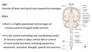
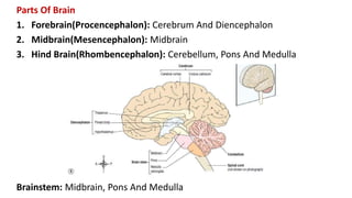
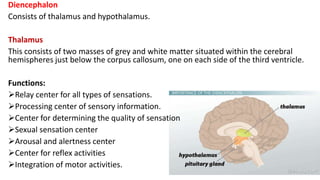
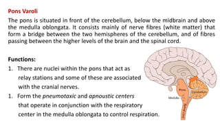
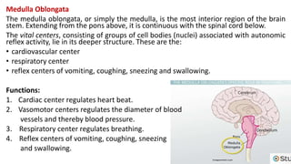
The central nervous system consists of the brain and spinal cord, which are covered by three layers of meninges. The brain is divided into the forebrain, midbrain, and hindbrain. The forebrain includes the cerebrum and diencephalon. The cerebrum is made up of four lobes that control functions like movement, sensation, thought, and memory. The diencephalon contains the thalamus and hypothalamus, which relay sensory information and control autonomic functions respectively. The midbrain relays information between the brain and spinal cord. The hindbrain contains the cerebellum, pons, and medulla, which coordinate movement, relay information, and control vital functions. The spinal
three types: skeletal, cardiac, smooth
Muscle cells are called muscle fibers
Contraction depends on two kinds of Myofilaments
Actin
Myosin
Prefixes to know: myo, mys, or sarco – word relates to muscle
Each muscle is a discrete organ
Muscle Type Overview
Skeletal Muscle tissue
Skeletal
Striated
Voluntary
Cardiac Muscle tissue
Cardiac
Striated
Involuntary
Smooth Muscle tissue
Visceral
Non-striated
Involuntary
Muscle Functions
1. Producing movement
2. Maintaining posture
3. Stabilizing joints
4. Generating heat
Functional Characteristics of Muscles
Excitability (or Irritability) = ability to receive and respond to stimuli
Contractility = ability to shorten forcibly
Extensibility = ability to be stretched or extended beyond resting length
Elasticity = ability to resume resting length after stretchingMuscle (organ)
Fascicle (a portion of the muscle)
Muscle Fiber (a cell)
These levels are supracellular
Connective Tissue Layer
Epimysium
Perimysium
Endomysium
Anatomy of a Muscle
Typical ex. is a skeletal muscle
The following are all subcellular.
Myofibril = or fibril, complex organelle composed of bundles of
myofilaments
Myofilament = macromolecular structure of contractile proteins
Sarcomere = the smallest, single contracting unit of a myofibril, a segment
Gross Anatomy
Deep fascia = binds large groups of muscles into functional groups
Muscle = hundreds of fascicles bound together by epimysium
Fascicle = thousands of muscle fibers bound into discrete units by
perimysium
Muscle fiber = single muscle cell surrounded by endomysium
Generous blood and nerve supply
Microscopic Anatomy of a Muscle Fiber
Muscle Fiber = elongated, cylindrical, multinucleated muscle cell
Sarcolemma = plasma (cell) membrane of a muscle cell
Sarcoplasm = cytoplasm of muscle cell with large amounts of glycogen and
classification of joints. example of different types of joints. different types of joints on the basis of axis of movements. clinical aspects of joints. different between arthritis.
تلخيصات بسيطه تخص طلاب اسنان في ماده الاناتومي
تابعونا علي الصفحه
https://www.facebook.com/dentology7/?ref=bookmarks
او
https://www.facebook.com/Doctor.Hossam.A
THE SPINAL CORD
White Matter of the Spinal Cord
Gray Matter of the Spinal Cord and Spinal Roots
THE BRAIN
Basic Parts and Organizationof the Brain
Parietal Lobe
Occipital Lobe
Cerebellum
Brain Stem
The Brain stem
Medulla Oblongata
Midbrain
Pons
THE SPINAL CORD
White Matter of the Spinal Cord
Gray Matter of the Spinal Cord and Spinal Roots
THE BRAIN
Basic Parts and Organizationof the Brain
The Brain stem
- Video recording of this lecture in English language: https://youtu.be/lK81BzxMqdo
- Video recording of this lecture in Arabic language: https://youtu.be/Ve4P0COk9OI
- Link to download the book free: https://nephrotube.blogspot.com/p/nephrotube-nephrology-books.html
- Link to NephroTube website: www.NephroTube.com
- Link to NephroTube social media accounts: https://nephrotube.blogspot.com/p/join-nephrotube-on-social-media.html
Lung Cancer: Artificial Intelligence, Synergetics, Complex System Analysis, S...Oleg Kshivets
RESULTS: Overall life span (LS) was 2252.1±1742.5 days and cumulative 5-year survival (5YS) reached 73.2%, 10 years – 64.8%, 20 years – 42.5%. 513 LCP lived more than 5 years (LS=3124.6±1525.6 days), 148 LCP – more than 10 years (LS=5054.4±1504.1 days).199 LCP died because of LC (LS=562.7±374.5 days). 5YS of LCP after bi/lobectomies was significantly superior in comparison with LCP after pneumonectomies (78.1% vs.63.7%, P=0.00001 by log-rank test). AT significantly improved 5YS (66.3% vs. 34.8%) (P=0.00000 by log-rank test) only for LCP with N1-2. Cox modeling displayed that 5YS of LCP significantly depended on: phase transition (PT) early-invasive LC in terms of synergetics, PT N0—N12, cell ratio factors (ratio between cancer cells- CC and blood cells subpopulations), G1-3, histology, glucose, AT, blood cell circuit, prothrombin index, heparin tolerance, recalcification time (P=0.000-0.038). Neural networks, genetic algorithm selection and bootstrap simulation revealed relationships between 5YS and PT early-invasive LC (rank=1), PT N0—N12 (rank=2), thrombocytes/CC (3), erythrocytes/CC (4), eosinophils/CC (5), healthy cells/CC (6), lymphocytes/CC (7), segmented neutrophils/CC (8), stick neutrophils/CC (9), monocytes/CC (10); leucocytes/CC (11). Correct prediction of 5YS was 100% by neural networks computing (area under ROC curve=1.0; error=0.0).
CONCLUSIONS: 5YS of LCP after radical procedures significantly depended on: 1) PT early-invasive cancer; 2) PT N0--N12; 3) cell ratio factors; 4) blood cell circuit; 5) biochemical factors; 6) hemostasis system; 7) AT; 8) LC characteristics; 9) LC cell dynamics; 10) surgery type: lobectomy/pneumonectomy; 11) anthropometric data. Optimal diagnosis and treatment strategies for LC are: 1) screening and early detection of LC; 2) availability of experienced thoracic surgeons because of complexity of radical procedures; 3) aggressive en block surgery and adequate lymph node dissection for completeness; 4) precise prediction; 5) adjuvant chemoimmunoradiotherapy for LCP with unfavorable prognosis.
Tom Selleck Health: A Comprehensive Look at the Iconic Actor’s Wellness Journeygreendigital
Tom Selleck, an enduring figure in Hollywood. has captivated audiences for decades with his rugged charm, iconic moustache. and memorable roles in television and film. From his breakout role as Thomas Magnum in Magnum P.I. to his current portrayal of Frank Reagan in Blue Bloods. Selleck's career has spanned over 50 years. But beyond his professional achievements. fans have often been curious about Tom Selleck Health. especially as he has aged in the public eye.
Follow us on: Pinterest
Introduction
Many have been interested in Tom Selleck health. not only because of his enduring presence on screen but also because of the challenges. and lifestyle choices he has faced and made over the years. This article delves into the various aspects of Tom Selleck health. exploring his fitness regimen, diet, mental health. and the challenges he has encountered as he ages. We'll look at how he maintains his well-being. the health issues he has faced, and his approach to ageing .
Early Life and Career
Childhood and Athletic Beginnings
Tom Selleck was born on January 29, 1945, in Detroit, Michigan, and grew up in Sherman Oaks, California. From an early age, he was involved in sports, particularly basketball. which played a significant role in his physical development. His athletic pursuits continued into college. where he attended the University of Southern California (USC) on a basketball scholarship. This early involvement in sports laid a strong foundation for his physical health and disciplined lifestyle.
Transition to Acting
Selleck's transition from an athlete to an actor came with its physical demands. His first significant role in "Magnum P.I." required him to perform various stunts and maintain a fit appearance. This role, which he played from 1980 to 1988. necessitated a rigorous fitness routine to meet the show's demands. setting the stage for his long-term commitment to health and wellness.
Fitness Regimen
Workout Routine
Tom Selleck health and fitness regimen has evolved. adapting to his changing roles and age. During his "Magnum, P.I." days. Selleck's workouts were intense and focused on building and maintaining muscle mass. His routine included weightlifting, cardiovascular exercises. and specific training for the stunts he performed on the show.
Selleck adjusted his fitness routine as he aged to suit his body's needs. Today, his workouts focus on maintaining flexibility, strength, and cardiovascular health. He incorporates low-impact exercises such as swimming, walking, and light weightlifting. This balanced approach helps him stay fit without putting undue strain on his joints and muscles.
Importance of Flexibility and Mobility
In recent years, Selleck has emphasized the importance of flexibility and mobility in his fitness regimen. Understanding the natural decline in muscle mass and joint flexibility with age. he includes stretching and yoga in his routine. These practices help prevent injuries, improve posture, and maintain mobilit
These simplified slides by Dr. Sidra Arshad present an overview of the non-respiratory functions of the respiratory tract.
Learning objectives:
1. Enlist the non-respiratory functions of the respiratory tract
2. Briefly explain how these functions are carried out
3. Discuss the significance of dead space
4. Differentiate between minute ventilation and alveolar ventilation
5. Describe the cough and sneeze reflexes
Study Resources:
1. Chapter 39, Guyton and Hall Textbook of Medical Physiology, 14th edition
2. Chapter 34, Ganong’s Review of Medical Physiology, 26th edition
3. Chapter 17, Human Physiology by Lauralee Sherwood, 9th edition
4. Non-respiratory functions of the lungs https://academic.oup.com/bjaed/article/13/3/98/278874
Title: Sense of Smell
Presenter: Dr. Faiza, Assistant Professor of Physiology
Qualifications:
MBBS (Best Graduate, AIMC Lahore)
FCPS Physiology
ICMT, CHPE, DHPE (STMU)
MPH (GC University, Faisalabad)
MBA (Virtual University of Pakistan)
Learning Objectives:
Describe the primary categories of smells and the concept of odor blindness.
Explain the structure and location of the olfactory membrane and mucosa, including the types and roles of cells involved in olfaction.
Describe the pathway and mechanisms of olfactory signal transmission from the olfactory receptors to the brain.
Illustrate the biochemical cascade triggered by odorant binding to olfactory receptors, including the role of G-proteins and second messengers in generating an action potential.
Identify different types of olfactory disorders such as anosmia, hyposmia, hyperosmia, and dysosmia, including their potential causes.
Key Topics:
Olfactory Genes:
3% of the human genome accounts for olfactory genes.
400 genes for odorant receptors.
Olfactory Membrane:
Located in the superior part of the nasal cavity.
Medially: Folds downward along the superior septum.
Laterally: Folds over the superior turbinate and upper surface of the middle turbinate.
Total surface area: 5-10 square centimeters.
Olfactory Mucosa:
Olfactory Cells: Bipolar nerve cells derived from the CNS (100 million), with 4-25 olfactory cilia per cell.
Sustentacular Cells: Produce mucus and maintain ionic and molecular environment.
Basal Cells: Replace worn-out olfactory cells with an average lifespan of 1-2 months.
Bowman’s Gland: Secretes mucus.
Stimulation of Olfactory Cells:
Odorant dissolves in mucus and attaches to receptors on olfactory cilia.
Involves a cascade effect through G-proteins and second messengers, leading to depolarization and action potential generation in the olfactory nerve.
Quality of a Good Odorant:
Small (3-20 Carbon atoms), volatile, water-soluble, and lipid-soluble.
Facilitated by odorant-binding proteins in mucus.
Membrane Potential and Action Potential:
Resting membrane potential: -55mV.
Action potential frequency in the olfactory nerve increases with odorant strength.
Adaptation Towards the Sense of Smell:
Rapid adaptation within the first second, with further slow adaptation.
Psychological adaptation greater than receptor adaptation, involving feedback inhibition from the central nervous system.
Primary Sensations of Smell:
Camphoraceous, Musky, Floral, Pepperminty, Ethereal, Pungent, Putrid.
Odor Detection Threshold:
Examples: Hydrogen sulfide (0.0005 ppm), Methyl-mercaptan (0.002 ppm).
Some toxic substances are odorless at lethal concentrations.
Characteristics of Smell:
Odor blindness for single substances due to lack of appropriate receptor protein.
Behavioral and emotional influences of smell.
Transmission of Olfactory Signals:
From olfactory cells to glomeruli in the olfactory bulb, involving lateral inhibition.
Primitive, less old, and new olfactory systems with different path
Pulmonary Thromboembolism - etilogy, types, medical- Surgical and nursing man...VarunMahajani
Disruption of blood supply to lung alveoli due to blockage of one or more pulmonary blood vessels is called as Pulmonary thromboembolism. In this presentation we will discuss its causes, types and its management in depth.
Title: Sense of Taste
Presenter: Dr. Faiza, Assistant Professor of Physiology
Qualifications:
MBBS (Best Graduate, AIMC Lahore)
FCPS Physiology
ICMT, CHPE, DHPE (STMU)
MPH (GC University, Faisalabad)
MBA (Virtual University of Pakistan)
Learning Objectives:
Describe the structure and function of taste buds.
Describe the relationship between the taste threshold and taste index of common substances.
Explain the chemical basis and signal transduction of taste perception for each type of primary taste sensation.
Recognize different abnormalities of taste perception and their causes.
Key Topics:
Significance of Taste Sensation:
Differentiation between pleasant and harmful food
Influence on behavior
Selection of food based on metabolic needs
Receptors of Taste:
Taste buds on the tongue
Influence of sense of smell, texture of food, and pain stimulation (e.g., by pepper)
Primary and Secondary Taste Sensations:
Primary taste sensations: Sweet, Sour, Salty, Bitter, Umami
Chemical basis and signal transduction mechanisms for each taste
Taste Threshold and Index:
Taste threshold values for Sweet (sucrose), Salty (NaCl), Sour (HCl), and Bitter (Quinine)
Taste index relationship: Inversely proportional to taste threshold
Taste Blindness:
Inability to taste certain substances, particularly thiourea compounds
Example: Phenylthiocarbamide
Structure and Function of Taste Buds:
Composition: Epithelial cells, Sustentacular/Supporting cells, Taste cells, Basal cells
Features: Taste pores, Taste hairs/microvilli, and Taste nerve fibers
Location of Taste Buds:
Found in papillae of the tongue (Fungiform, Circumvallate, Foliate)
Also present on the palate, tonsillar pillars, epiglottis, and proximal esophagus
Mechanism of Taste Stimulation:
Interaction of taste substances with receptors on microvilli
Signal transduction pathways for Umami, Sweet, Bitter, Sour, and Salty tastes
Taste Sensitivity and Adaptation:
Decrease in sensitivity with age
Rapid adaptation of taste sensation
Role of Saliva in Taste:
Dissolution of tastants to reach receptors
Washing away the stimulus
Taste Preferences and Aversions:
Mechanisms behind taste preference and aversion
Influence of receptors and neural pathways
Impact of Sensory Nerve Damage:
Degeneration of taste buds if the sensory nerve fiber is cut
Abnormalities of Taste Detection:
Conditions: Ageusia, Hypogeusia, Dysgeusia (parageusia)
Causes: Nerve damage, neurological disorders, infections, poor oral hygiene, adverse drug effects, deficiencies, aging, tobacco use, altered neurotransmitter levels
Neurotransmitters and Taste Threshold:
Effects of serotonin (5-HT) and norepinephrine (NE) on taste sensitivity
Supertasters:
25% of the population with heightened sensitivity to taste, especially bitterness
Increased number of fungiform papillae
Anti ulcer drugs and their Advance pharmacology ||
Anti-ulcer drugs are medications used to prevent and treat ulcers in the stomach and upper part of the small intestine (duodenal ulcers). These ulcers are often caused by an imbalance between stomach acid and the mucosal lining, which protects the stomach lining.
||Scope: Overview of various classes of anti-ulcer drugs, their mechanisms of action, indications, side effects, and clinical considerations.
2. CNS:
Consists of brain and spinal cord covered by meninges.
Brain
Brain is a highly specialized central organ of
nervous system encaged inside cranium.
It is the central controlling and coordinating center
of nervous system.it plays central role in control
of most bodily functions, including awareness,
movement, sensation, thought, speech and memory.
3. Parts Of Brain
1. Forebrain(Procencephalon): Cerebrum And Diencephalon
2. Midbrain(Mesencephalon): Midbrain
3. Hind Brain(Rhombencephalon): Cerebellum, Pons And Medulla
Brainstem: Midbrain, Pons And Medulla
4. Cerebrum:
Divided into two hemispheres by longitudinal fissure
Connected by corpus callosum
Cavity called lateral ventricle is present.
Outer grey matter(collection of cell bodies)
inner white matter(collection of neurites)
Numerous sulcus(depression) and
gyrus(elevation) present
Divided into four lobes: frontal, parietal,
temporal and occipital lobes.
5. Functions Of Cerebrum:
1. Mental activities involving memory, intelligence, sense of
responsibility, moral sense, thinking, reasoning, learning, etc.
2. Sensory perceptions including perception of pain, touch,
temperature, pressure, sight, hearing, taste, smell, etc.
3. Motor activities including initiation and control of voluntary
muscle contractions for the body movement.
6. Functional Areas Of Cerebrum
Motor Areas
Motor Area
Premotor Area
Motor Speech Area
Frontal Area
Sensory Areas
Sensory Area
Parietal Area
Sensory Speech Area
Auditory Area
Olfactory Area
Taste Area
Visual Area
7. Frontal lobe:
Also called motor cortex.
Lies in front of central sulcus.
Areas: motor area
premotor area
prefrontal cortex
motor speech area(Broca’s area)
Function: associated with reasoning, planning, parts of speech,
movement, emotions and problem solving.
8. Parietal lobe:
Lies behind the central sulcus.
Also called sensory cortex.
Areas: sensory area
sensory association area
Function: it is associated with movement, orientation, recognition,
perception of stimuli.
9. Temporal lobe:
Areas: olfactory area
olfactory association area
auditory area
auditory association area
sensory speech area
Function: it is associated with perception and recognition of auditory
stimuli, memory and speech.
10. Occipital lobe:
Located in the posterior region of cerebral cortex
Areas: primary visual area
visual association area
Function: it is associated with visual processing.
11. Diencephalon
Consists of thalamus and hypothalamus.
Thalamus
This consists of two masses of grey and white matter situated within the cerebral
hemispheres just below the corpus callosum, one on each side of the third ventricle.
Functions:
Relay center for all types of sensations.
Processing center of sensory information.
Center for determining the quality of sensation
Sexual sensation center
Arousal and alertness center
Center for reflex activities
Integration of motor activities.
12. Hypothalamus
It is situated below and in front of the thalamus, immediately above the pituitary
gland. The hypothalamus is linked to the posterior lobe of the pituitary gland by
nerve fibers and to the anterior lobe by a complex system of blood vessels. Through
these connections, the hypothalamus controls the output of hormones from both
lobes of the pituitary gland.
Functions:
Other functions of the hypothalamus include control of:
the autonomic nervous system
appetite and satiety
thirst and water balance
body temperature
emotional reactions, e.g. pleasure, fear, rage
sexual behavior and child rearing
sleeping and waking cycles.
13. Mid Brain
The midbrain is the area of the brain situated around the cerebral aqueduct
between the cerebrum above and the pons below. It consists of nuclei and
nerve fibres (tracts), which connect the cerebrum with lower parts of the brain
and with the spinal cord. It has two pair of colliculus( superior and inferior)
and two crura.
Functions:
1. Integration of different activities mainly visual(superior colliculi) and
auditory reflexes(inferior colliculi).
2. Acts as relay center to information from spinal cord and cerebellum
towards cerebrum and via crus cerebri.
14. Pons Varoli
The pons is situated in front of the cerebellum, below the midbrain and above
the medulla oblongata. It consists mainly of nerve fibres (white matter) that
form a bridge between the two hemispheres of the cerebellum, and of fibres
passing between the higher levels of the brain and the spinal cord.
Functions:
1. There are nuclei within the pons that act as
relay stations and some of these are associated
with the cranial nerves.
1. Form the pneumotaxic and apnoustic centers
that operate in conjunction with the respiratory
center in the medulla oblongata to control respiration.
15. Medulla Oblongata
The medulla oblongata, or simply the medulla, is the most interior region of the brain
stem. Extending from the pons above, it is continuous with the spinal cord below.
The vital centers, consisting of groups of cell bodies (nuclei) associated with autonomic
reflex activity, lie in its deeper structure. These are the:
• cardiovascular center
• respiratory center
• reflex centers of vomiting, coughing, sneezing and swallowing.
Functions:
1. Cardiac center regulates heart beat.
2. Vasomotor centers regulates the diameter of blood
vessels and thereby blood pressure.
3. Respiratory center regulates breathing.
4. Reflex centers of vomiting, coughing, sneezing
and swallowing.
16. Cerebellum
The cerebellum is situated behind the pons and immediately below the
posterior portion of the cerebrum occupying the posterior cranial fossa. It is
ovoid in shape and has two hemispheres, separated by a narrow median strip
called the vermis. Grey matter forms the surface of the cerebellum, and the
white matter lies deeply.
Functions:
1. Coordination and regulation of muscle tone.
2. Appropriate trajectory and endpoint of
movements.
3. Maintenance of posture and equilibrium.
4. Cerebellar activity is not under voluntary control.
17. Spinal Cord
Long cylindrical lower part of nervous system
Occupies Upper 2/3 of vertebral canal
Enclosed in three meninges
Gives rise to 31 pairs of spinal nerves
45 cm in adult male; 42 cm in adult female
Extends from upper border of atlas vertebra to
lower of L1 vertebra(L3 vertebra in case of children)
Continuous with medulla oblongata superiorly
Terminates as conus medullaris inferiorly
20. External Features of Spinal Cord
Divided into two halves by:
Anterior median fissure
posterior median sulcus
Each half divided into 3 parts
(anterior, lateral & posterior) by:
anterolateral sulci
posterolateral sulci
21. Internal Structure of Spinal Cord:
Outer white matter
Inner grey matter:
H shaped
anterior horn
posterior horn
Lateral horn in T1-L2 and S2-S4.
Central canal
22. Variation In Shape And Size Of Horn In Different Segments
Segments of
spinal cord
Posterior
horn
Lateral horn Anterior Horn
Cervical, oval Slender Absent Narrow in C1-C3;
Broad in C4-C8;
Supply upper
limb
Thoracic,
circular
Slender Present;
thoracolumbar
flow
Broad in T1;
Slender in T2-T12
Lumbar,
circular
Bulbous Present
(only in L1 )
Bulbous;
Supply lower
limbs
Sacral,
circular but
smaller
Thick Present
(S2-S4);
Sacral outflow
Bulbous;
Supply lower
limbs
23. Functions Of Spinal Cord
1. To transmit impulses to and from the brain and to house spinal reflex.
2. Tracts carrying sensory information to brain are ascending tracts.
3. Tracts carrying motor information from brain are descending tracts.
4. Many spinal reflexes also passes through the spinal cord.
25. Meninges
The connective tissue membrane that covers the brain and spinal cord are
called meninges.
Three Layers:
Dura Mater
Arachnoid Mater
Pia Mater
26. Dura Mater
Outermost tough and fibrous layer.
Attached to bony surface of skull and vertebral canal.
Covers whole of brain and extends through foramen magnum to lower
border of S2 vertebrae, covers filum terminale and gets fused with
periosteum of coccyx.
Supports and protects soft and delicate nervous tissue of brain and spinal
cord from mechanical forces.
27. Arachnoid Mater
Middle, thin, transparent web like strands of connective tissue which covers
brain and spinal cord.
Extends down to S2 vertebrae
Pia Mater
Inner and very thin membrane on the surface of brain and spinal cord.
It dips in all sulci and fissures.
Extends to lower border of L1 (up to L3 in children)
28. Spaces In Between The Meninges
Epidural/Extradural Space: Small space below the bone and above the dura mater
Subdural Space: Small space below the dura mater and above arachnoid mater
Subarachnoid Space: Space below the arachnoid mater and above pia mater.
It consists CSF.
29. Ventricles Of Brain
Communicating network of cavities filled with CSF which is located within
brain parenchyma. It contains capillary network called choroid plexus.
Types Of Ventricle:
Lateral Ventricle: these are cavities within cerebral hemisphere. there are two
lateral ventricles. Each lateral ventricle is separated from each other by septum
pellucidum.it communicates with third ventricle through interventricular
foramen.
Third Ventricle: it is space situated below the lateral ventricle and in between
thalamus.it communicates with fourth ventricle through cerebral aqueduct.
Fourth Ventricle: it is diamond shaped space below and behind third ventricle
between cerebellum and pons. It is continuous below with central canal of
spinal cord.
31. Cerebro Spinal Fluid (CSF)
CSF is clear, colorless fluid which is composed mainly of water and amino acids
and ions. Formed by choroid plexus in ventricles and circulates in subarachnoid
space. It is about 80-150 ml in adult.
Functions of CSF:
1. It serves as cushion between CNS and supporting bones.
2. It acts as shock absorber.
3. It supports brain and spinal cord and maintains uniform pressure upon them.
4. It nourishes CNS.
5. It removes metabolites( waste products).
6. It serves as pathway for pineal secretion to reach pituitary gland.
32. Formation of CSF
About two third or more of this fluid originates as secretion from choroid
plexus in the four ventricles, mainly lateral ventricle. Additional small amount
are secreted by ependymal surface of all ventricles and arachnoid
membranes; and a small amount comes from brain itself through the
perivascular spaces that surrounds the blood vessels passing through the
brain.
Composition of CSF
Water, inorganic salts(Na, K, Cl, Mg), protein, glucose, urea, uric acid,
creatinine, few lymphocytes.
33. Circulation of CSF
1. The fluid secreted by lateral ventricle passes to third ventricle through
inter ventricular foramen.
2. Small amount of fluid is added in the third ventricle, it flows downwards
along cerebral aqueduct to the fourth ventricle.
3. In fourth ventricle another small amount of fluid is added. Finally the
fluid passes out of fourth ventricle through three openings: two lateral
foramen of Lushka and a midline foramen of Magendie, entering the
cisterna magna, a fluid filled space that lies behind medulla and beneath
cerebellum.
4. The cisterna magna is continuous with the subarachnoid space that
surrounds the entire brain and spinal cord.
Absorption of CSF
CSF is absorbed by arachnoid villi that projects into the large sagittal venous
sinus and other venous sinuses of cerebrum.
35. CSF is secreted from
I. Choroid plexus
II. Arachnoid villi
III. Pia mater
IV. Corpus callosum
CSF is present in
I. Subrachnoid space
II. Subdural space
III. Blood
IV. All of above
CSF
I. afloat the brain
II. Is blood filtrate
III. Acts as shock absorber
IV. All of above
The immediate covering of brain is
I. Dura mater
II. Arachnoid mater
III. Pia mater
IV. None of above