Downloaded 42 times
![Connective Tissue
Dr. Shahbaz Ahmad PT
DPT[UIPT][UOL]
MS-MSK-PT [UIPT][UOL]*
Lecturer [LIHS][LCPS]](https://image.slidesharecdn.com/3-connectivetissue-191211155831/85/connective-tissue-1-320.jpg)
![Connective Tissue
Dr. Shahbaz Ahmad PT
DPT[UIPT][UOL]
MS-MSK-PT [UIPT][UOL]*
Lecturer [LIHS][LCPS]](https://image.slidesharecdn.com/3-connectivetissue-191211155831/75/connective-tissue-1-2048.jpg)
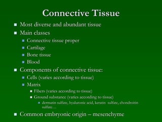
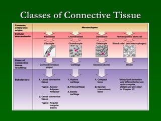
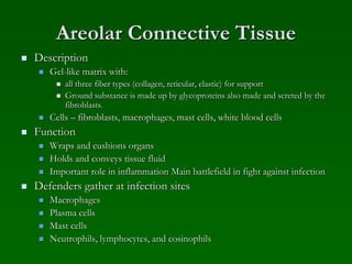
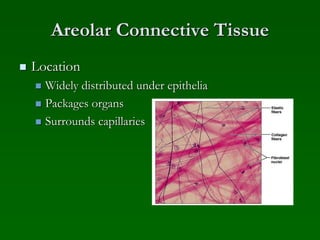
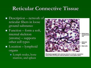


Connective tissue is the most diverse and abundant tissue in the body. It includes connective tissue proper, cartilage, bone, blood, and is characterized by cells embedded in an extracellular matrix containing fibers and ground substance. The main classes of connective tissue are connective tissue proper (loose and dense tissues), cartilage, and bone. Areolar connective tissue is a loose connective tissue found throughout the body that supports and cushions other tissues.


