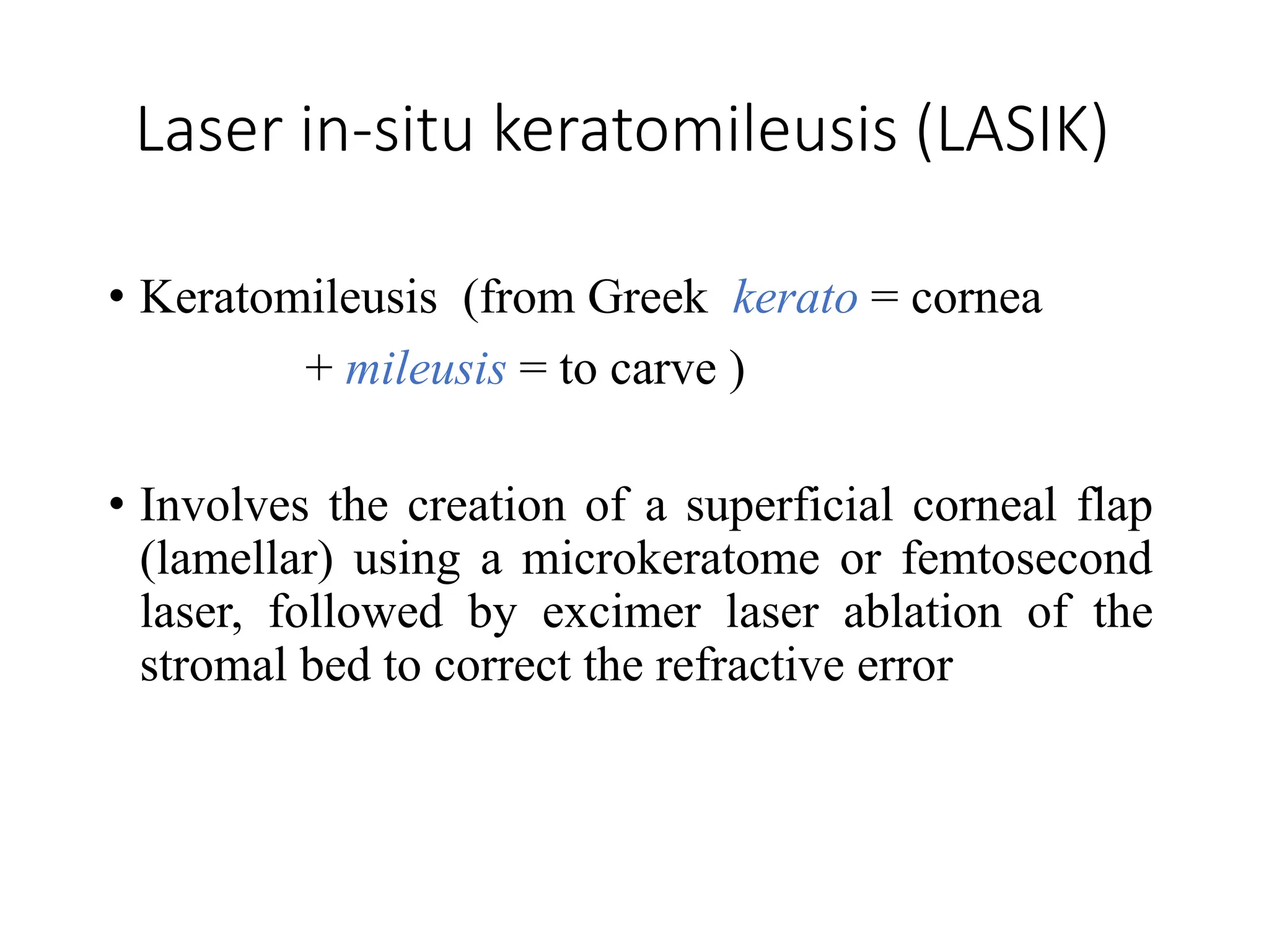
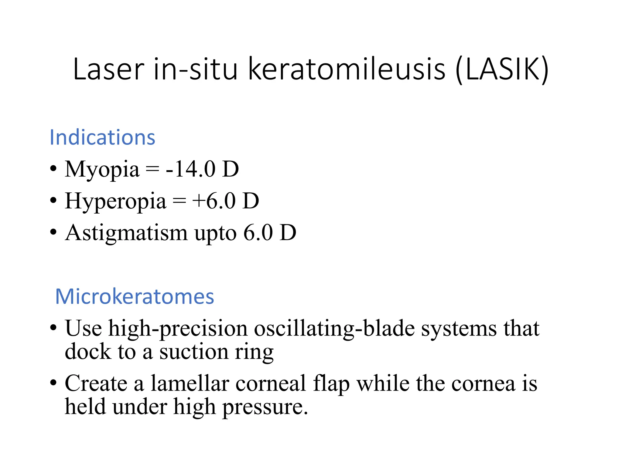
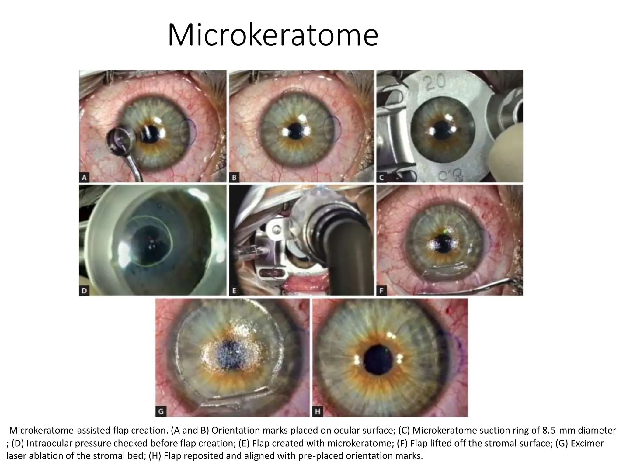
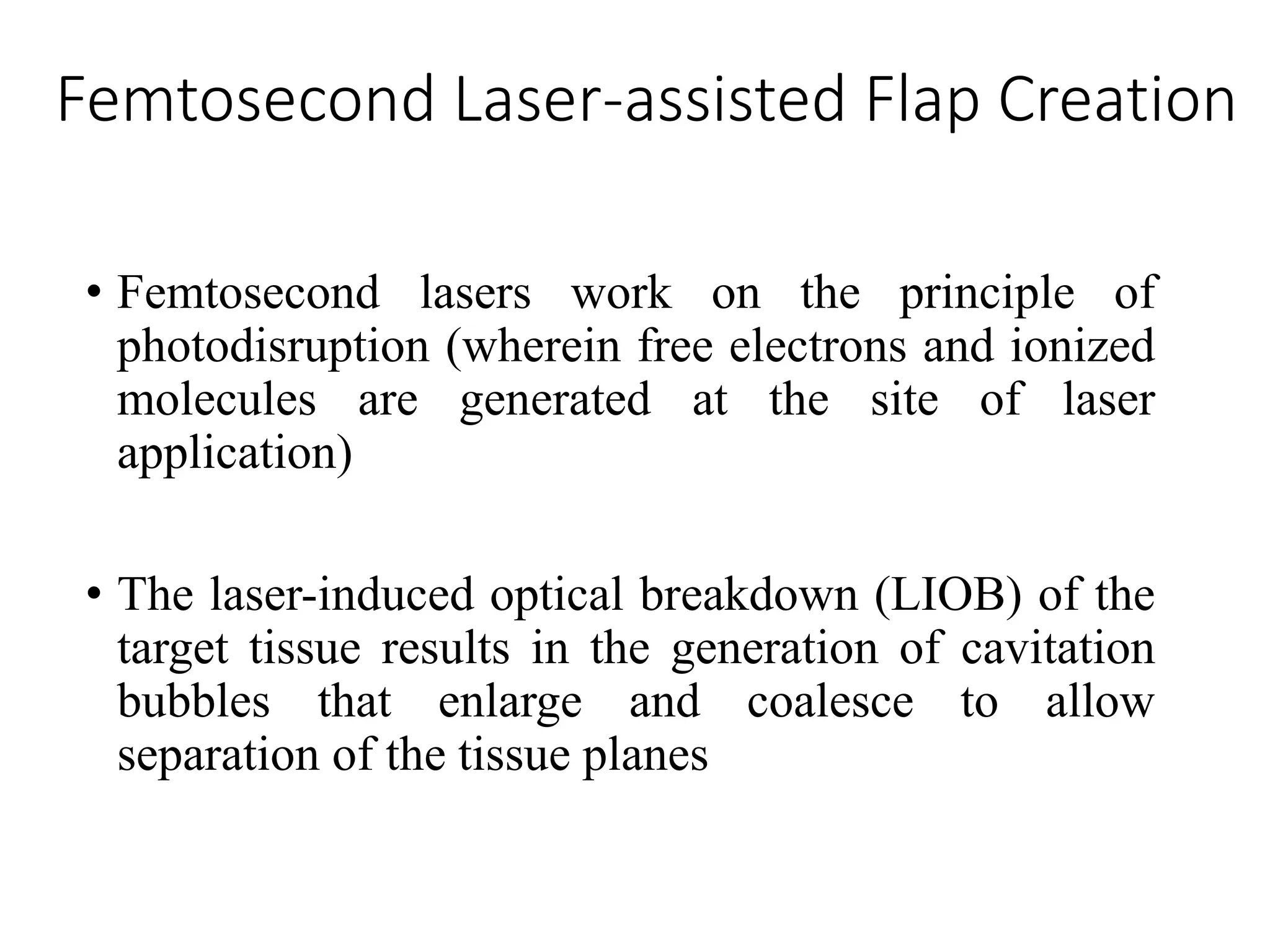
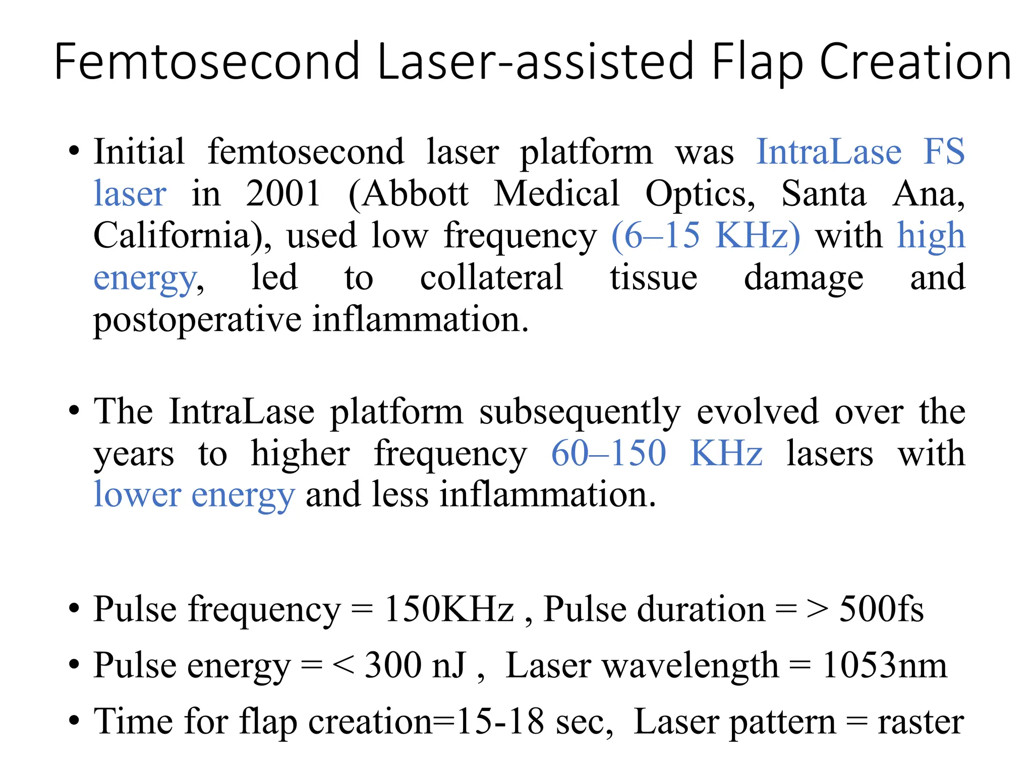
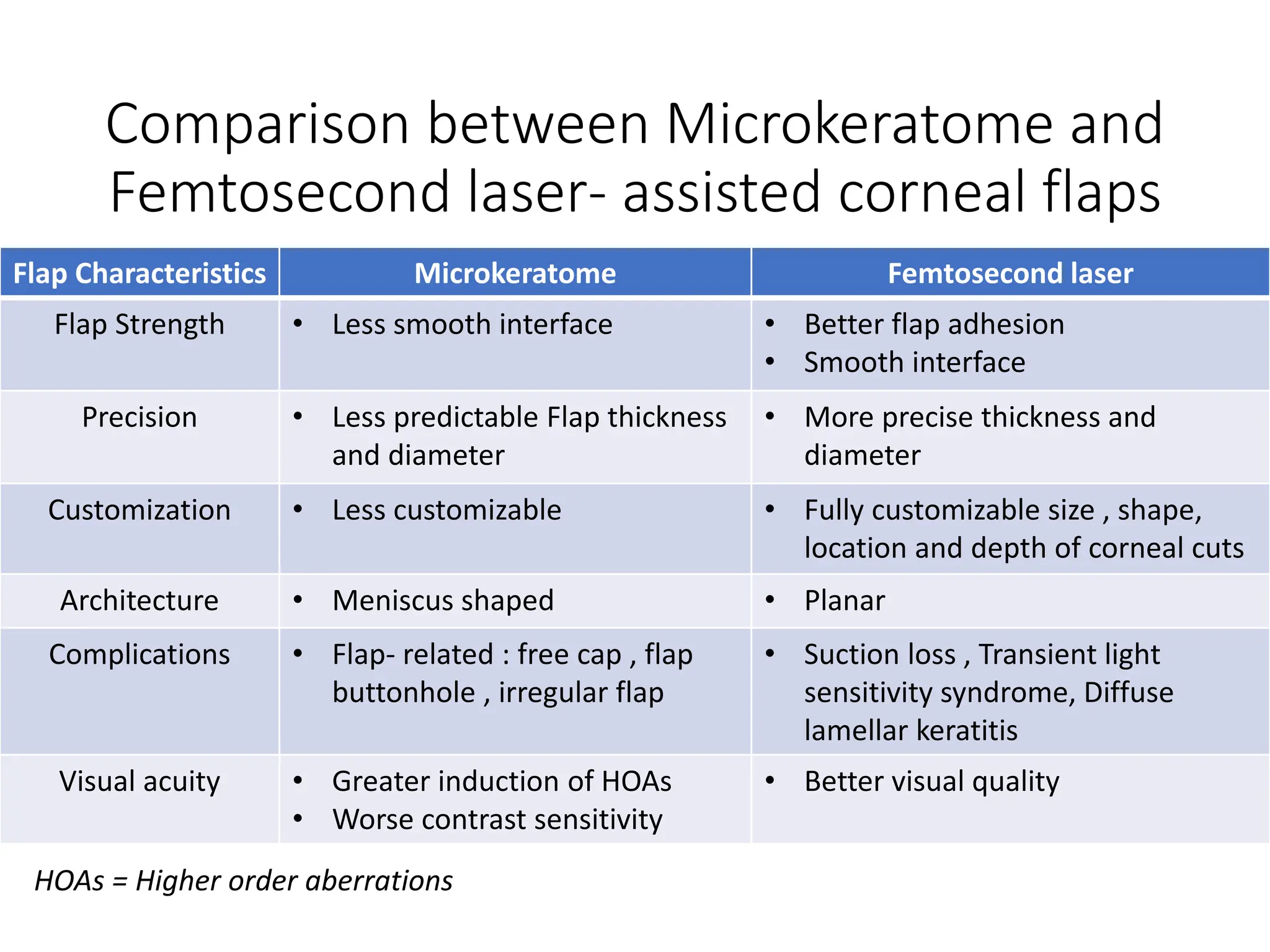
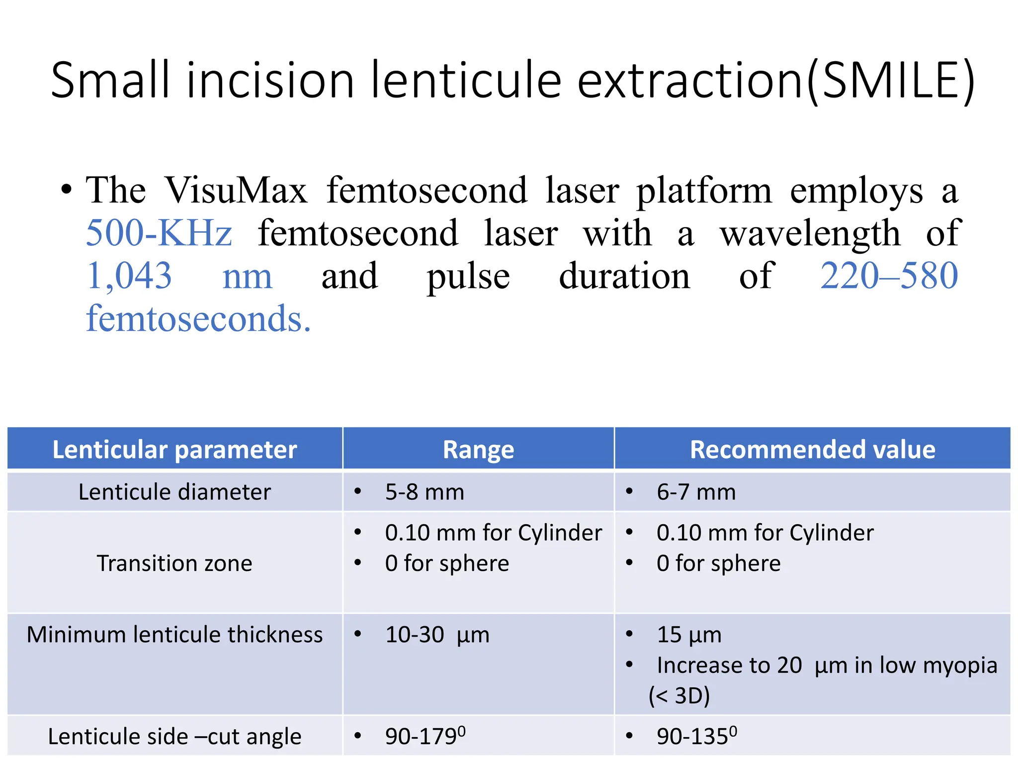
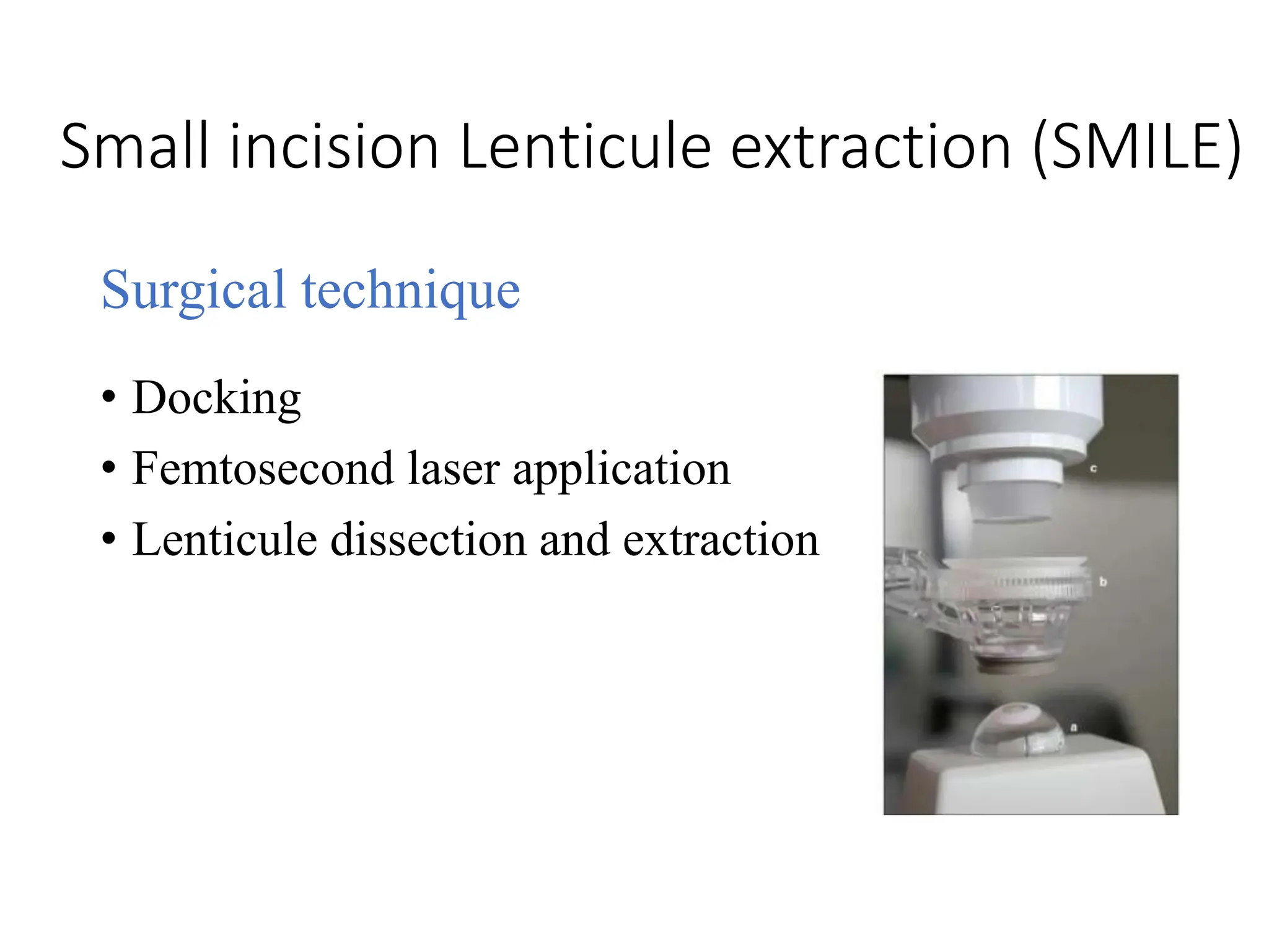
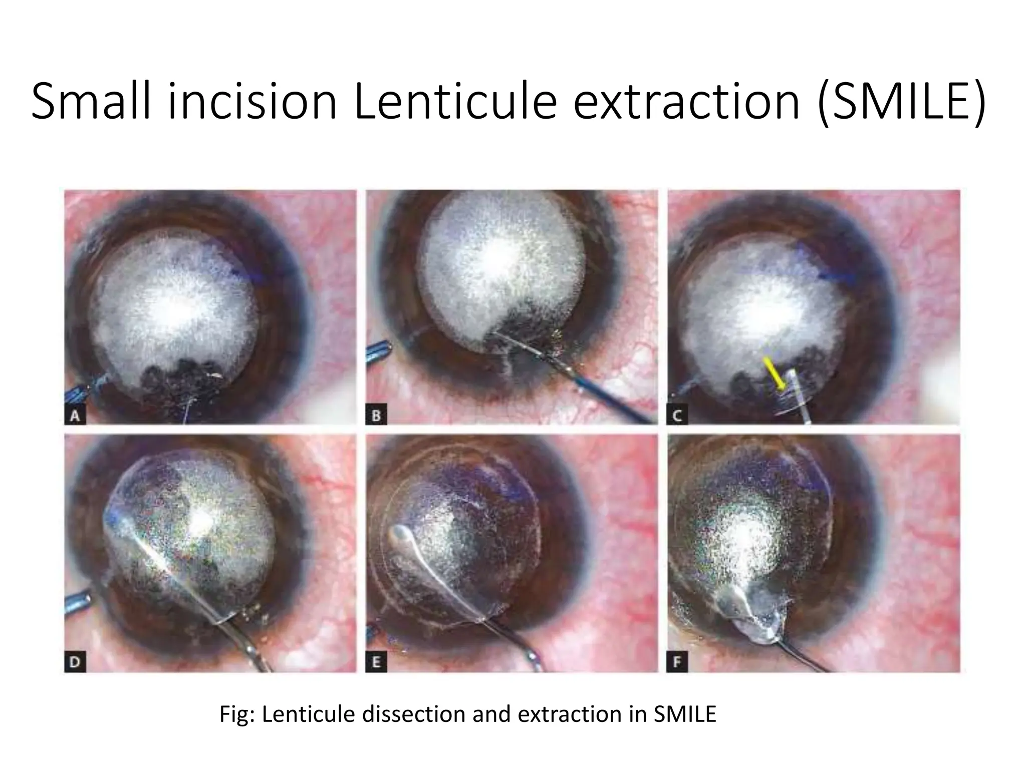
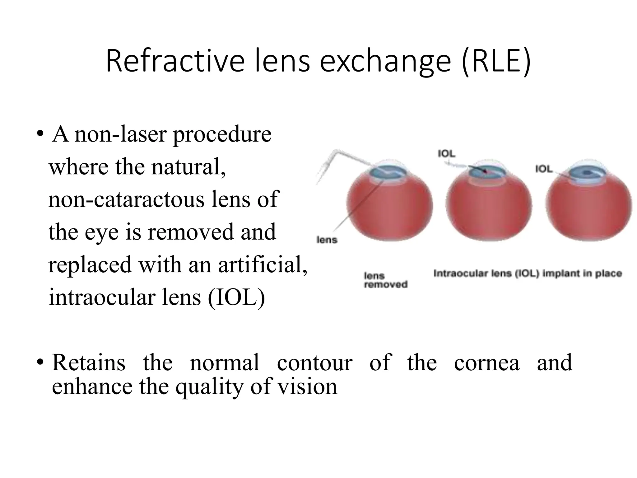
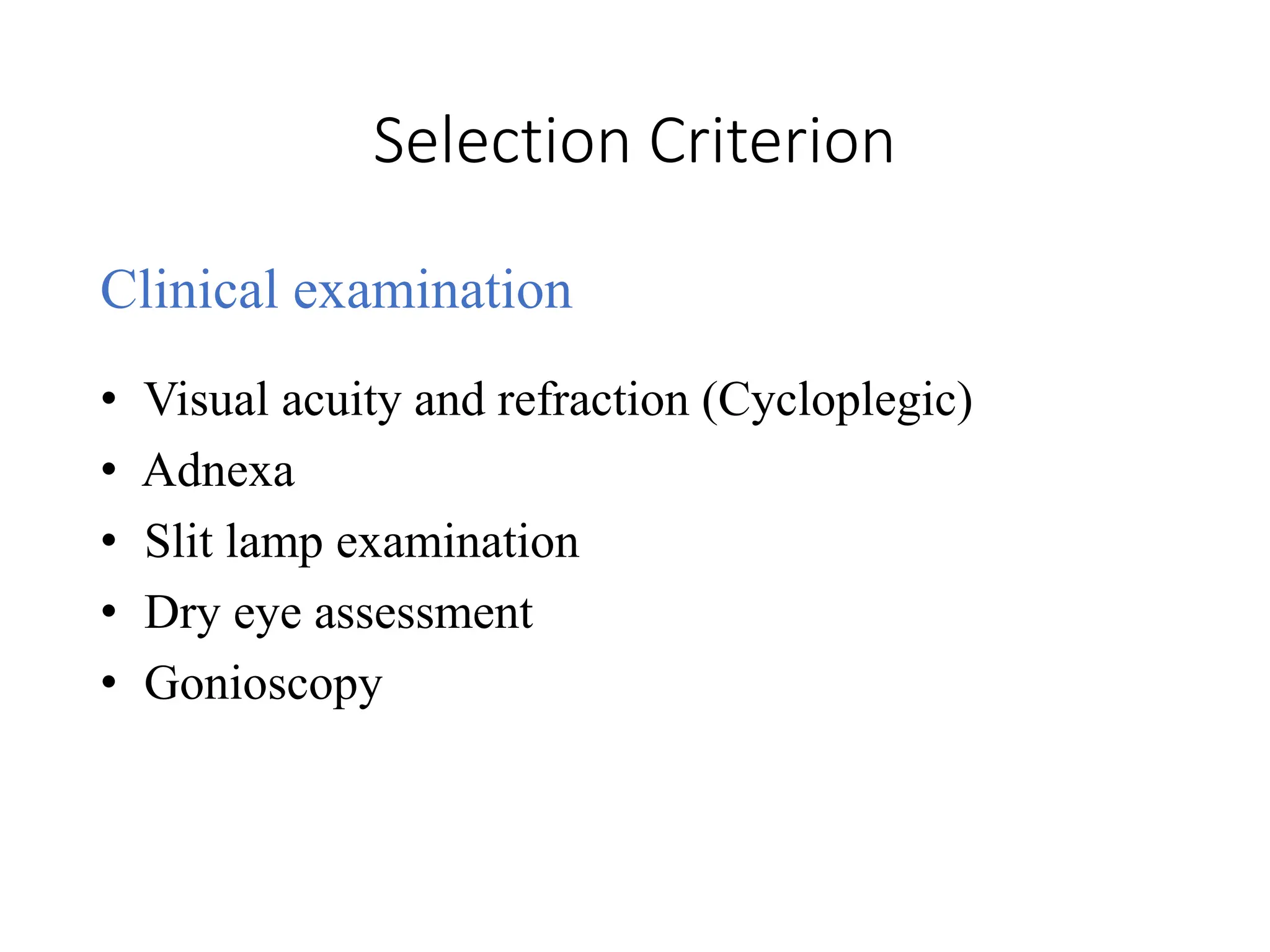
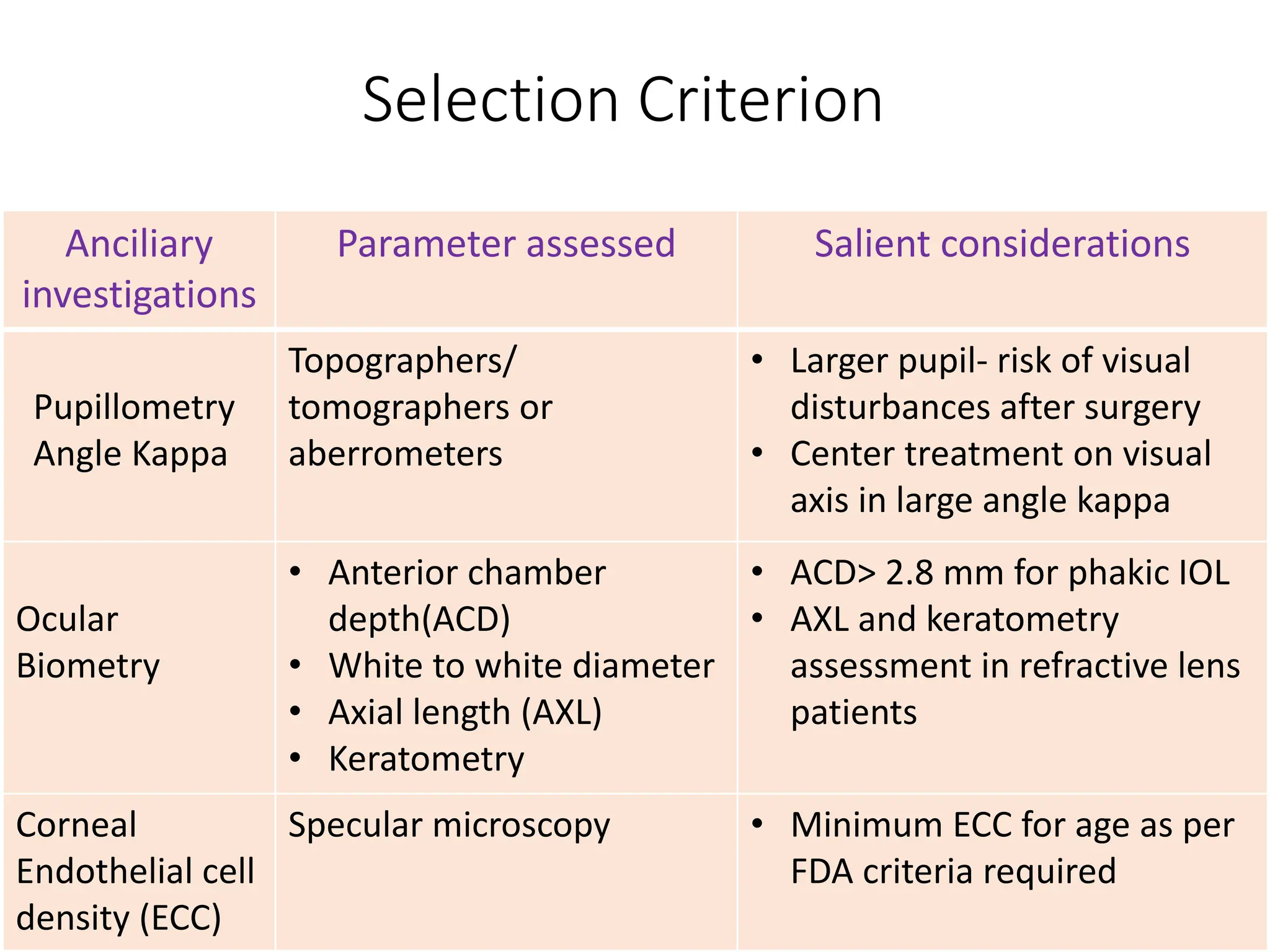
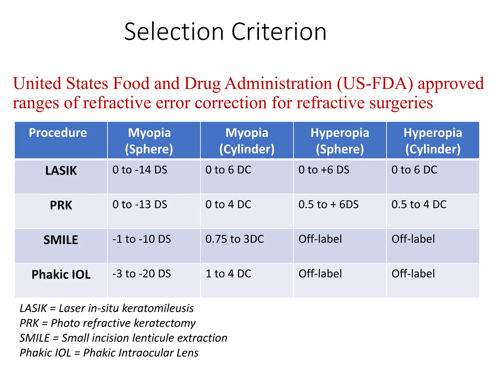
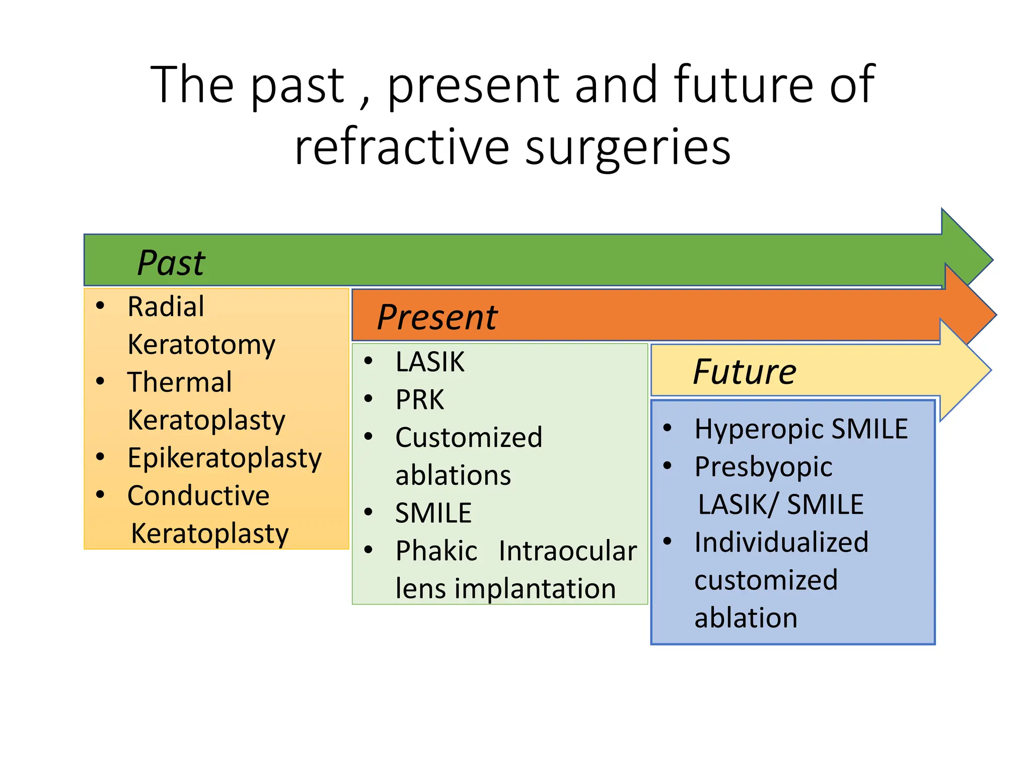
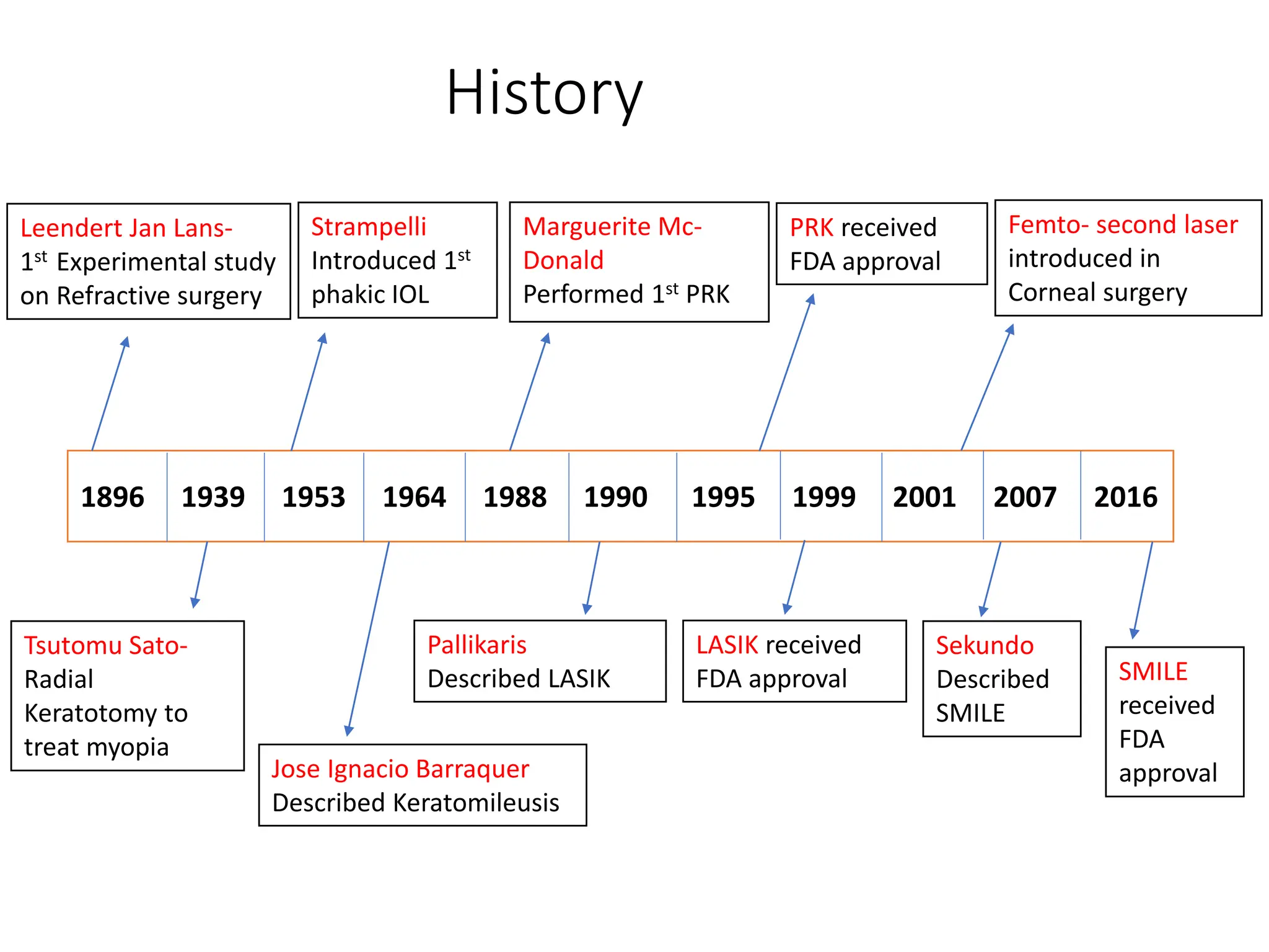
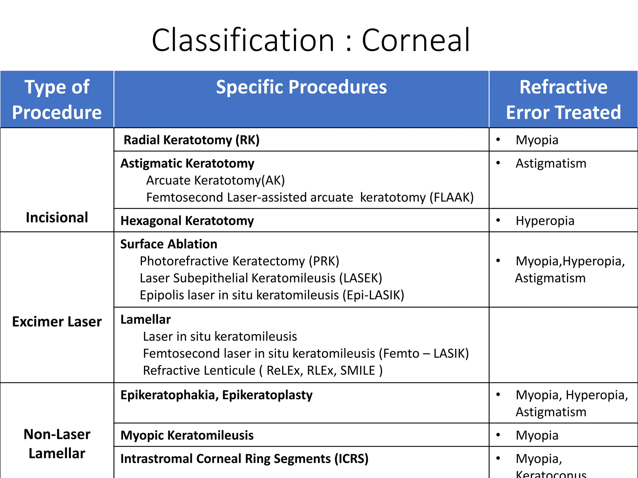
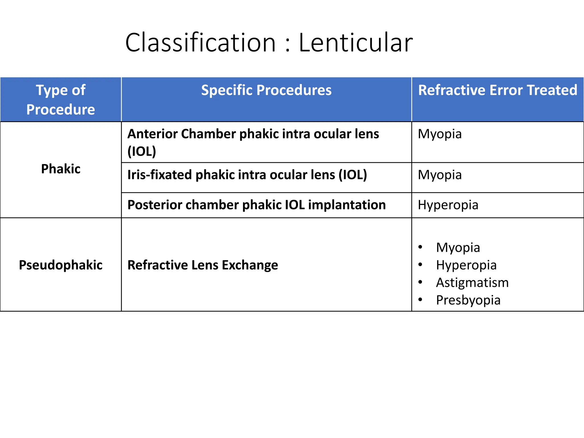
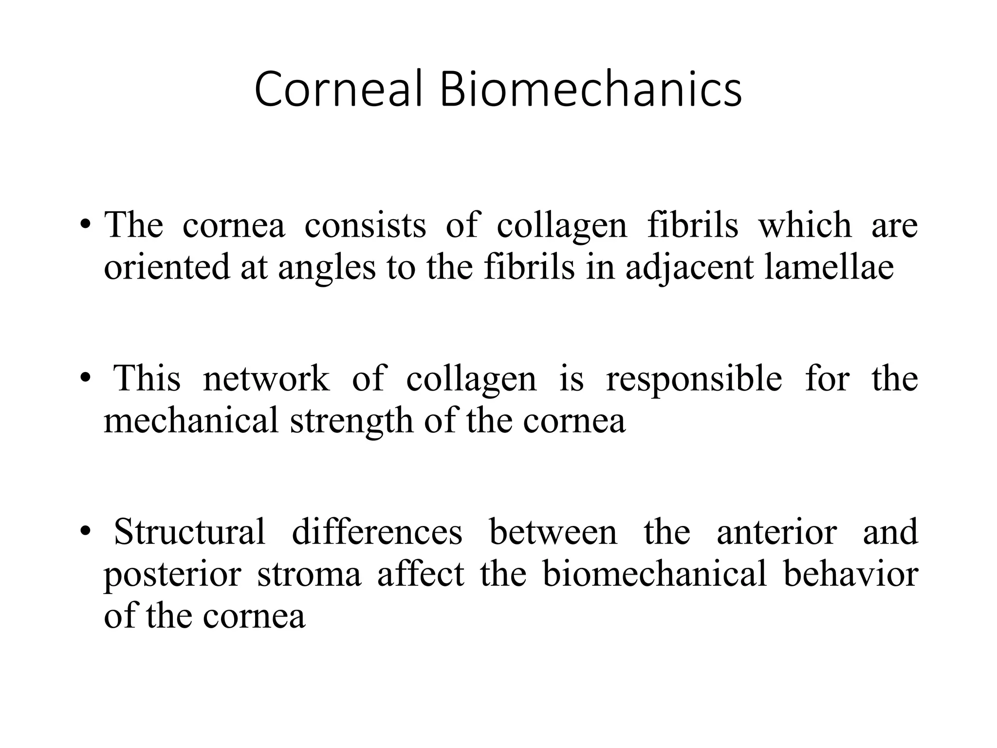
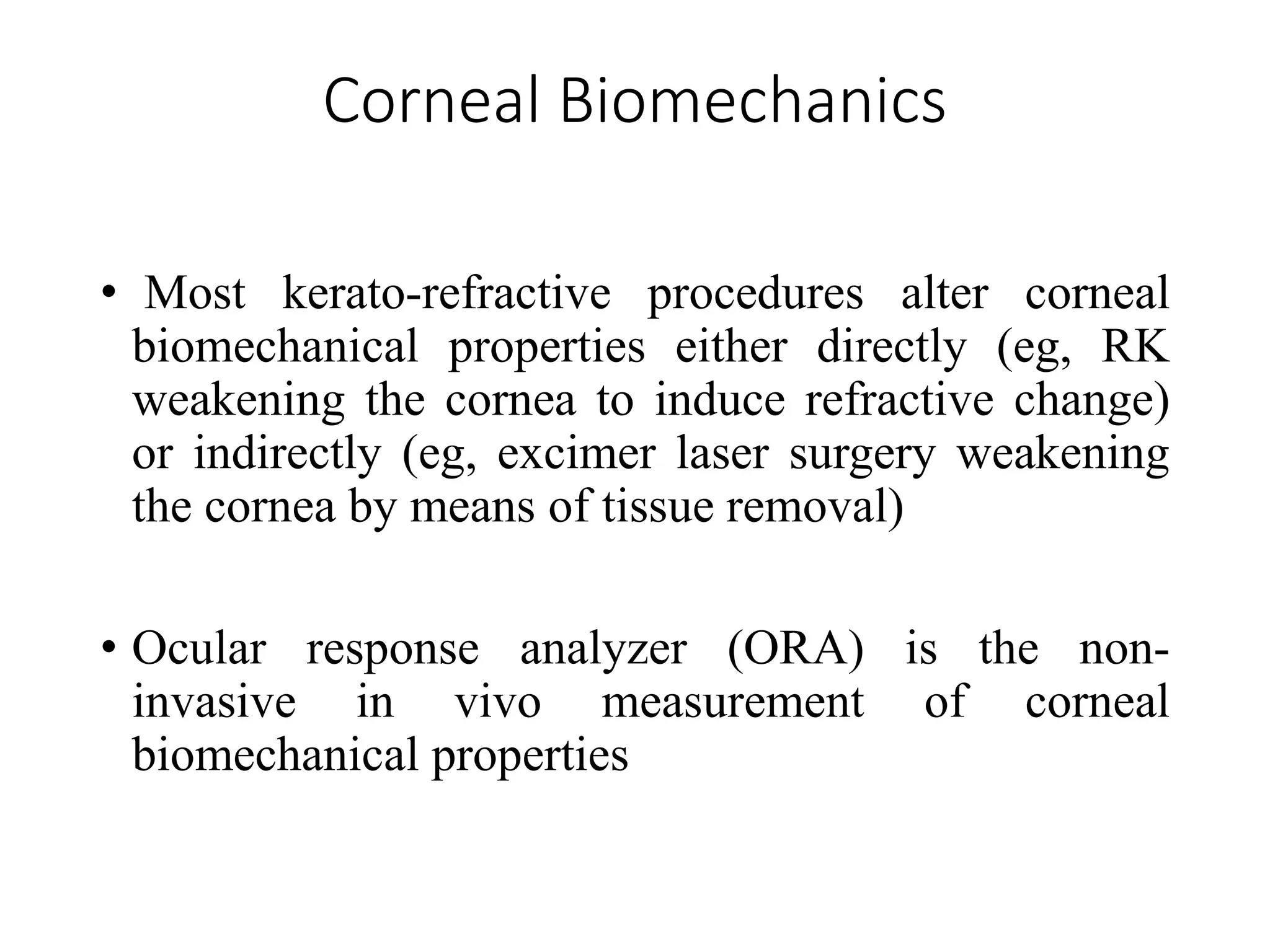
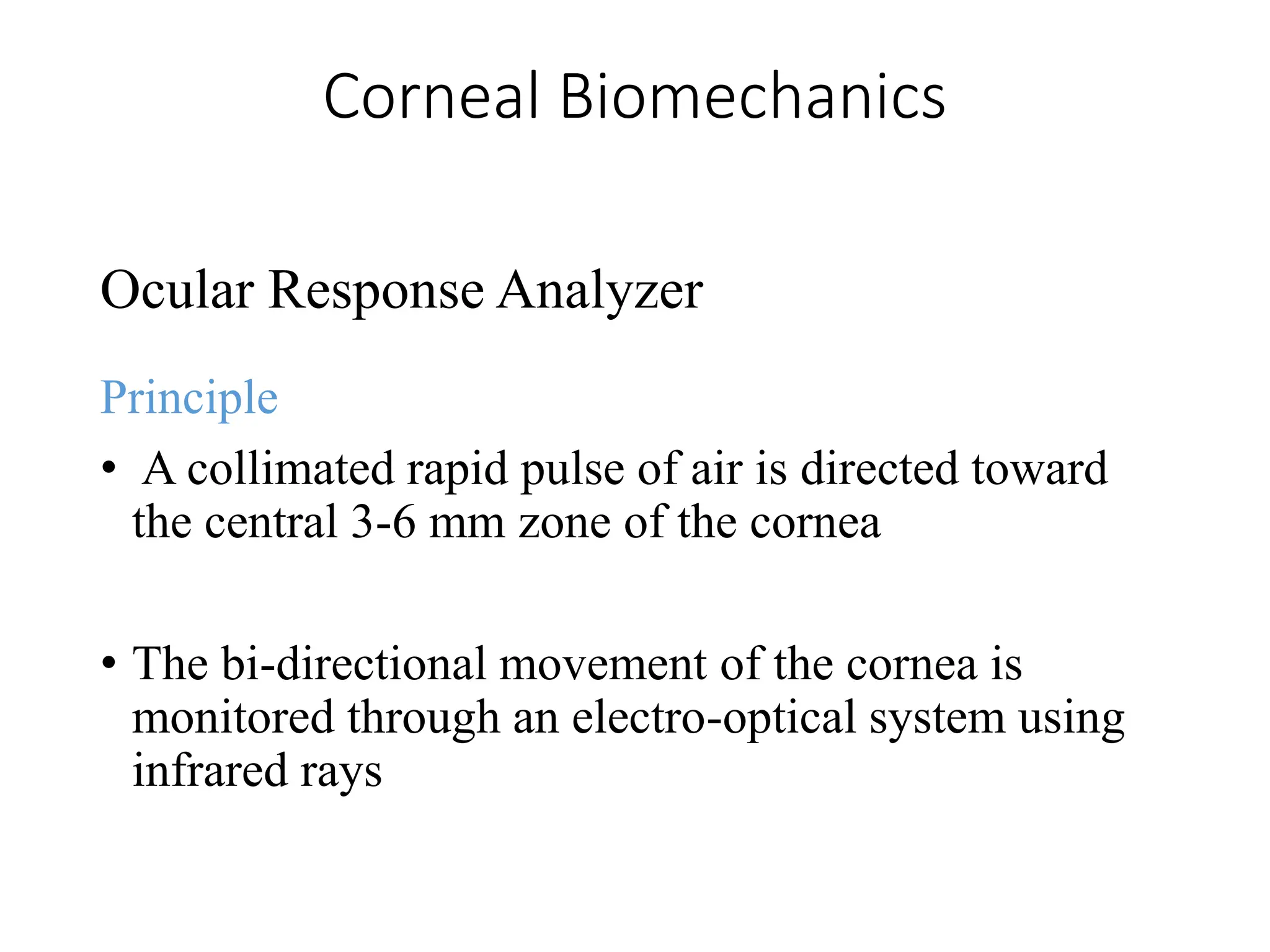
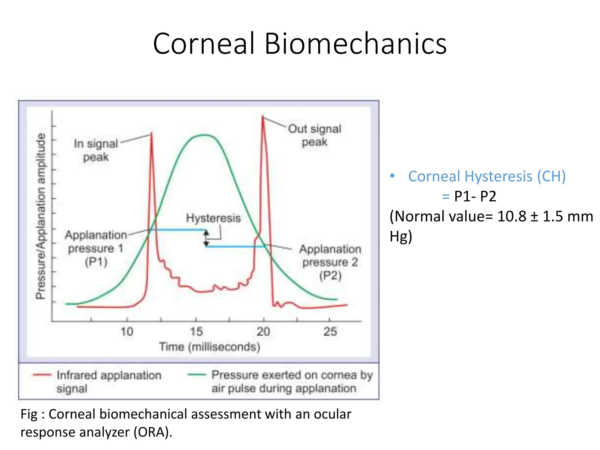
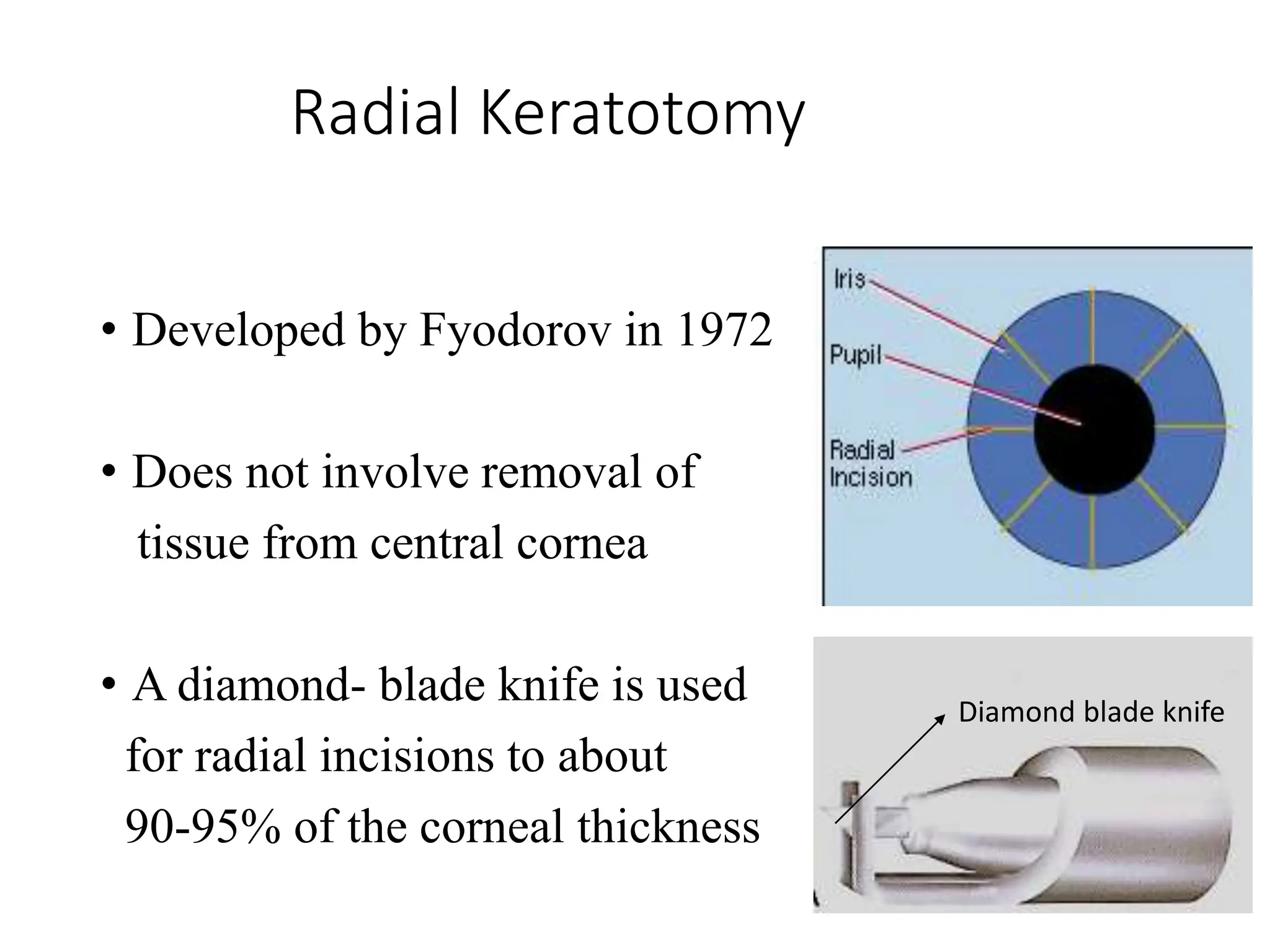
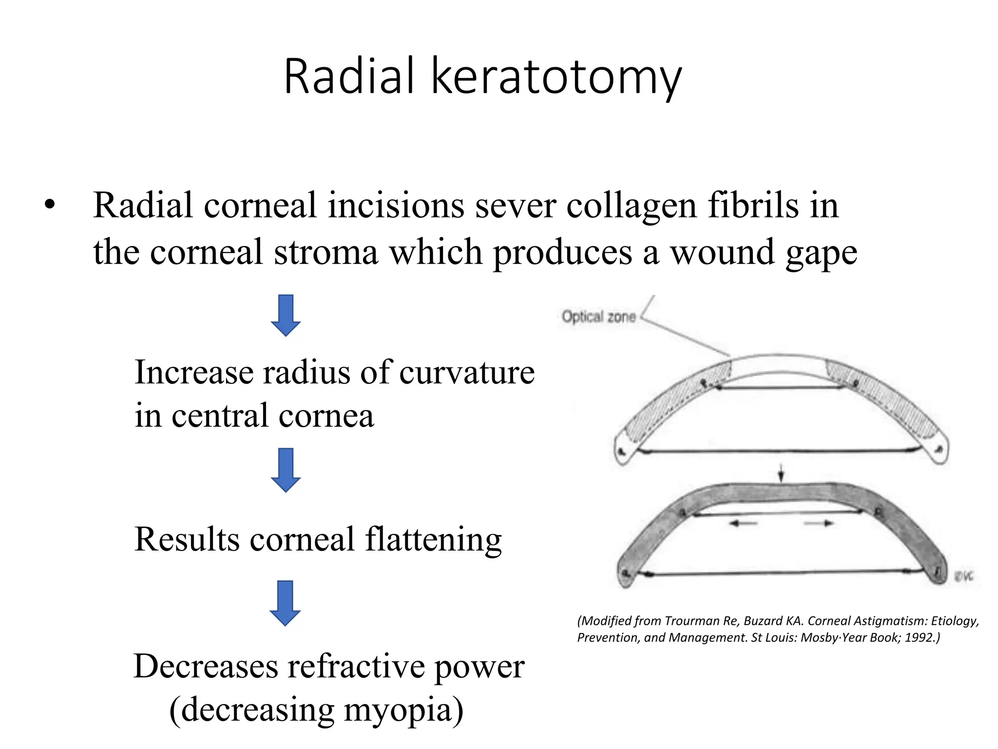
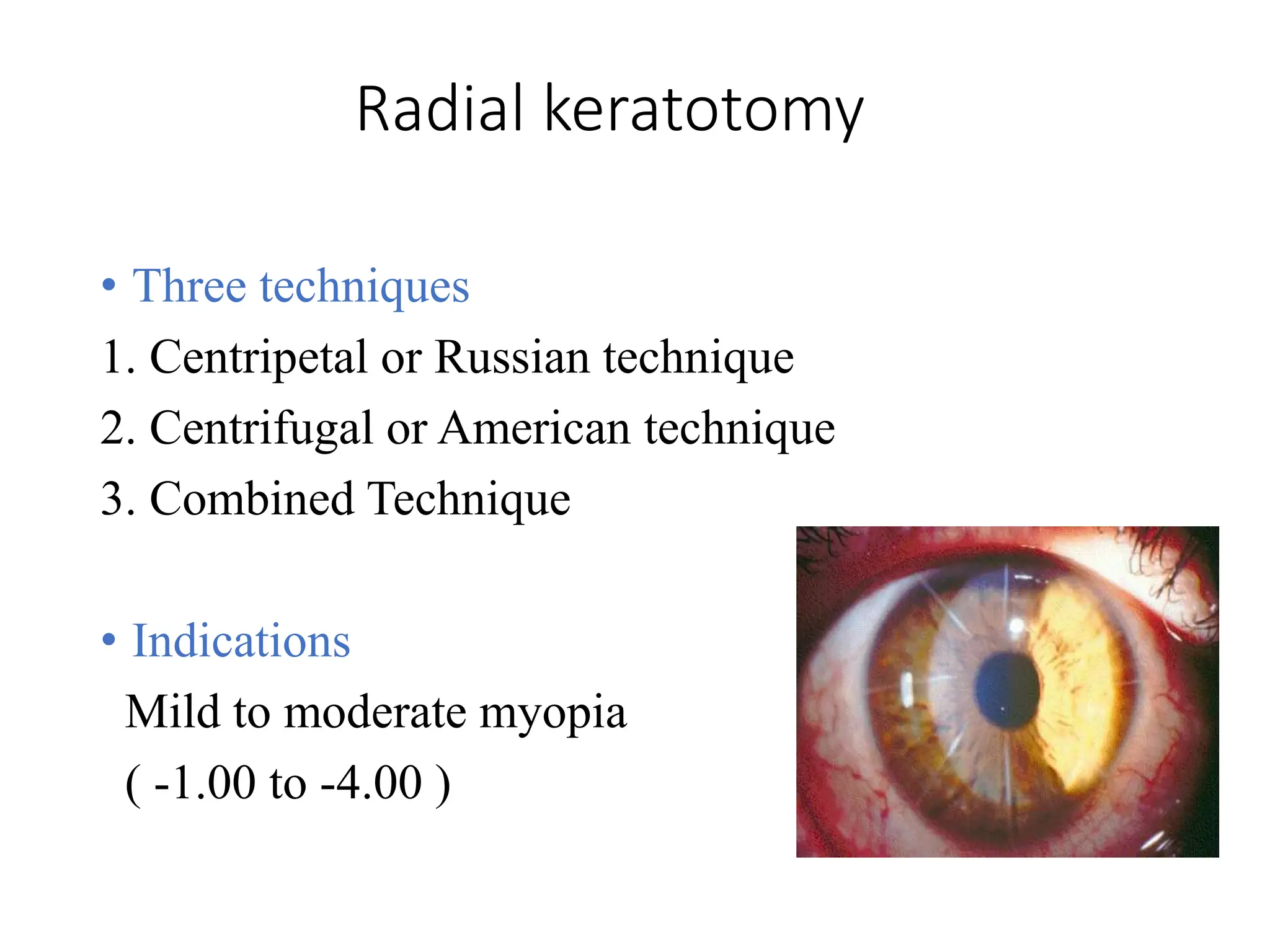
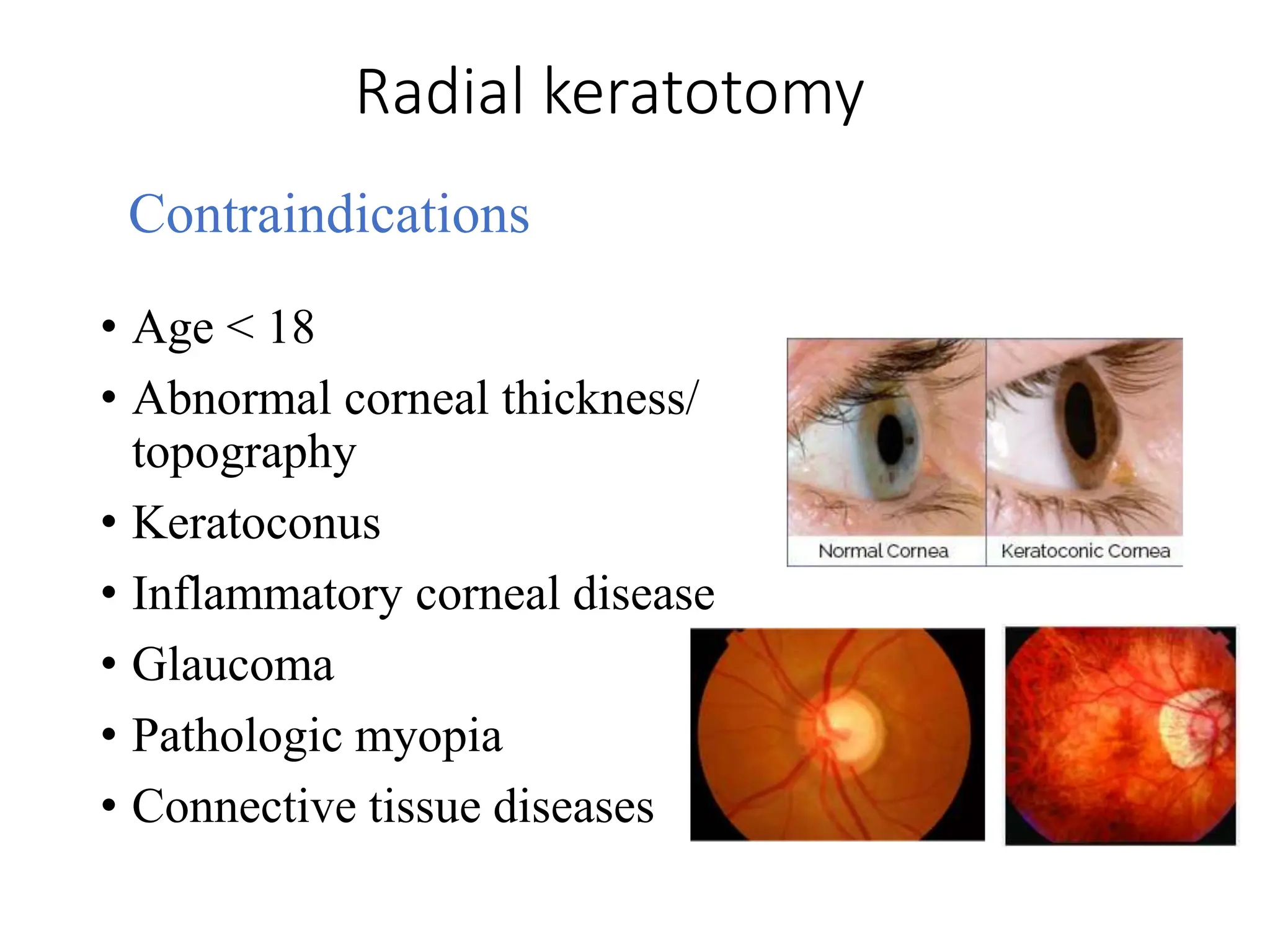
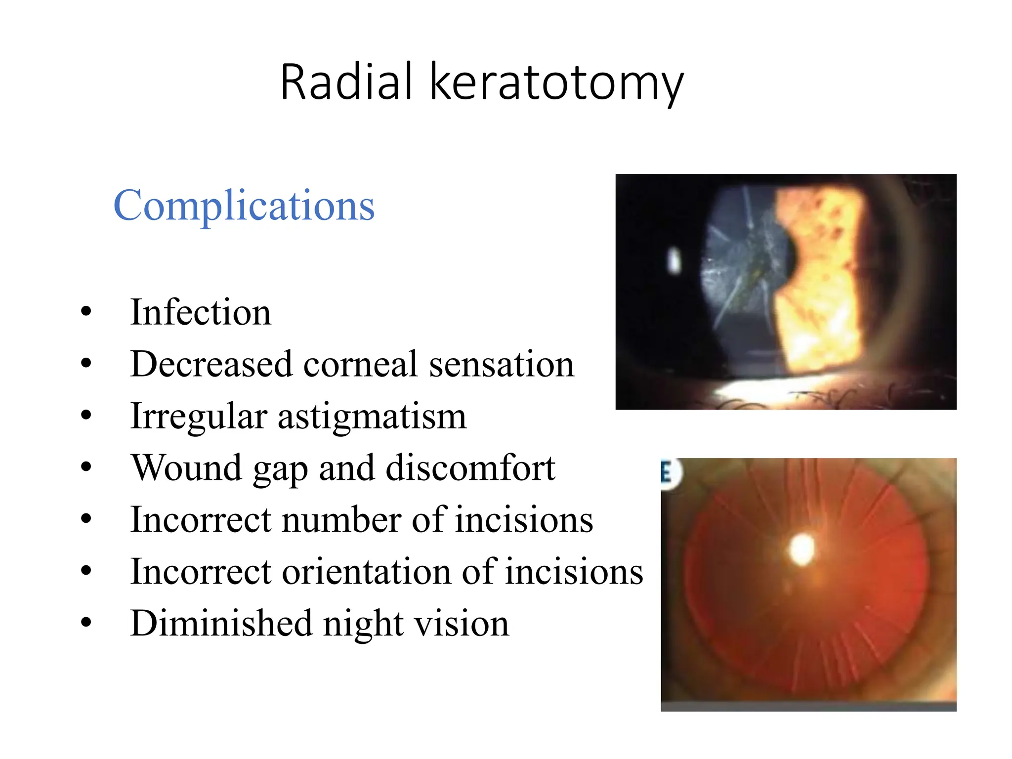
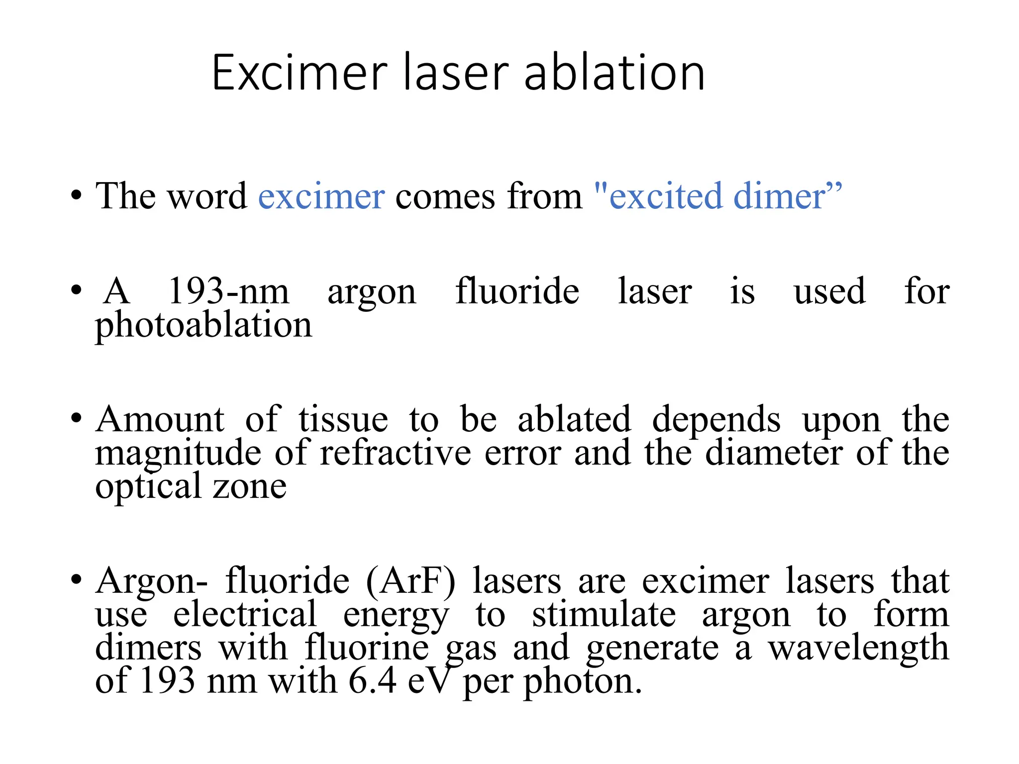
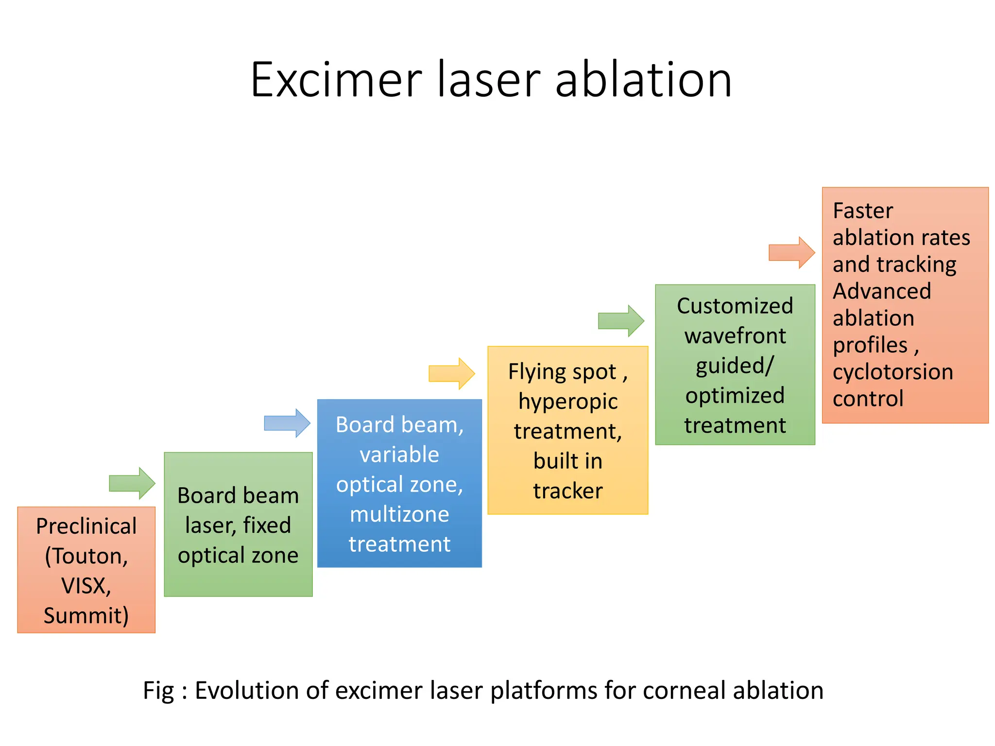
REFRACTIVE SURGERIES:
Principle, Selection of eyes, Instruments,
Procedures for myopia
Moderators
Dr. Sanjeev Bhattarai
Suraj Chhetri
Presenter
Archana Sharma
B.Optometry
Third Year
Objectives
• To explain the principle of refractive surgery
• To describe different types and procedures of
refractive surgery
• To describe criteria for the selection of eyes in
refractive surgery
Presentation Layout
• History
• Introduction to refractive surgery
• Classification
• Principle of different procedures
• Procedures for myopia
• Selection of eyes
• Summary
History
1896 1939 1953 1964 1988 1990 1995 1999 2001 2007 2016
Leendert Jan Lans-
1st Experimental study
on Refractive surgery
Tsutomu Sato-
Radial
Keratotomy to
treat myopia
Strampelli
Introduced 1st
phakic IOL
Jose Ignacio Barraquer
Described Keratomileusis
Marguerite Mc-
Donald
Performed 1st PRK
Pallikaris
Described LASIK
PRK received
FDA approval
LASIK received
FDA approval
Sekundo
Described
SMILE
Femto- second laser
introduced in
Corneal surgery
SMILE
received
FDA
approval
Introduction
• Defined as the surgical correction of refractive errors
of the human eye
• Refractive surgical techniques are used to correct or
reduce refractive errors by reshaping of cornea using
a laser or implantation addition IOL or extraction of
clear crystalline lens
• Purpose
- Alter refractive state of eye to enable patient to
see without visual aids
Introduction
• Till date, more than 40 million laser-assisted in situ
keratomileusis (LASIK) and 2 million SMILE
procedures have been performed worldwide 1
• Two currently established surgical methods for the
correction of refractive error
- Refractive corneal surgery
- Refractive lens surgery
1Aristeidou A, Taniguchi EV, Tsatsos M, Muller R, McAlinden C, Pineda R, Paschalis EI. The
evolution of corneal and refractive surgery with the femtosecond laser. Eye and Vision. 2015
Dec;2:1-4.
Principle
• To make the central cornea flatter and periphery
steeper ( For myopia )
Methods for refractive surgery
Corneal Procedures
Radial keratotomy (RK)
Photo refractive keratectomy (PRK)
Laser in-situ keratomileusis (LASIK)
Laser sub-epithelial keratomileusis (LASEK)
Small incision lenticule extraction
(SMILE)
Implantation of plastic intrastromal corneal ring
segments (ICRS or INTACS)
Laser thermal keratoplasty (LTK)
Lenticular Procedures
Refractive lens exchange (RLE)
Phakic IOL implantation
Type of
Procedure
Specific Procedures Refractive
Error Treated
Incisional
Radial Keratotomy (RK) • Myopia
Astigmatic Keratotomy
Arcuate Keratotomy(AK)
Femtosecond Laser-assisted arcuate keratotomy (FLAAK)
• Astigmatism
Hexagonal Keratotomy • Hyperopia
Excimer Laser
Surface Ablation
Photorefractive Keratectomy (PRK)
Laser Subepithelial Keratomileusis (LASEK)
Epipolis laser in situ keratomileusis (Epi-LASIK)
• Myopia,Hyperopia,
Astigmatism
Lamellar
Laser in situ keratomileusis
Femtosecond laser in situ keratomileusis (Femto – LASIK)
Refractive Lenticule ( ReLEx, RLEx, SMILE )
Non-Laser
Lamellar
Epikeratophakia, Epikeratoplasty • Myopia, Hyperopia,
Astigmatism
Myopic Keratomileusis • Myopia
Intrastromal Corneal Ring Segments (ICRS) • Myopia,
Classification : Corneal
Classification : Lenticular
• Type of Procedure
Type of
Procedure
Specific Procedures Refractive Error Treated
Phakic
Anterior Chamber phakic intra ocular lens
(IOL)
Myopia
Iris-fixated phakic intra ocular lens (IOL) Myopia
Posterior chamber phakic IOL implantation Hyperopia
Pseudophakic Refractive Lens Exchange
• Myopia
• Hyperopia
• Astigmatism
• Presbyopia
Corneal Biomechanics
• The cornea consists of collagen fibrils which are
oriented at angles to the fibrils in adjacent lamellae
• This network of collagen is responsible for the
mechanical strength of the cornea
• Structural differences between the anterior and
posterior stroma affect the biomechanical behavior
of the cornea
Corneal Biomechanics
• Most kerato-refractive procedures alter corneal
biomechanical properties either directly (eg, RK
weakening the cornea to induce refractive change)
or indirectly (eg, excimer laser surgery weakening
the cornea by means of tissue removal)
• Ocular response analyzer (ORA) is the non-
invasive in vivo measurement of corneal
biomechanical properties
Corneal Biomechanics
Ocular Response Analyzer
Principle
• A collimated rapid pulse of air is directed toward
the central 3-6 mm zone of the cornea
• The bi-directional movement of the cornea is
monitored through an electro-optical system using
infrared rays
Corneal Biomechanics
Fig : Corneal biomechanical assessment with an ocular
response analyzer (ORA).
• Corneal Hysteresis (CH)
= P1- P2
(Normal value= 10.8 ± 1.5 mm
Hg)
Radial Keratotomy
• Developed by Fyodorov in 1972
• Does not involve removal of
tissue from central cornea
• A diamond- blade knife is used
for radial incisions to about
90-95% of the corneal thickness
Diamond blade knife
Radial keratotomy
(Modified from Trourman Re, Buzard KA. Corneal Astigmatism: Etiology,
Prevention, and Management. St Louis: Mosby·Year Book; 1992.)
• Radial corneal incisions sever collagen fibrils in
the corneal stroma which produces a wound gape
Decreases refractive power
(decreasing myopia)
Results corneal flattening
Increase radius of curvature
in central cornea
Radial keratotomy
• Three techniques
1. Centripetal or Russian technique
2. Centrifugal or American technique
3. Combined Technique
• Indications
Mild to moderate myopia
( -1.00 to -4.00 )
Radial keratotomy
Contraindications
• Age < 18
• Abnormal corneal thickness/
topography
• Keratoconus
• Inflammatory corneal disease
• Glaucoma
• Pathologic myopia
• Connective tissue diseases
Complications
• Infection
• Decreased corneal sensation
• Irregular astigmatism
• Wound gap and discomfort
• Incorrect number of incisions
• Incorrect orientation of incisions
• Diminished night vision
Radial keratotomy
Surface Ablation
• Surface ablation techniques involve the removal of
the epithelium followed by excimer laser ablation of
the stromal bed
• This includes :
1. Photorefractive keratectomy (PRK)
2. Laser subepithelial keratomileusis (LASEK)
3. Epithelial laser in situ keratomileusis
(epi-LASIK)
1. Photorefractive Keratectomy (PRK)
• Involves the removal of corneal
epithelium either mechanically
or with the help of excimer laser
itself (transepithelial)
• Followed by excimer laser
ablation of the stromal bed
• A bandage contact lens is placed at the end of
surgery till re-epithelialization is completed
Photorefractive Keratectomy(PRK)
Technique of epithelial removal Instrument /method
Mechanical debridement
(Fig A)
• Blunt spatula
• Sharp blade
• Battery – operated rotating brushes
Chemical de-epithelization
(Fig B)
• Preoperative topical anesthetics
• 18-25 % ethanol for 20 to 40 seconds
Photoablative de-epithelization • Transepithelial PRK- excimer laser pulses
Photorefractive Keratectomy(PRK)
Indications
• Myopia =-13.00 D
• Hypermetropia = +6.00 D
• Astigmatism upto 3D
• Thin cornea
• Epithelial irregularities/ dystrophies
• LASIK complications in contralateral eye
Photorefractive Keratectomy(PRK)
https://youtu.be/oBn0DXmYefc?si=Q7rAW-MlsMvofkJR
2. Laser Sub-epithelial Keratomileusis
(LASEK)
• The epithelium is loosened with alcohol and lifted
off from the corneal stroma as a hinged flap
• Excimer laser ablation is performed, and the
epithelial flap is repositioned at the end of surgery
Indications
• Low to moderate Myopia & astigmatism
• Risk of ocular trauma
• Thin cornea
• Steep cornea
• Flat cornea
Laser Sub-epithelial Keratomileusis(LASEK)
Metal handle as alcohol
reservoir
Epithelial flap formation
With Vannas scissors
Ablation with excimer
laser
Epithelial sheet is repositioned Bandage contact lens is placed
3. Epipolis-laser in Situ Keratomileusis
(epi-LASIK)
• Involves the creation of an epithelial flap with the
help of an epikeratome.
• A hinged epithelial flap is created, which is reflected
off the corneal surface to allow excimer laser
ablation of the stromal bed.
• The epithelial flap is repositioned at the end of
surgery
Epipolis-laser in Situ Keratomileusis
(epi-LASIK)
Indication
Less steep corneas (low myopia)
Advantage over LASEK
• Less pain
• Faster healing
• Less corneal haze
Excimer laser ablation
• The word excimer comes from "excited dimer”
• A 193-nm argon fluoride laser is used for
photoablation
• Amount of tissue to be ablated depends upon the
magnitude of refractive error and the diameter of the
optical zone
• Argon- fluoride (ArF) lasers are excimer lasers that
use electrical energy to stimulate argon to form
dimers with fluorine gas and generate a wavelength
of 193 nm with 6.4 eV per photon.
Preclinical
(Touton,
VISX,
Summit)
Board beam
laser, fixed
optical zone
Board beam,
variable
optical zone,
multizone
treatment
Flying spot ,
hyperopic
treatment,
built in
tracker
Customized
wavefront
guided/
optimized
treatment
Faster
ablation rates
and tracking
Advanced
ablation
profiles ,
cyclotorsion
control
Fig : Evolution of excimer laser platforms for corneal ablation
Excimer laser ablation
Depth of ablation (µm) = [diameter of optical
zone(mm)] 2 × 1/3 power(D)
• An estimate of the ablation
depth during LASIK surgery
is provided by the
Munnerlyn’s equation
• Discovered by Charles
Munnerlyn
Laser in-situ keratomileusis (LASIK)
• Keratomileusis (from Greek kerato = cornea
+ mileusis = to carve )
• Involves the creation of a superficial corneal flap
(lamellar) using a microkeratome or femtosecond
laser, followed by excimer laser ablation of the
stromal bed to correct the refractive error
Laser in-situ keratomileusis (LASIK)
Indications
• Myopia = -14.0 D
• Hyperopia = +6.0 D
• Astigmatism upto 6.0 D
Microkeratomes
• Use high-precision oscillating-blade systems that
dock to a suction ring
• Create a lamellar corneal flap while the cornea is
held under high pressure.
Microkeratome
Microkeratome-assisted flap creation. (A and B) Orientation marks placed on ocular surface; (C) Microkeratome suction ring of 8.5-mm diameter
; (D) Intraocular pressure checked before flap creation; (E) Flap created with microkeratome; (F) Flap lifted off the stromal surface; (G) Excimer
laser ablation of the stromal bed; (H) Flap reposited and aligned with pre-placed orientation marks.
Femtosecond Laser-assisted Flap Creation
• Femtosecond lasers work on the principle of
photodisruption (wherein free electrons and ionized
molecules are generated at the site of laser
application)
• The laser-induced optical breakdown (LIOB) of the
target tissue results in the generation of cavitation
bubbles that enlarge and coalesce to allow
separation of the tissue planes
Femtosecond Laser-assisted Flap Creation
• Initial femtosecond laser platform was IntraLase FS
laser in 2001 (Abbott Medical Optics, Santa Ana,
California), used low frequency (6–15 KHz) with high
energy, led to collateral tissue damage and
postoperative inflammation.
• The IntraLase platform subsequently evolved over the
years to higher frequency 60–150 KHz lasers with
lower energy and less inflammation.
• Pulse frequency = 150KHz , Pulse duration = > 500fs
• Pulse energy = < 300 nJ , Laser wavelength = 1053nm
• Time for flap creation=15-18 sec, Laser pattern = raster
Laser in-situ keratomileusis (LASIK)
https://youtu.be/bFWFZQVKCc0?si=GRsRxxXFSEU8baG7
Comparison between Microkeratome and
Femtosecond laser- assisted corneal flaps
Flap Characteristics Microkeratome Femtosecond laser
Flap Strength • Less smooth interface • Better flap adhesion
• Smooth interface
Precision • Less predictable Flap thickness
and diameter
• More precise thickness and
diameter
Customization • Less customizable • Fully customizable size , shape,
location and depth of corneal cuts
Architecture • Meniscus shaped • Planar
Complications • Flap- related : free cap , flap
buttonhole , irregular flap
• Suction loss , Transient light
sensitivity syndrome, Diffuse
lamellar keratitis
Visual acuity • Greater induction of HOAs
• Worse contrast sensitivity
• Better visual quality
HOAs = Higher order aberrations
Customized corneal ablation
• First customized corneal ablation (wavefront guided)
was performed by Theo Seiler in 1999
• Conventional corneal ablative profiles were based on
the subjective refractive error of the patient; myopic
correction entailed greater ablation of central cornea
than the periphery
• Three types :
(1) Corneal wavefront-optimized (WO)
(2) Corneal wavefront-guided (WG)
(3) Topography guided (TG)
Types of customized corneal ablative
procedures
Characteri
-stics
Wavefront-
optimized
ablation
Wavefront- guided
ablation
Topography- guided
ablation
Treatment
Priniciple
• Prevent induction
of new HOAs
• Treat pre-existing
ocular aberrations
and minimize
induction of new
HOAs
• Treat pre-existing
corneal aberrations
and minimize
induction of new
HOAs
Patient
selection
• For regular
corneal
astigmatism and
HOAs within the
normal range
• High preoperative
HOAs (>0.4 microns)
• Moderate refractive
errors
• Reliable ocular
wavefront data with
regular cornea
• Reliable topography
scans with significant
corneal HOAs
• Large angle kappa
Types of customized corneal
ablative procedures
ablation
Topography- guided ablation
Characteristi
-cs
Wavefront-
optimized ablation
Wavefront-
guided ablation
Topography- guided
ablation
Advantages
• Does not require
preoperative
aberrometry
• Allows creation of
larger OZ than
conventional
treatments
• Aims to correct
all pre-existing
HOAs
• Takes into
account entire
ocular aberration
profile
• Treats HOAs based
on corneal
aberration profile
• Surgeon can specify
the target
asphericity
Disadvantages
• Does not corrected
pre-existing HOAs
• Greater tissue
ablated in
periphery
• Not feasible in
highly aberrated
corneas
• Expensive
aberrometers
required
• Does not take into
account internal
aberrations
• Treatment accuracy
relies on repeatable
and reliable
topography scans
Small incision lenticule extraction (SMILE)
• Involves creation of an intrastromal
lenticule and its extraction from
a small side incision with a width
of 2–5 mm
• Indications 1
Myopia = -1.0 to -10.0 D
Myopic astigmatism = -0.75 D to -3.0 D
Manifest refraction spherical equivalent = -10.0 D
• The use of SMILE for hyperopic corrections is off-
label and under investigation in clinical trials
1Ang M, Mehta JS, Chan C, Htoon HM, Koh JCW, Tan DT. Refractive lenticule extraction: transition and comparison of 3 surgical
techniques. J Cataract Refract Surg. 2014; 40(9):1415-24.
Lenticule
Small incision lenticule extraction (SMILE)
• At present, the VisuMax
femtosecond laser
(Carl Zeiss Meditec, Germany)
is the only commercially
available laser platform
approved for ReLEx.
ReLEx = Refractive lenticule extraction
Small incision lenticule extraction(SMILE)
• The VisuMax femtosecond laser platform employs a
500-KHz femtosecond laser with a wavelength of
1,043 nm and pulse duration of 220–580
femtoseconds.
Lenticular parameter Range Recommended value
Lenticule diameter • 5-8 mm • 6-7 mm
Transition zone
• 0.10 mm for Cylinder
• 0 for sphere
• 0.10 mm for Cylinder
• 0 for sphere
Minimum lenticule thickness • 10-30 µm • 15 µm
• Increase to 20 µm in low myopia
(< 3D)
Lenticule side –cut angle • 90-1790 • 90-1350
Small incision Lenticule extraction (SMILE)
Surgical technique
• Docking
• Femtosecond laser application
• Lenticule dissection and extraction
Small incision Lenticule extraction (SMILE)
Fig: Lenticule dissection and extraction in SMILE
Small incision Lenticule extraction(SMILE)
https://youtu.be/wW4W7QqfzLo?si=HcHoCLaIugj5PpKL
Phakic Intraocular lens (PIOLs)
• PIOLs are implanted into the human eye in addition
to the natural ocular lens
• A reversible procedure in which an artificial lens is
implanted into the anterior or posterior chamber of
the eye in addition to the natural lens in order to
correct high refractive errors
Phakic Intraocular lens (PIOLs)
• At present, only two pIOLs have received FDA
approval for the correction of myopia and myopic
astigmatism
-Verisyse (Abbott Medical Optics, Inc.), which is an
iris-supported pIOL
-Visian implantable collamer lens (ICL, STAAR
Surgical), which is a posterior chamber pIOL
Types of phakic IOLs (PIOLs)
1. Anterior chamber angle supported piols
Functions with haptics positioned in
the angle where the iris and cornea meet
2. Anterior chamber iris-fixated piols
Uses small “lobster claws” to enclavate
iris-tissue and position the piol in front
of the pupil
3. Posterior chamber piols
Uses plate haptics to support and
position the lens in the posterior chamber
Phakic Intraocular lens (PIOLs)
Indications
• Age > 21 years
• Stable refractive error within
6-12 months
• Unsatisfactory vision with
contact lenses / spectacles
• Iridocorneal angle at least
300
• No ocular pathology
Contraindications
• Age < 21 years
• Recurrent or chronic
uveitis
• IOP > 21 mmHg
• Previous ocular or
intraocular surgery
• Systemic disease
Refractive lens exchange (RLE)
• A non-laser procedure
where the natural,
non-cataractous lens of
the eye is removed and
replaced with an artificial,
intraocular lens (IOL)
• Retains the normal contour of the cornea and
enhance the quality of vision
Refractive lens exchange (RLE)
Indication
• Moderate to high myopia
• Hyperopia
• Patients who are not LASIK candidates
Selection Criterion
• Selection of the appropriate procedure is based on
1. Clinical history and examination
2. Risk management
3. Patient counseling
Selection Criterion
Clinical history
• Age (Minimum age criteria):
≥ 18 years for LASIK/PRK
≥ 22 years for SMILE
≥ 21 years for phakic IOL
>40 years for presbyopic LASIK
• Patient motivation / expectations
• Occupational demands
• Refractive error
• Past ocular disorders
• Systemic Conditions
Selection Criterion
Clinical examination
• Visual acuity and refraction (Cycloplegic)
• Adnexa
• Slit lamp examination
• Dry eye assessment
• Gonioscopy
Selection Criterion
Anciliary
investigations
Parameter
assessed
Salient considerations
Topography/
Tomography
Videokeratography
Pentacam/Sirius/
Galilei
High risk parameters for ectasia on Pentacam:
• Km > 47 D
• Inferior steepening > 1.4 D
• Skewing of axis > 22°
• Raised anterior/posterior elevation
• Thinnest pachymetry < 470 microns
• Increased postoperative aberrations
, if K < 34 D; >50 D
Pachymetry
• Scheimpflug
Tomographers
• ASOCT
Risk of ectasia:
• Residual stromal bed thickness (RSBT)
< 250 microns
• Percentage tissue altered (PTA) > 40%
Wavefront
Analysis
Aberrometry – total,
corneal and internal
aberrations
• WG ablation , if HOAs > 0.4 microns
• TG ablation , if significant corneal
aberrations present
Selection Criterion
Anciliary
investigations
Parameter assessed Salient considerations
Pupillometry
Angle Kappa
Topographers/
tomographers or
aberrometers
• Larger pupil- risk of visual
disturbances after surgery
• Center treatment on visual
axis in large angle kappa
Ocular
Biometry
• Anterior chamber
depth(ACD)
• White to white diameter
• Axial length (AXL)
• Keratometry
• ACD> 2.8 mm for phakic IOL
• AXL and keratometry
assessment in refractive lens
patients
Corneal
Endothelial cell
density (ECC)
Specular microscopy • Minimum ECC for age as per
FDA criteria required
Selection Criterion
United States Food and Drug Administration (US-FDA) approved
ranges of refractive error correction for refractive surgeries
Procedure Myopia
(Sphere)
Myopia
(Cylinder)
Hyperopia
(Sphere)
Hyperopia
(Cylinder)
LASIK 0 to -14 DS 0 to 6 DC 0 to +6 DS 0 to 6 DC
PRK 0 to -13 DS 0 to 4 DC 0.5 to + 6DS 0.5 to 4 DC
SMILE -1 to -10 DS 0.75 to 3DC Off-label Off-label
Phakic IOL -3 to -20 DS 1 to 4 DC Off-label Off-label
LASIK = Laser in-situ keratomileusis
PRK = Photo refractive keratectomy
SMILE = Small incision lenticule extraction
Phakic IOL = Phakic Intraocular Lens
• Radial
Keratotomy
• Thermal
Keratoplasty
• Epikeratoplasty
• Conductive
Keratoplasty
The past , present and future of
refractive surgeries
• LASIK
• PRK
• Customized
ablations
• SMILE
• Phakic Intraocular
lens implantation
• Hyperopic SMILE
• Presbyopic
LASIK/ SMILE
• Individualized
customized
ablation
Past
Present
Future
Summary
• The journey of refractive surgery has evolved from
corneal incisional surgeries to excimer – laser based
corneal ablative procedures to present day
minimally invasive femtosecond laser- based
techniques such as SMILE.
• Every procedure has its own advantages and
disadvantages , and the pros and cons must be
weighted on an individual basis.
• Pre- operative work up is essential for a successful
refractive practice.
Summary
• Patients should be made aware about the choice of
procedure suited for them along with possible visual
outcomes and complications.
• Laser-assisted in situ keratomileusis is the most
commonly performed refractive surgery worldwide.
• SMILE has similar efficacy, predictability, and safety
as femtosecond-LASIK.
Summary
For LASIK/ PRK/ SMILE/ phakic IOL
• Age < 40 years
• Myopia < 14 D
• Residual stromal bed thickness > 250-300 microns
• Post-operative K = 34-50 D
References
THANK YOU!

Refractive Surgery- Principle, Selection of eyes, Instruments, Procedures for myopia

  • 1.
    REFRACTIVE SURGERIES: Principle, Selectionof eyes, Instruments, Procedures for myopia Moderators Dr. Sanjeev Bhattarai Suraj Chhetri Presenter Archana Sharma B.Optometry Third Year
  • 2.
    Objectives • To explainthe principle of refractive surgery • To describe different types and procedures of refractive surgery • To describe criteria for the selection of eyes in refractive surgery
  • 3.
    Presentation Layout • History •Introduction to refractive surgery • Classification • Principle of different procedures • Procedures for myopia • Selection of eyes • Summary
  • 4.
    History 1896 1939 19531964 1988 1990 1995 1999 2001 2007 2016 Leendert Jan Lans- 1st Experimental study on Refractive surgery Tsutomu Sato- Radial Keratotomy to treat myopia Strampelli Introduced 1st phakic IOL Jose Ignacio Barraquer Described Keratomileusis Marguerite Mc- Donald Performed 1st PRK Pallikaris Described LASIK PRK received FDA approval LASIK received FDA approval Sekundo Described SMILE Femto- second laser introduced in Corneal surgery SMILE received FDA approval
  • 5.
    Introduction • Defined asthe surgical correction of refractive errors of the human eye • Refractive surgical techniques are used to correct or reduce refractive errors by reshaping of cornea using a laser or implantation addition IOL or extraction of clear crystalline lens • Purpose - Alter refractive state of eye to enable patient to see without visual aids
  • 6.
    Introduction • Till date,more than 40 million laser-assisted in situ keratomileusis (LASIK) and 2 million SMILE procedures have been performed worldwide 1 • Two currently established surgical methods for the correction of refractive error - Refractive corneal surgery - Refractive lens surgery 1Aristeidou A, Taniguchi EV, Tsatsos M, Muller R, McAlinden C, Pineda R, Paschalis EI. The evolution of corneal and refractive surgery with the femtosecond laser. Eye and Vision. 2015 Dec;2:1-4.
  • 7.
    Principle • To makethe central cornea flatter and periphery steeper ( For myopia )
  • 8.
    Methods for refractivesurgery Corneal Procedures Radial keratotomy (RK) Photo refractive keratectomy (PRK) Laser in-situ keratomileusis (LASIK) Laser sub-epithelial keratomileusis (LASEK) Small incision lenticule extraction (SMILE) Implantation of plastic intrastromal corneal ring segments (ICRS or INTACS) Laser thermal keratoplasty (LTK) Lenticular Procedures Refractive lens exchange (RLE) Phakic IOL implantation
  • 9.
    Type of Procedure Specific ProceduresRefractive Error Treated Incisional Radial Keratotomy (RK) • Myopia Astigmatic Keratotomy Arcuate Keratotomy(AK) Femtosecond Laser-assisted arcuate keratotomy (FLAAK) • Astigmatism Hexagonal Keratotomy • Hyperopia Excimer Laser Surface Ablation Photorefractive Keratectomy (PRK) Laser Subepithelial Keratomileusis (LASEK) Epipolis laser in situ keratomileusis (Epi-LASIK) • Myopia,Hyperopia, Astigmatism Lamellar Laser in situ keratomileusis Femtosecond laser in situ keratomileusis (Femto – LASIK) Refractive Lenticule ( ReLEx, RLEx, SMILE ) Non-Laser Lamellar Epikeratophakia, Epikeratoplasty • Myopia, Hyperopia, Astigmatism Myopic Keratomileusis • Myopia Intrastromal Corneal Ring Segments (ICRS) • Myopia, Classification : Corneal
  • 10.
    Classification : Lenticular •Type of Procedure Type of Procedure Specific Procedures Refractive Error Treated Phakic Anterior Chamber phakic intra ocular lens (IOL) Myopia Iris-fixated phakic intra ocular lens (IOL) Myopia Posterior chamber phakic IOL implantation Hyperopia Pseudophakic Refractive Lens Exchange • Myopia • Hyperopia • Astigmatism • Presbyopia
  • 11.
    Corneal Biomechanics • Thecornea consists of collagen fibrils which are oriented at angles to the fibrils in adjacent lamellae • This network of collagen is responsible for the mechanical strength of the cornea • Structural differences between the anterior and posterior stroma affect the biomechanical behavior of the cornea
  • 12.
    Corneal Biomechanics • Mostkerato-refractive procedures alter corneal biomechanical properties either directly (eg, RK weakening the cornea to induce refractive change) or indirectly (eg, excimer laser surgery weakening the cornea by means of tissue removal) • Ocular response analyzer (ORA) is the non- invasive in vivo measurement of corneal biomechanical properties
  • 13.
    Corneal Biomechanics Ocular ResponseAnalyzer Principle • A collimated rapid pulse of air is directed toward the central 3-6 mm zone of the cornea • The bi-directional movement of the cornea is monitored through an electro-optical system using infrared rays
  • 14.
    Corneal Biomechanics Fig :Corneal biomechanical assessment with an ocular response analyzer (ORA). • Corneal Hysteresis (CH) = P1- P2 (Normal value= 10.8 ± 1.5 mm Hg)
  • 15.
    Radial Keratotomy • Developedby Fyodorov in 1972 • Does not involve removal of tissue from central cornea • A diamond- blade knife is used for radial incisions to about 90-95% of the corneal thickness Diamond blade knife
  • 16.
    Radial keratotomy (Modified fromTrourman Re, Buzard KA. Corneal Astigmatism: Etiology, Prevention, and Management. St Louis: Mosby·Year Book; 1992.) • Radial corneal incisions sever collagen fibrils in the corneal stroma which produces a wound gape Decreases refractive power (decreasing myopia) Results corneal flattening Increase radius of curvature in central cornea
  • 17.
    Radial keratotomy • Threetechniques 1. Centripetal or Russian technique 2. Centrifugal or American technique 3. Combined Technique • Indications Mild to moderate myopia ( -1.00 to -4.00 )
  • 18.
    Radial keratotomy Contraindications • Age< 18 • Abnormal corneal thickness/ topography • Keratoconus • Inflammatory corneal disease • Glaucoma • Pathologic myopia • Connective tissue diseases
  • 19.
    Complications • Infection • Decreasedcorneal sensation • Irregular astigmatism • Wound gap and discomfort • Incorrect number of incisions • Incorrect orientation of incisions • Diminished night vision Radial keratotomy
  • 20.
    Surface Ablation • Surfaceablation techniques involve the removal of the epithelium followed by excimer laser ablation of the stromal bed • This includes : 1. Photorefractive keratectomy (PRK) 2. Laser subepithelial keratomileusis (LASEK) 3. Epithelial laser in situ keratomileusis (epi-LASIK)
  • 21.
    1. Photorefractive Keratectomy(PRK) • Involves the removal of corneal epithelium either mechanically or with the help of excimer laser itself (transepithelial) • Followed by excimer laser ablation of the stromal bed • A bandage contact lens is placed at the end of surgery till re-epithelialization is completed
  • 22.
    Photorefractive Keratectomy(PRK) Technique ofepithelial removal Instrument /method Mechanical debridement (Fig A) • Blunt spatula • Sharp blade • Battery – operated rotating brushes Chemical de-epithelization (Fig B) • Preoperative topical anesthetics • 18-25 % ethanol for 20 to 40 seconds Photoablative de-epithelization • Transepithelial PRK- excimer laser pulses
  • 23.
    Photorefractive Keratectomy(PRK) Indications • Myopia=-13.00 D • Hypermetropia = +6.00 D • Astigmatism upto 3D • Thin cornea • Epithelial irregularities/ dystrophies • LASIK complications in contralateral eye
  • 24.
  • 25.
    2. Laser Sub-epithelialKeratomileusis (LASEK) • The epithelium is loosened with alcohol and lifted off from the corneal stroma as a hinged flap • Excimer laser ablation is performed, and the epithelial flap is repositioned at the end of surgery Indications • Low to moderate Myopia & astigmatism • Risk of ocular trauma • Thin cornea • Steep cornea • Flat cornea
  • 26.
    Laser Sub-epithelial Keratomileusis(LASEK) Metalhandle as alcohol reservoir Epithelial flap formation With Vannas scissors Ablation with excimer laser Epithelial sheet is repositioned Bandage contact lens is placed
  • 27.
    3. Epipolis-laser inSitu Keratomileusis (epi-LASIK) • Involves the creation of an epithelial flap with the help of an epikeratome. • A hinged epithelial flap is created, which is reflected off the corneal surface to allow excimer laser ablation of the stromal bed. • The epithelial flap is repositioned at the end of surgery
  • 28.
    Epipolis-laser in SituKeratomileusis (epi-LASIK) Indication Less steep corneas (low myopia) Advantage over LASEK • Less pain • Faster healing • Less corneal haze
  • 29.
    Excimer laser ablation •The word excimer comes from "excited dimer” • A 193-nm argon fluoride laser is used for photoablation • Amount of tissue to be ablated depends upon the magnitude of refractive error and the diameter of the optical zone • Argon- fluoride (ArF) lasers are excimer lasers that use electrical energy to stimulate argon to form dimers with fluorine gas and generate a wavelength of 193 nm with 6.4 eV per photon.
  • 30.
    Preclinical (Touton, VISX, Summit) Board beam laser, fixed opticalzone Board beam, variable optical zone, multizone treatment Flying spot , hyperopic treatment, built in tracker Customized wavefront guided/ optimized treatment Faster ablation rates and tracking Advanced ablation profiles , cyclotorsion control Fig : Evolution of excimer laser platforms for corneal ablation Excimer laser ablation
  • 31.
    Excimer laser ablation Depthof ablation (µm) = [diameter of optical zone(mm)] 2 × 1/3 power(D) • An estimate of the ablation depth during LASIK surgery is provided by the Munnerlyn’s equation • Discovered by Charles Munnerlyn
  • 32.
    Laser in-situ keratomileusis(LASIK) • Keratomileusis (from Greek kerato = cornea + mileusis = to carve ) • Involves the creation of a superficial corneal flap (lamellar) using a microkeratome or femtosecond laser, followed by excimer laser ablation of the stromal bed to correct the refractive error
  • 33.
    Laser in-situ keratomileusis(LASIK) Indications • Myopia = -14.0 D • Hyperopia = +6.0 D • Astigmatism upto 6.0 D Microkeratomes • Use high-precision oscillating-blade systems that dock to a suction ring • Create a lamellar corneal flap while the cornea is held under high pressure.
  • 34.
    Microkeratome Microkeratome-assisted flap creation.(A and B) Orientation marks placed on ocular surface; (C) Microkeratome suction ring of 8.5-mm diameter ; (D) Intraocular pressure checked before flap creation; (E) Flap created with microkeratome; (F) Flap lifted off the stromal surface; (G) Excimer laser ablation of the stromal bed; (H) Flap reposited and aligned with pre-placed orientation marks.
  • 35.
    Femtosecond Laser-assisted FlapCreation • Femtosecond lasers work on the principle of photodisruption (wherein free electrons and ionized molecules are generated at the site of laser application) • The laser-induced optical breakdown (LIOB) of the target tissue results in the generation of cavitation bubbles that enlarge and coalesce to allow separation of the tissue planes
  • 36.
    Femtosecond Laser-assisted FlapCreation • Initial femtosecond laser platform was IntraLase FS laser in 2001 (Abbott Medical Optics, Santa Ana, California), used low frequency (6–15 KHz) with high energy, led to collateral tissue damage and postoperative inflammation. • The IntraLase platform subsequently evolved over the years to higher frequency 60–150 KHz lasers with lower energy and less inflammation. • Pulse frequency = 150KHz , Pulse duration = > 500fs • Pulse energy = < 300 nJ , Laser wavelength = 1053nm • Time for flap creation=15-18 sec, Laser pattern = raster
  • 37.
    Laser in-situ keratomileusis(LASIK) https://youtu.be/bFWFZQVKCc0?si=GRsRxxXFSEU8baG7
  • 38.
    Comparison between Microkeratomeand Femtosecond laser- assisted corneal flaps Flap Characteristics Microkeratome Femtosecond laser Flap Strength • Less smooth interface • Better flap adhesion • Smooth interface Precision • Less predictable Flap thickness and diameter • More precise thickness and diameter Customization • Less customizable • Fully customizable size , shape, location and depth of corneal cuts Architecture • Meniscus shaped • Planar Complications • Flap- related : free cap , flap buttonhole , irregular flap • Suction loss , Transient light sensitivity syndrome, Diffuse lamellar keratitis Visual acuity • Greater induction of HOAs • Worse contrast sensitivity • Better visual quality HOAs = Higher order aberrations
  • 39.
    Customized corneal ablation •First customized corneal ablation (wavefront guided) was performed by Theo Seiler in 1999 • Conventional corneal ablative profiles were based on the subjective refractive error of the patient; myopic correction entailed greater ablation of central cornea than the periphery • Three types : (1) Corneal wavefront-optimized (WO) (2) Corneal wavefront-guided (WG) (3) Topography guided (TG)
  • 40.
    Types of customizedcorneal ablative procedures Characteri -stics Wavefront- optimized ablation Wavefront- guided ablation Topography- guided ablation Treatment Priniciple • Prevent induction of new HOAs • Treat pre-existing ocular aberrations and minimize induction of new HOAs • Treat pre-existing corneal aberrations and minimize induction of new HOAs Patient selection • For regular corneal astigmatism and HOAs within the normal range • High preoperative HOAs (>0.4 microns) • Moderate refractive errors • Reliable ocular wavefront data with regular cornea • Reliable topography scans with significant corneal HOAs • Large angle kappa
  • 41.
    Types of customizedcorneal ablative procedures ablation Topography- guided ablation Characteristi -cs Wavefront- optimized ablation Wavefront- guided ablation Topography- guided ablation Advantages • Does not require preoperative aberrometry • Allows creation of larger OZ than conventional treatments • Aims to correct all pre-existing HOAs • Takes into account entire ocular aberration profile • Treats HOAs based on corneal aberration profile • Surgeon can specify the target asphericity Disadvantages • Does not corrected pre-existing HOAs • Greater tissue ablated in periphery • Not feasible in highly aberrated corneas • Expensive aberrometers required • Does not take into account internal aberrations • Treatment accuracy relies on repeatable and reliable topography scans
  • 42.
    Small incision lenticuleextraction (SMILE) • Involves creation of an intrastromal lenticule and its extraction from a small side incision with a width of 2–5 mm • Indications 1 Myopia = -1.0 to -10.0 D Myopic astigmatism = -0.75 D to -3.0 D Manifest refraction spherical equivalent = -10.0 D • The use of SMILE for hyperopic corrections is off- label and under investigation in clinical trials 1Ang M, Mehta JS, Chan C, Htoon HM, Koh JCW, Tan DT. Refractive lenticule extraction: transition and comparison of 3 surgical techniques. J Cataract Refract Surg. 2014; 40(9):1415-24. Lenticule
  • 43.
    Small incision lenticuleextraction (SMILE) • At present, the VisuMax femtosecond laser (Carl Zeiss Meditec, Germany) is the only commercially available laser platform approved for ReLEx. ReLEx = Refractive lenticule extraction
  • 44.
    Small incision lenticuleextraction(SMILE) • The VisuMax femtosecond laser platform employs a 500-KHz femtosecond laser with a wavelength of 1,043 nm and pulse duration of 220–580 femtoseconds. Lenticular parameter Range Recommended value Lenticule diameter • 5-8 mm • 6-7 mm Transition zone • 0.10 mm for Cylinder • 0 for sphere • 0.10 mm for Cylinder • 0 for sphere Minimum lenticule thickness • 10-30 µm • 15 µm • Increase to 20 µm in low myopia (< 3D) Lenticule side –cut angle • 90-1790 • 90-1350
  • 45.
    Small incision Lenticuleextraction (SMILE) Surgical technique • Docking • Femtosecond laser application • Lenticule dissection and extraction
  • 46.
    Small incision Lenticuleextraction (SMILE) Fig: Lenticule dissection and extraction in SMILE
  • 47.
    Small incision Lenticuleextraction(SMILE) https://youtu.be/wW4W7QqfzLo?si=HcHoCLaIugj5PpKL
  • 48.
    Phakic Intraocular lens(PIOLs) • PIOLs are implanted into the human eye in addition to the natural ocular lens • A reversible procedure in which an artificial lens is implanted into the anterior or posterior chamber of the eye in addition to the natural lens in order to correct high refractive errors
  • 49.
    Phakic Intraocular lens(PIOLs) • At present, only two pIOLs have received FDA approval for the correction of myopia and myopic astigmatism -Verisyse (Abbott Medical Optics, Inc.), which is an iris-supported pIOL -Visian implantable collamer lens (ICL, STAAR Surgical), which is a posterior chamber pIOL
  • 50.
    Types of phakicIOLs (PIOLs) 1. Anterior chamber angle supported piols Functions with haptics positioned in the angle where the iris and cornea meet 2. Anterior chamber iris-fixated piols Uses small “lobster claws” to enclavate iris-tissue and position the piol in front of the pupil 3. Posterior chamber piols Uses plate haptics to support and position the lens in the posterior chamber
  • 51.
    Phakic Intraocular lens(PIOLs) Indications • Age > 21 years • Stable refractive error within 6-12 months • Unsatisfactory vision with contact lenses / spectacles • Iridocorneal angle at least 300 • No ocular pathology Contraindications • Age < 21 years • Recurrent or chronic uveitis • IOP > 21 mmHg • Previous ocular or intraocular surgery • Systemic disease
  • 52.
    Refractive lens exchange(RLE) • A non-laser procedure where the natural, non-cataractous lens of the eye is removed and replaced with an artificial, intraocular lens (IOL) • Retains the normal contour of the cornea and enhance the quality of vision
  • 53.
    Refractive lens exchange(RLE) Indication • Moderate to high myopia • Hyperopia • Patients who are not LASIK candidates
  • 54.
    Selection Criterion • Selectionof the appropriate procedure is based on 1. Clinical history and examination 2. Risk management 3. Patient counseling
  • 55.
    Selection Criterion Clinical history •Age (Minimum age criteria): ≥ 18 years for LASIK/PRK ≥ 22 years for SMILE ≥ 21 years for phakic IOL >40 years for presbyopic LASIK • Patient motivation / expectations • Occupational demands • Refractive error • Past ocular disorders • Systemic Conditions
  • 56.
    Selection Criterion Clinical examination •Visual acuity and refraction (Cycloplegic) • Adnexa • Slit lamp examination • Dry eye assessment • Gonioscopy
  • 57.
    Selection Criterion Anciliary investigations Parameter assessed Salient considerations Topography/ Tomography Videokeratography Pentacam/Sirius/ Galilei Highrisk parameters for ectasia on Pentacam: • Km > 47 D • Inferior steepening > 1.4 D • Skewing of axis > 22° • Raised anterior/posterior elevation • Thinnest pachymetry < 470 microns • Increased postoperative aberrations , if K < 34 D; >50 D Pachymetry • Scheimpflug Tomographers • ASOCT Risk of ectasia: • Residual stromal bed thickness (RSBT) < 250 microns • Percentage tissue altered (PTA) > 40% Wavefront Analysis Aberrometry – total, corneal and internal aberrations • WG ablation , if HOAs > 0.4 microns • TG ablation , if significant corneal aberrations present
  • 58.
    Selection Criterion Anciliary investigations Parameter assessedSalient considerations Pupillometry Angle Kappa Topographers/ tomographers or aberrometers • Larger pupil- risk of visual disturbances after surgery • Center treatment on visual axis in large angle kappa Ocular Biometry • Anterior chamber depth(ACD) • White to white diameter • Axial length (AXL) • Keratometry • ACD> 2.8 mm for phakic IOL • AXL and keratometry assessment in refractive lens patients Corneal Endothelial cell density (ECC) Specular microscopy • Minimum ECC for age as per FDA criteria required
  • 59.
    Selection Criterion United StatesFood and Drug Administration (US-FDA) approved ranges of refractive error correction for refractive surgeries Procedure Myopia (Sphere) Myopia (Cylinder) Hyperopia (Sphere) Hyperopia (Cylinder) LASIK 0 to -14 DS 0 to 6 DC 0 to +6 DS 0 to 6 DC PRK 0 to -13 DS 0 to 4 DC 0.5 to + 6DS 0.5 to 4 DC SMILE -1 to -10 DS 0.75 to 3DC Off-label Off-label Phakic IOL -3 to -20 DS 1 to 4 DC Off-label Off-label LASIK = Laser in-situ keratomileusis PRK = Photo refractive keratectomy SMILE = Small incision lenticule extraction Phakic IOL = Phakic Intraocular Lens
  • 60.
    • Radial Keratotomy • Thermal Keratoplasty •Epikeratoplasty • Conductive Keratoplasty The past , present and future of refractive surgeries • LASIK • PRK • Customized ablations • SMILE • Phakic Intraocular lens implantation • Hyperopic SMILE • Presbyopic LASIK/ SMILE • Individualized customized ablation Past Present Future
  • 61.
    Summary • The journeyof refractive surgery has evolved from corneal incisional surgeries to excimer – laser based corneal ablative procedures to present day minimally invasive femtosecond laser- based techniques such as SMILE. • Every procedure has its own advantages and disadvantages , and the pros and cons must be weighted on an individual basis. • Pre- operative work up is essential for a successful refractive practice.
  • 62.
    Summary • Patients shouldbe made aware about the choice of procedure suited for them along with possible visual outcomes and complications. • Laser-assisted in situ keratomileusis is the most commonly performed refractive surgery worldwide. • SMILE has similar efficacy, predictability, and safety as femtosecond-LASIK.
  • 63.
    Summary For LASIK/ PRK/SMILE/ phakic IOL • Age < 40 years • Myopia < 14 D • Residual stromal bed thickness > 250-300 microns • Post-operative K = 34-50 D
  • 64.
  • 65.

Editor's Notes

  • #9 • Cataract surgery or refractive lens exchange (RLE) with implantation of a monofocal, toric, multifocal, accommodative, or extended depth of focus intraocular lens.
  • #12 These include differences in glycosaminoglycans as well as more lamellar interweaving in the anterior corneal stroma thus theanteriorcornea swells far less than the posterior cornea does. The 193nm excimer laser energy is well absorbed by the proteins, glycosaminoglycans, and nucleic acids that make up the cornea.
  • #15 The pressure exerted on the cornea during applanating events is depicted by the green line on the ORA signal plot. The amplitude of the infrared signal is depicted by the red line. The first (“in” signal peak) and second (“out” signal peak) peaks occur during the inward and outward applanation events.
  • #16 oblate cornea - flatter in the center and steeper in the periphery Rather there is redistribution of power from center to periphery
  • #17 Effect of radial incisions. A, 8-incision radial keratotomy (RK) with circular central optical zone (dashed line), which shows the limit of the inner incision length. B, Crosssectional view of the cornea, showing RK incisions (shaded areas). C, Flattening is induced in the central cornea
  • #30 Photoablation occurs because the cornea has an extremely high absorption coefficient at 193 nm single 193-nm photon has sufficient energy to directly break carboncarbon and carbon- nitrogen bonds that form the peptide backbone of the corneal collagen molecules. Excimer laser radiation ruptures the collagen polymer into small fragments, expelling a discrete volume and depth of corneal tissue from the surface with each pulse of the laser without Significantly damaging adjacent tissue
  • #31 The older platforms with lower frequency took 7–10 seconds per diopter of ablation performed. In contrast, the new 500-Hz laser platforms take 4 seconds per diopter of ablation for an optical zone of 6.5 mm.
  • #32 For example, to change refraction by 4 diopters with an optical zone of 3 mm would require ablation of 12 μm Central corneal thickness - thickness of fl ap - depth of ablation ~ RSBT .
  • #34 creates a localized flattening of the cornea, as it advances with a rise in the intraocular pressure (IOP). The flap hinge is created opposite to the point of entry of the blade The flap may be lifted with a spatula for subsequent excimer laser ablation
  • #36 employ neodymium:glass lasers with a near-infrared wavelength of 1,000–1,053 nm to generate focused ultrashort pulses to the tune of 10−15 seconds. LASIK process of ablative photodecomposition, which involves laser absorption, bond breaking, and ablation
  • #40 hyperopic correction entailed predominant ablation of mid-peripheral cornea.
  • #43 contact sports or at risk of flap dislodgement. SMILE may also be preferred in cases with large pupils, as the induced higher order and spherical aberrations are less with SMILE
  • #44 faster regeneration of sub-basal nerve fiber layer with better postoperative ocular surface stability,
  • #46 A 500-KHz femtosecond laser is used to create the refractive lenticule. total duration of femtosecond laser delivery is 25–28 seconds and is not influenced by the magnitude of refractive error
  • #47 : Lenticule dissection and extraction in small incision lenticule extraction (SMILE). (A) Cap side cut opened; (B) Anterior lamellar channel created; (C) Posterior lamellar channel created and meniscus sign identified (arrow); (D) Anterior lamellar plane dissected; (E) Posterior lamellar plane dissected; (F) Lenticule extracted.
  • #49 patients with thin or abnormal corneas,
  • #52 Autoimmune disorder , connective tissue disease ,dm
  • #55 A comprehensive preoperative work-up is essential to ensure good visual outcomes, safety, and patient satisfaction
  • #63 however, there is a faster restoration of ocular surface stability and recovery of corneal sensations after SMILE