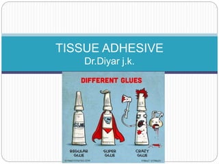
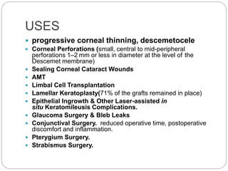
TISSUE ADHESIVE
Dr.Diyar j.k.
Tissue adhesive sealants
ď‚— have been used as substitutes for sutures in
ophthalmic surgery in recent years since the latter
may cause irritation, inflammation and infection.
ď‚— Tissue adhesives were developed as suture adjuncts
and alternatives for sealing wounded tissues. They
are gaining popularity for their ease of use and
postoperative comfort.
ď‚— Its high efficacy, relative ease of application, and its
ability to delay an otherwise emergent surgical repair
in the operating room.
ď‚— can immediately restore structural integrity to the
globe.In these situations, penetrating keratoplasty or
other more permanent procedures can be avoided or
at least delayed until a time when the eye is quiet &
surgical intervention has a better chance of success.
TYPES
ď‚— synthetic adhesives (e.g.,cyanoacrylate
derivatives) .
ď‚— biologic adhesives (e.g., fibrin-based
adhesives),biodendrimers ,Other adhesives in
development include acrylic-based adhesives,
polyethylene glycol hydrogels, chondroitin sulfate,
riboflavin-fibrinogen compounds, photoactivated
serum albumin solder and photo-polymerized
hyaluronic acid compounds
• the tissueglue can arrest keratolysis by
blocking leukocytic proteases from
the corneal wound.
USES
ď‚— progressive corneal thinning, descemetocele
ď‚— Corneal Perforations (small, central to mid-peripheral
perforations 1–2 mm or less in diameter at the level of the
Descemet membrane)
ď‚— Sealing Corneal Cataract Wounds
ď‚— AMT
ď‚— Limbal Cell Transplantation
ď‚— Lamellar Keratoplasty(71% of the grafts remained in place)
ď‚— Epithelial Ingrowth & Other Laser-assisted in
situ Keratomileusis Complications.
ď‚— Glaucoma Surgery & Bleb Leaks
ď‚— Conjunctival Surgery. reduced operative time, postoperative
discomfort and inflammation.
ď‚— Pterygium Surgery.
ď‚— Strabismus Surgery.
Cyanoacrylate tissue adhesive
'Krazy glues'
ď‚— N-butyl-2-cyanoacrylate is approved for
dermatologic use
ď‚— the tensile strength of bonding of these polymers
has been observed to be the highest among all
glues. It also has bacteriostatic property. Eiferman
and Snyder reported antibacterial activity
of butyl-2-cyanoacrylate against Gram-
positive organisms
 “superglue,” methyl-2-cyanoacrylate,
appears to be more toxic to
the cornea than the adhesives
mentioned previously
ď‚— Sharma et al. compared the effectiveness of fibrin
glue and N-cyanoacrylate for treating small
corneal perforations of 3 mm diameter.[20]Both
adhesives resulted in effective closure of corneal
perforations <3 mm in diameter. The investigators
demonstrated that adhesive plug formation with
cyanoacrylate was faster than that of fibrin glue.
However, fibrin glue-assisted corneal perforation
closure resulted in faster healing and less
vascularization.
 Nobe et al.39 reported that, for
both infectious and traumatic
perforations, corneal transplantation
had a better chance of
remaining clear if keratoplastycould
be delayed
ď‚— Kenyon6 and others39 have emphasized
the importance of early application
of corneal glue in anynoninfected,
progressive thinning disorder before
perforation
Fibrin glue
ď‚— ( Fbrin sealant) mimics natural Fibrin formation,
ultimately resulting in the formation of a fibrin clot.
Several fibrin glues have been approved by
(FDA) and are commercially available. These
products include TISSEEL Fibrin Sealant ,
EVICEL Fibrin Sealant and BioGlue Surgical
Adhesive .
ď‚— Another option is the CryoSeal FS System
(ThermoGenesis Corp, Rancho Cordova, CA),
which can be used in the automated preparation
of fibrin sealant from the patient’s own plasma.
Also, because both pooled human plasma and
bovine products are used to obtain some
components of these sealants, the clinician
should keep in mind the potential for disease
Advantages
ď‚— They tend to solidify less quickly than cyanoacrylate
glue, which may make application easier and more
accurate.
ď‚— they are softer and smoother than cyanoacrylate glue,
can be used over and under a superficial covering
layer (amniotic membrane or conjunctiva) & tend to
cause less discomfort and fewer symptoms.
disadvantages
ď‚— the need to store some brands frozen and thaw them
prior to use
ď‚— the lower tensile strength than cyanoacrylate, the
unknown length of time they remain in place,
ď‚— the promotion of microbial growth and the potential
for transmission of disease and allergy as some of
these glues are made from human and animal
products
ď‚— Use in the corneal surface reconstruction
procedures, amniotic membrane placement,
and in the management of small corneal
perforations.
Method Of Application
ď‚— Careful slit lamp examination and photographs or
drawings should be performed before the
application of any tissue adhesive,because
after gluing & placement of a bandage soft
contact lens (BSCL), subsequent
examination of the perforation site may be
difficult. size and extent of the perforation,
as well as the status of the lens and
the presence of uveal prolapse.
ď‚— Although the procedure often cannot be
performedwith strict sterile technique, care
should be taken to keep the field as
aseptic as possible.
SURGICAL TECHNIQUE
Cyanoacrylate glue can usually be applied on an
outpatient basis using topical anesthetics.
An eyelid speculum , necrotic tissue and corneal
epithelium should be removed from the involved area
out to a 2-mm zone. The area should then be dried with
a cellulose sponge. The easiest way to apply glue is
under biomicroscopic magnication. A small drop of the
uid adhesive is applied to the corneal wound with the tip
of a 30-gauge needle or anterior chamber cannula. The
glue does not polymerize on plastic.
The glue can then be applied to the surface of the
cornea in as thin a layer as possible using the plastic
handle of a cellulose sponge or the wooden stick of a
cottontipped applicator. The glue polymerizes
ď‚— STAY SAFE
ď‚— EAT CAKE
ReSure Sealant
Postoperative management
ď‚— All patients should be placed on topical and/or systemic
aqueous suppressants if medically tolerable (
PERFORATION)
ď‚— prophylactic broad-spectrum topical antibiotics
ď‚— Protective shield or glasses should be placed on the eye at
all times. Preservative-free artificial tears should be used
at least 4-8 times a day.
ď‚— The need for hospital admission and the use of
intravenous antibiotics in the case of a sterile perforation is
controversial.
ď‚— As a rule, we tend to leave the glue in place until it loosens
excessively or becomes spontaneously dislodged. If the
stroma appears healed, the glue can theoretically be
removed after several months. However, the risk of
perforation still exists. Tissue adhesive has remained on
the eye for up to 660 days before spontaneously
Complications
Although tissue adhesives are well tolerated by the
eye, several complications have been reported.
These include :
ď‚— cataract formation. (thought to be due to direct
contact of glue with the lens)
ď‚— corneal infiltration (infectious and noninfectious)
ď‚— glaucoma, giantpapillary conjunctivitis, retinal
toxicity
ď‚— granulomatous keratitis and symblepharon
formation
It is often difficult, however, to determine
whether the complication was due to the
THANK YOU
THANK YOU

Tissue Adhesive In Ophthalmology

  • 1.
  • 2.
    Tissue adhesive sealants ď‚—have been used as substitutes for sutures in ophthalmic surgery in recent years since the latter may cause irritation, inflammation and infection. ď‚— Tissue adhesives were developed as suture adjuncts and alternatives for sealing wounded tissues. They are gaining popularity for their ease of use and postoperative comfort. ď‚— Its high efficacy, relative ease of application, and its ability to delay an otherwise emergent surgical repair in the operating room. ď‚— can immediately restore structural integrity to the globe.In these situations, penetrating keratoplasty or other more permanent procedures can be avoided or at least delayed until a time when the eye is quiet & surgical intervention has a better chance of success.
  • 3.
    TYPES ď‚— synthetic adhesives(e.g.,cyanoacrylate derivatives) . ď‚— biologic adhesives (e.g., fibrin-based adhesives),biodendrimers ,Other adhesives in development include acrylic-based adhesives, polyethylene glycol hydrogels, chondroitin sulfate, riboflavin-fibrinogen compounds, photoactivated serum albumin solder and photo-polymerized hyaluronic acid compounds
  • 4.
    • the tissuegluecan arrest keratolysis by blocking leukocytic proteases from the corneal wound.
  • 6.
    USES  progressive cornealthinning, descemetocele  Corneal Perforations (small, central to mid-peripheral perforations 1–2 mm or less in diameter at the level of the Descemet membrane)  Sealing Corneal Cataract Wounds  AMT  Limbal Cell Transplantation  Lamellar Keratoplasty(71% of the grafts remained in place)  Epithelial Ingrowth & Other Laser-assisted in situ Keratomileusis Complications.  Glaucoma Surgery & Bleb Leaks  Conjunctival Surgery. reduced operative time, postoperative discomfort and inflammation.  Pterygium Surgery.  Strabismus Surgery.
  • 7.
    Cyanoacrylate tissue adhesive 'Krazyglues' ď‚— N-butyl-2-cyanoacrylate is approved for dermatologic use ď‚— the tensile strength of bonding of these polymers has been observed to be the highest among all glues. It also has bacteriostatic property. Eiferman and Snyder reported antibacterial activity of butyl-2-cyanoacrylate against Gram- positive organisms
  • 8.
     “superglue,” methyl-2-cyanoacrylate, appearsto be more toxic to the cornea than the adhesives mentioned previously
  • 9.
    ď‚— Sharma etal. compared the effectiveness of fibrin glue and N-cyanoacrylate for treating small corneal perforations of 3 mm diameter.[20]Both adhesives resulted in effective closure of corneal perforations <3 mm in diameter. The investigators demonstrated that adhesive plug formation with cyanoacrylate was faster than that of fibrin glue. However, fibrin glue-assisted corneal perforation closure resulted in faster healing and less vascularization.
  • 10.
     Nobe et al.39reported that, for both infectious and traumatic perforations, corneal transplantation had a better chance of remaining clear if keratoplastycould be delayed  Kenyon6 and others39 have emphasized the importance of early application of corneal glue in anynoninfected, progressive thinning disorder before perforation
  • 11.
    Fibrin glue  (Fbrin sealant) mimics natural Fibrin formation, ultimately resulting in the formation of a fibrin clot. Several fibrin glues have been approved by (FDA) and are commercially available. These products include TISSEEL Fibrin Sealant , EVICEL Fibrin Sealant and BioGlue Surgical Adhesive .  Another option is the CryoSeal FS System (ThermoGenesis Corp, Rancho Cordova, CA), which can be used in the automated preparation of fibrin sealant from the patient’s own plasma. Also, because both pooled human plasma and bovine products are used to obtain some components of these sealants, the clinician should keep in mind the potential for disease
  • 12.
    Advantages ď‚— They tendto solidify less quickly than cyanoacrylate glue, which may make application easier and more accurate. ď‚— they are softer and smoother than cyanoacrylate glue, can be used over and under a superficial covering layer (amniotic membrane or conjunctiva) & tend to cause less discomfort and fewer symptoms. disadvantages ď‚— the need to store some brands frozen and thaw them prior to use ď‚— the lower tensile strength than cyanoacrylate, the unknown length of time they remain in place, ď‚— the promotion of microbial growth and the potential for transmission of disease and allergy as some of these glues are made from human and animal products
  • 14.
    ď‚— Use inthe corneal surface reconstruction procedures, amniotic membrane placement, and in the management of small corneal perforations.
  • 15.
    Method Of Application ď‚—Careful slit lamp examination and photographs or drawings should be performed before the application of any tissue adhesive,because after gluing & placement of a bandage soft contact lens (BSCL), subsequent examination of the perforation site may be difficult. size and extent of the perforation, as well as the status of the lens and the presence of uveal prolapse. ď‚— Although the procedure often cannot be performedwith strict sterile technique, care should be taken to keep the field as aseptic as possible.
  • 16.
    SURGICAL TECHNIQUE Cyanoacrylate gluecan usually be applied on an outpatient basis using topical anesthetics. An eyelid speculum , necrotic tissue and corneal epithelium should be removed from the involved area out to a 2-mm zone. The area should then be dried with a cellulose sponge. The easiest way to apply glue is under biomicroscopic magnication. A small drop of the uid adhesive is applied to the corneal wound with the tip of a 30-gauge needle or anterior chamber cannula. The glue does not polymerize on plastic. The glue can then be applied to the surface of the cornea in as thin a layer as possible using the plastic handle of a cellulose sponge or the wooden stick of a cottontipped applicator. The glue polymerizes
  • 17.
  • 18.
  • 19.
    Postoperative management ď‚— Allpatients should be placed on topical and/or systemic aqueous suppressants if medically tolerable ( PERFORATION) ď‚— prophylactic broad-spectrum topical antibiotics ď‚— Protective shield or glasses should be placed on the eye at all times. Preservative-free artificial tears should be used at least 4-8 times a day. ď‚— The need for hospital admission and the use of intravenous antibiotics in the case of a sterile perforation is controversial. ď‚— As a rule, we tend to leave the glue in place until it loosens excessively or becomes spontaneously dislodged. If the stroma appears healed, the glue can theoretically be removed after several months. However, the risk of perforation still exists. Tissue adhesive has remained on the eye for up to 660 days before spontaneously
  • 20.
    Complications Although tissue adhesivesare well tolerated by the eye, several complications have been reported. These include : ď‚— cataract formation. (thought to be due to direct contact of glue with the lens) ď‚— corneal infiltration (infectious and noninfectious) ď‚— glaucoma, giantpapillary conjunctivitis, retinal toxicity ď‚— granulomatous keratitis and symblepharon formation It is often difficult, however, to determine whether the complication was due to the
  • 21.
  • 22.

Editor's Notes

  • #3 biocompatible and biodegradable materials.  with chemical-, thermal- and photo-activated CXL capabilities. The added advantages of mechanical and degradation rate tunability make dendrimers attractive candidates for ophthalmic adhesives.
  • #8 polymer is a large molecule, or macromolecule, composed of many repeated subunits. Due to their broad range of properties, both synthetic and natural polymers play essential and ubiquitous roles in everyday life
  • #10 the adhesive was also resorbed by three weeks. They reported 50% success with fibrin glue alone, with adhesion being maintained for 4—6 daysOne disadvantage of utilizing fibrin glue for flap reattachment is the resulting opacity of the glue that makes it difficult to determine inflammatory response.
  • #11 Not only is the procedure much easier to perform on a nonperforated eye, but tissue adhesive has been shown to arrest the process of ulceration
  • #19  20 seconds , Composed of a synthetic, polyethylene glycol hydrogel, the product is approximately 90% water after polymerization, The hydrogel contains a blue visualizationÂ