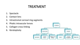
Keratoconus: Overview and Update on Treatment
PRESENTER:DR. ATIF RAHMAN RAINI
INTRODUCTION
• Greek word (kerato: Cornea; konos: Cone),meaning cone-shaped
protrusion of the cornea.
• Non-inflammatory, progressive thinning of the cornea that is usually
bilateral and involves the central two-thirds of the cornea.
EPIDEMIOLOGY
• 50 to 230 per 100,000
• Affects all races and sexes equally
• Onset around puberty
ETIOLOGY
• Etiology unknown and most likely multifactorial
• Strongly associated with eye rubbing
• Contact lens wear
• Positive family history has been reported in 6-8%.
• Unaffected first-degree relatives have a higher rate of abnormal
topography
• High concordance rate in monozygotic twins
• Autosomal-dominant inherited keratoconus
PATHOLOGY
• Keratoconus can involve each layer of the cornea.
• Early degeneration of basal epithelial cells disruption of the
basement membrane growth of epithelium posterior to the
Bowman’s layer and collagen anterior to the epithelium breaks in
the Bowman’s layer.
HISTO-PATHOLOGY
• Scarring of the Bowman’s layer and the anterior stroma
• stroma has normal-sized collagen fibers but low numbers of collagen
lamellae stromal thinning
• Endothelial cell pleomorphism and polymegathism may be
manifested
CLINICAL PRESENTATION
• Usually present in teenage years or twenties
• Visual blur
• Distortion secondary to myopia and high astigmatism
• Photophobia
• Glare
• Monocular diplopia
• Retinoscopy : scissors reflex
• Rizzutti’s sign : a conical reflection on the nasal cornea when light is
shone temporally
• Fleischer ring
• Vogt lines : Fine and roughly parallel striations
• Munson’s sign : advanced keratoconus
• Acute hydrops
Scissor reflex
Rizzutti’s sign
Fleischer ring
Vogt lines
Munson’s sign
DIAGNOSTIC EVALUATION
• Corneal topography is a valuable diagnostic tool for –
1) diagnosing subclinical keratoconus
2) tracking the progression of the disease
• Rabinowitz has suggested four quantitative videokeratographic
indices for screening keratoconus –
central corneal power 47.2 D
inferior-superior dioptric asymmetry over 1.2 D
 Sim-K astigmatism 1.5 D
 skewed radial axes 21
• Maeda and Klyce developed a topographic classifier using eight
indices to diagnose keratoconus.
• KPI ( Keratoconus Predictive Index) > 0.23 indicative of keratoconus.
• Rabinowitz and Rasheed described KISA% index.
• KISA% index is derived from the product of four indices-
1)K-value- expression of central corneal steepening
2) I-S value- expression of the inferior-superior dioptric
asymmetry
3) corneal astigmatism index - quantifies the degree of regular
corneal astigmatism (SimK1-Sim K2)
4) skewed radial axis (SRAX) index- an expression of irregular
astigmatism occurring in keratoconus
KISA%= (K) x (I-S) x (AST) x (SRAX) x 1/3
• When the KISA% is 100% or higher in an eye with no other pathology,
the patient is very likely to have clinically detectable keratoconus.
• Values ranging from 60 to 100% indicative of keratoconus suspects
Topographic devices
• Orbscan (Bausch and Lomb Inc., Rochester, NY, USA) provides
data on
 anterior and posterior elevation
 best-fit sphere
 corneal pachymetry map
• used as a tool for screening keratoconus suspects
• Pentacam (Oculus, Lynnwood, WA, USA)- uses a rotating Scheimpflug
camera.
• Scheimpflug system determines—
net corneal power
elevation maps
 anterior chamber depth
 corneal wavefront
TREATMENT
1. Spectacle
2. Contact lens
3. Intrastromal corneal ring segments
4. Phakic intraocular lenses
5. Collagen cross-linking
6. Keratoplasty
Spectacles
• Initially managed with spectacles.
• contact lenses – when spectacles fails
Contact lens
• For mild or moderate irregularities – soft, soft- toric
• For severe irregularities- RIGID GAS PERMEABLE (RGP)
• Super Cone and Rose K – special RGP contact lens with a steep
central posterior curve to vault over the cone and flatter
peripheral curves.
• Hybrid contact lenses- comprise a rigid center and a soft skirt –
SynergEyes-KC (SynergEyes Inc.,Carlsbad, CA, USA)
• Piggyback contact lens- soft lens fitted to the cornea and an RGP lens
placed on top.
• Gas-permeable scleral contact lenses - highly irregular corneas
Intrastromal corneal ring segments
• Intacs; Addition Technology Inc., Sunnyvale, CA, USA – originally
developed for low-myopia correction .
 now approved for reduction of myopia and irregular
astigmatism associated with keratoconus.
• Ring segments are inserted through mechanical and femtosecond
laser-assisted corneal tunnels.
• Hypothesized that ICRS induces a displacement of the local anterior
surface, steepening of the peripheral cornea and a flattening of the
central portion of the anterior cornea by adding extra material at the
corneal midperiphery and provides biomechanical support for the
thin ectatic cornea.
Complication associated with ICRS
• 35% visually significant complication rate.
• Complication-
 epithelial defects
 anterior and posterior perforation during channel creation
 extention of incision towards visual axis
 uneven placement of implants
 impant decentration
• Shallow segment depth - associated with
infectious keratitis
 segment superficialization and exposure
stromal thinning
 epithelial breakdown
corneal melting
• Lamellar channel deposits - primarily consist of intrastromal lipid
accumulations and keratocytes.
• thought to arise in response to corneal injury.
• Intrastromal deposits do not alter the optical performance of Intacs
or the anatomical and physiological changes in the cornea.
• Ruckhofer et al reported a 74% overall incidence of intrastromal
deposits.
Variation of INTACS
Intacs SK – used for Severe Keratoconus
 Newer design of ICRS with a smaller 6mm optical zone to correct higher
grades of keratectasia with an elliptical cross-section to minimize the glare.
• Rodriguez et al. showed a clinically significant reduction in
keratectasia with improvement in uncorrected visual acuity (UCVA),
spherical equivalent and keratometry in patients with severe post-
Lasik ectasia after placement of SK.
• Ferrara ring (Keravision Inc., Fremont, CA, USA)- made of polymethyl
methacrylate-Perspex CQ acrylic segments.
• These segments also vary in thickness (0.15, 0.20, 0.30 and 0.35 mm)
• Segments have 160of arc and optic zone of 5 mm.
Phakic intraocular lenses
• Progression of keratoconus leading to refractive change is a concern
after implantation of any type of Phakic IOL.
• Phakic IOL implantation should not be performed until refraction and
keratometry are stable.
Indication of Phakic IOL implantation
• BCVA of 20/50 or better
• clear central cornea
• keratometric values 52.00D
• stable refraction (cylinder 3.00 D) for 2 years
If these criteria are not met, penetrating keratoplasty or collagen cross-
linking (CXL) may provide better visual outcomes.
• Used for correction of residual refractive error in the post-ICRS
keratoconus patients.
• Use of anterior or posterior phakic IOLs, including toric lenses, either
alone or after implantation of ICRS.
• In a prospective non-comparative interventional case series, angle-
supported Phakic IOLs (ZSAL-4, Morcher GmbH) were implanted in 12
eyes with stage I-II keratoconus, myopia from 6.5 to 14.00D and
astigmatism from 1.00 to 5.00D. At 1 year post-operatively, spherical
error was within 1.00 D,without any significant change in astigmatism
in all cases, and UCVA was 20/40 or better in all cases.
• Main concern with angle-supported phakic IOLs is endothelial cell
loss.
• Moshirfar et al. reported successful implantation of iris-supported
Verisyse phakic IOL (AMO, Santa Ana, CA, USA) in two cases and
found 4% endothelial cell loss at 3-months post-operatively in both
cases.
• Alfonso et al. implanted the myopic Phakic posterior chamber
Implantable Collamer Lens (ICL, STAAR, Monrovia, CA, USA) in 25 eyes
with myopia from 3.00 to 18.00 D and astigmatism from 0.5 to 3.00 D
and 12 months follow-up, and reported that the spherical equivalent
refraction was within 1.00 D of the desired refraction in all cases.
• El-Raggal et al. conducted a prospective evaluation of sequential
implantation of Intacs and Verisyse phakic IOL in eight eyes and
showed the safety, stability and effectiveness of the procedure.
Collagen cross-linking
• Disadvantage of the previously mentioned procedures is that none
adequately prevent keratoconus progression that occurs due to the
underlying biomechanical corneal changes.
• Technique uses photo-oxidative CXL technique using riboflavin and
ultraviolet-A (UVA) light.
• Developed to counteract the progressive corneal thinning and
consequently the progression of keratoconus.
• With cross-linking, additional covalent bonding between collagen
molecules can be achieved, which stabilizes the collagen scaffold and
changes several tissue properties.
• Raiskup-Wolf et al. retrospectively evaluated the long-term effect of
riboflavin and UVA in progressive keratoconus with maximum follow-
up of 6 years and showed that the steepening significantly decreased
by 2.68 D in the first year, 2.21 D in the second year and 4.84 D in the
third year, with an associated improvement in BCVA and stability.
CONCLUSION
• With the advent of corneal cross-linking technology to stabilize the
biomechanically weakened collagen in keratoconus, the spectrum of
keratoconus management now includes both the prevention and the
treatment of progression of disease.
THANK YOU……

Keratoconus 2

  • 1.
    Keratoconus: Overview andUpdate on Treatment PRESENTER:DR. ATIF RAHMAN RAINI
  • 2.
    INTRODUCTION • Greek word(kerato: Cornea; konos: Cone),meaning cone-shaped protrusion of the cornea. • Non-inflammatory, progressive thinning of the cornea that is usually bilateral and involves the central two-thirds of the cornea.
  • 3.
    EPIDEMIOLOGY • 50 to230 per 100,000 • Affects all races and sexes equally • Onset around puberty
  • 4.
    ETIOLOGY • Etiology unknownand most likely multifactorial • Strongly associated with eye rubbing • Contact lens wear • Positive family history has been reported in 6-8%. • Unaffected first-degree relatives have a higher rate of abnormal topography • High concordance rate in monozygotic twins • Autosomal-dominant inherited keratoconus
  • 5.
    PATHOLOGY • Keratoconus caninvolve each layer of the cornea. • Early degeneration of basal epithelial cells disruption of the basement membrane growth of epithelium posterior to the Bowman’s layer and collagen anterior to the epithelium breaks in the Bowman’s layer.
  • 6.
    HISTO-PATHOLOGY • Scarring ofthe Bowman’s layer and the anterior stroma • stroma has normal-sized collagen fibers but low numbers of collagen lamellae stromal thinning • Endothelial cell pleomorphism and polymegathism may be manifested
  • 7.
    CLINICAL PRESENTATION • Usuallypresent in teenage years or twenties • Visual blur • Distortion secondary to myopia and high astigmatism • Photophobia • Glare • Monocular diplopia • Retinoscopy : scissors reflex • Rizzutti’s sign : a conical reflection on the nasal cornea when light is shone temporally • Fleischer ring
  • 8.
    • Vogt lines: Fine and roughly parallel striations • Munson’s sign : advanced keratoconus • Acute hydrops
  • 9.
  • 10.
  • 11.
  • 12.
  • 13.
  • 14.
    DIAGNOSTIC EVALUATION • Cornealtopography is a valuable diagnostic tool for – 1) diagnosing subclinical keratoconus 2) tracking the progression of the disease
  • 15.
    • Rabinowitz hassuggested four quantitative videokeratographic indices for screening keratoconus – central corneal power 47.2 D inferior-superior dioptric asymmetry over 1.2 D  Sim-K astigmatism 1.5 D  skewed radial axes 21
  • 16.
    • Maeda andKlyce developed a topographic classifier using eight indices to diagnose keratoconus. • KPI ( Keratoconus Predictive Index) > 0.23 indicative of keratoconus. • Rabinowitz and Rasheed described KISA% index.
  • 17.
    • KISA% indexis derived from the product of four indices- 1)K-value- expression of central corneal steepening 2) I-S value- expression of the inferior-superior dioptric asymmetry 3) corneal astigmatism index - quantifies the degree of regular corneal astigmatism (SimK1-Sim K2) 4) skewed radial axis (SRAX) index- an expression of irregular astigmatism occurring in keratoconus KISA%= (K) x (I-S) x (AST) x (SRAX) x 1/3
  • 18.
    • When theKISA% is 100% or higher in an eye with no other pathology, the patient is very likely to have clinically detectable keratoconus. • Values ranging from 60 to 100% indicative of keratoconus suspects
  • 19.
    Topographic devices • Orbscan(Bausch and Lomb Inc., Rochester, NY, USA) provides data on  anterior and posterior elevation  best-fit sphere  corneal pachymetry map • used as a tool for screening keratoconus suspects
  • 20.
    • Pentacam (Oculus,Lynnwood, WA, USA)- uses a rotating Scheimpflug camera. • Scheimpflug system determines— net corneal power elevation maps  anterior chamber depth  corneal wavefront
  • 21.
    TREATMENT 1. Spectacle 2. Contactlens 3. Intrastromal corneal ring segments 4. Phakic intraocular lenses 5. Collagen cross-linking 6. Keratoplasty
  • 22.
    Spectacles • Initially managedwith spectacles. • contact lenses – when spectacles fails
  • 23.
    Contact lens • Formild or moderate irregularities – soft, soft- toric • For severe irregularities- RIGID GAS PERMEABLE (RGP) • Super Cone and Rose K – special RGP contact lens with a steep central posterior curve to vault over the cone and flatter peripheral curves.
  • 24.
    • Hybrid contactlenses- comprise a rigid center and a soft skirt – SynergEyes-KC (SynergEyes Inc.,Carlsbad, CA, USA) • Piggyback contact lens- soft lens fitted to the cornea and an RGP lens placed on top. • Gas-permeable scleral contact lenses - highly irregular corneas
  • 25.
    Intrastromal corneal ringsegments • Intacs; Addition Technology Inc., Sunnyvale, CA, USA – originally developed for low-myopia correction .  now approved for reduction of myopia and irregular astigmatism associated with keratoconus. • Ring segments are inserted through mechanical and femtosecond laser-assisted corneal tunnels.
  • 26.
    • Hypothesized thatICRS induces a displacement of the local anterior surface, steepening of the peripheral cornea and a flattening of the central portion of the anterior cornea by adding extra material at the corneal midperiphery and provides biomechanical support for the thin ectatic cornea.
  • 27.
    Complication associated withICRS • 35% visually significant complication rate. • Complication-  epithelial defects  anterior and posterior perforation during channel creation  extention of incision towards visual axis  uneven placement of implants  impant decentration
  • 28.
    • Shallow segmentdepth - associated with infectious keratitis  segment superficialization and exposure stromal thinning  epithelial breakdown corneal melting
  • 29.
    • Lamellar channeldeposits - primarily consist of intrastromal lipid accumulations and keratocytes. • thought to arise in response to corneal injury. • Intrastromal deposits do not alter the optical performance of Intacs or the anatomical and physiological changes in the cornea. • Ruckhofer et al reported a 74% overall incidence of intrastromal deposits.
  • 30.
    Variation of INTACS IntacsSK – used for Severe Keratoconus  Newer design of ICRS with a smaller 6mm optical zone to correct higher grades of keratectasia with an elliptical cross-section to minimize the glare. • Rodriguez et al. showed a clinically significant reduction in keratectasia with improvement in uncorrected visual acuity (UCVA), spherical equivalent and keratometry in patients with severe post- Lasik ectasia after placement of SK.
  • 31.
    • Ferrara ring(Keravision Inc., Fremont, CA, USA)- made of polymethyl methacrylate-Perspex CQ acrylic segments. • These segments also vary in thickness (0.15, 0.20, 0.30 and 0.35 mm) • Segments have 160of arc and optic zone of 5 mm.
  • 32.
    Phakic intraocular lenses •Progression of keratoconus leading to refractive change is a concern after implantation of any type of Phakic IOL. • Phakic IOL implantation should not be performed until refraction and keratometry are stable.
  • 33.
    Indication of PhakicIOL implantation • BCVA of 20/50 or better • clear central cornea • keratometric values 52.00D • stable refraction (cylinder 3.00 D) for 2 years If these criteria are not met, penetrating keratoplasty or collagen cross- linking (CXL) may provide better visual outcomes.
  • 34.
    • Used forcorrection of residual refractive error in the post-ICRS keratoconus patients. • Use of anterior or posterior phakic IOLs, including toric lenses, either alone or after implantation of ICRS.
  • 35.
    • In aprospective non-comparative interventional case series, angle- supported Phakic IOLs (ZSAL-4, Morcher GmbH) were implanted in 12 eyes with stage I-II keratoconus, myopia from 6.5 to 14.00D and astigmatism from 1.00 to 5.00D. At 1 year post-operatively, spherical error was within 1.00 D,without any significant change in astigmatism in all cases, and UCVA was 20/40 or better in all cases.
  • 36.
    • Main concernwith angle-supported phakic IOLs is endothelial cell loss. • Moshirfar et al. reported successful implantation of iris-supported Verisyse phakic IOL (AMO, Santa Ana, CA, USA) in two cases and found 4% endothelial cell loss at 3-months post-operatively in both cases.
  • 37.
    • Alfonso etal. implanted the myopic Phakic posterior chamber Implantable Collamer Lens (ICL, STAAR, Monrovia, CA, USA) in 25 eyes with myopia from 3.00 to 18.00 D and astigmatism from 0.5 to 3.00 D and 12 months follow-up, and reported that the spherical equivalent refraction was within 1.00 D of the desired refraction in all cases. • El-Raggal et al. conducted a prospective evaluation of sequential implantation of Intacs and Verisyse phakic IOL in eight eyes and showed the safety, stability and effectiveness of the procedure.
  • 38.
    Collagen cross-linking • Disadvantageof the previously mentioned procedures is that none adequately prevent keratoconus progression that occurs due to the underlying biomechanical corneal changes. • Technique uses photo-oxidative CXL technique using riboflavin and ultraviolet-A (UVA) light. • Developed to counteract the progressive corneal thinning and consequently the progression of keratoconus.
  • 39.
    • With cross-linking,additional covalent bonding between collagen molecules can be achieved, which stabilizes the collagen scaffold and changes several tissue properties. • Raiskup-Wolf et al. retrospectively evaluated the long-term effect of riboflavin and UVA in progressive keratoconus with maximum follow- up of 6 years and showed that the steepening significantly decreased by 2.68 D in the first year, 2.21 D in the second year and 4.84 D in the third year, with an associated improvement in BCVA and stability.
  • 40.
    CONCLUSION • With theadvent of corneal cross-linking technology to stabilize the biomechanically weakened collagen in keratoconus, the spectrum of keratoconus management now includes both the prevention and the treatment of progression of disease.
  • 41.