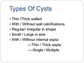
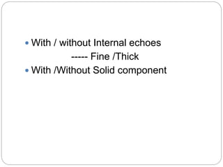
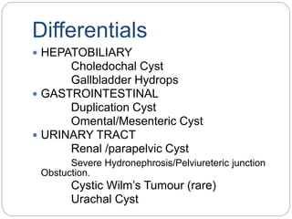
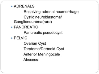
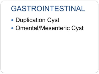
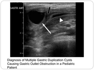
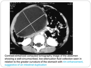
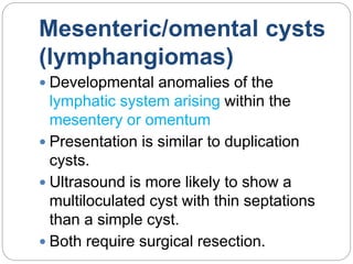
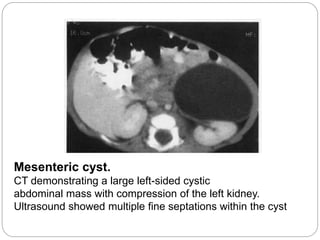
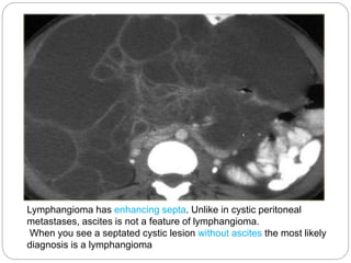
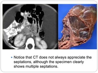
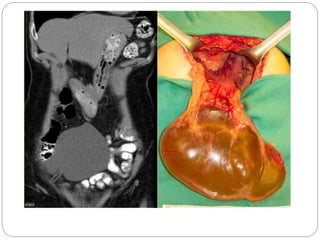
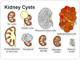
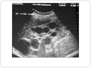
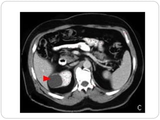
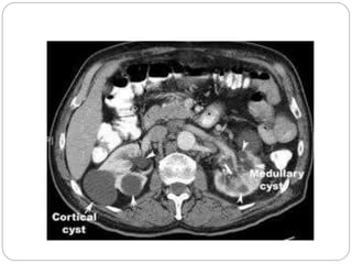
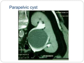
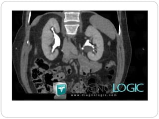
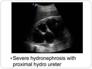
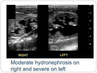
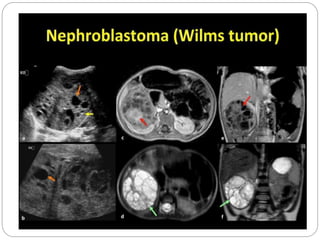
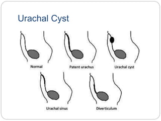
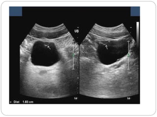
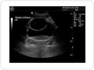
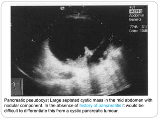
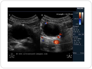
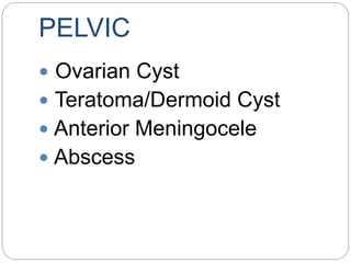
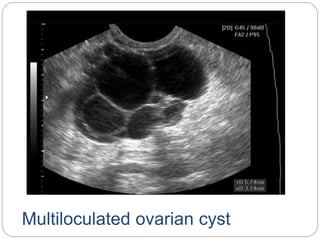
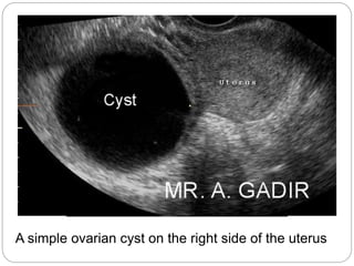
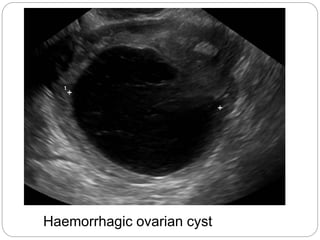
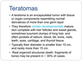
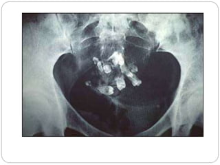
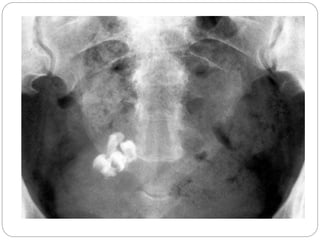
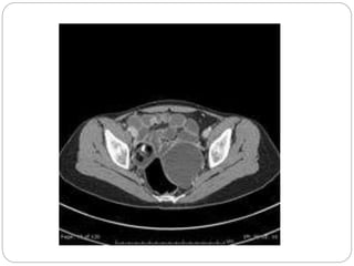
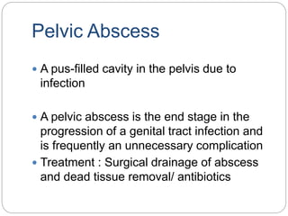
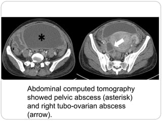
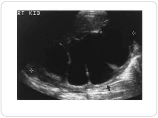
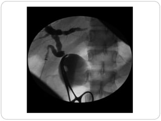
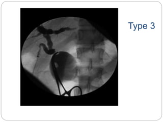
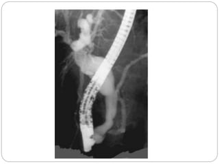
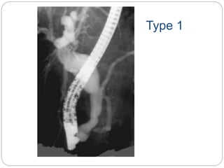
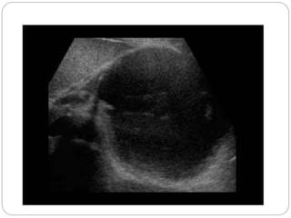
Intra abdominal cysts in Children
Objectives
 What is a cyst?
 Types of cysts
 Differentials
 Imaging appearances
CYST
A cyst is a closed pocket or
pouch of tissue. It can be filled
with air, fluid, pus, or other
material
 http://www.ncbi.nlm.nih.gov/pubmedhealth/PMH0003724/
Types Of Cysts
 Thin /Thick walled
 With / Without wall calcifications
 Regular/ Irregular in shape
 Small / Large in size
 With / Without internal septa
---Thin / Thick septa
----Single / Multiple
 With / without Internal echoes
----- Fine /Thick
 With /Without Solid component
Differentials
 HEPATOBILIARY
Choledochal Cyst
Gallbladder Hydrops
 GASTROINTESTINAL
Duplication Cyst
Omental/Mesenteric Cyst
 URINARY TRACT
Renal /parapelvic Cyst
Severe Hydronephrosis/Pelviureteric junction
Obstuction.
Cystic Wilm’s Tumour (rare)
Urachal Cyst
 ADRENALS
Resolving adrenal heamorrhage
Cystic neuroblastoma/
Ganglioneuroma(rare)
 PANCREATIC
Pancreatic pseudocyst
 PELVIC
Ovarian Cyst
Teratoma/Dermoid Cyst
Anterior Meningocele
Abscess
Hepatobiliary
 CHOLEDOCHAL CYST
 GALLBLADER HYDROPS
CHOLEDOCHAL CYST
Congenital dilatations of the biliary tree
• Most cause symptoms in childhood and
adult life.
TYPES (Todani ‘s )
 Type I- Cystic (51 %) or fusiform
(10.6%)
 TypeII- Diverticulum
 Type IlI- Choledochocele of
intraduodenal common bile duct
 Type IV- Extra- and intrahepatic cysts
(28.5%)
 Type V- Intrahepatic dilatations (4.6%)
Complications include cholangitis,
biliary calculi, pancreatitis and biliary
cirrhosis.
• On ultrasound or CT the biliary tree
dilatation or cyst can be seen.
• 99mTc-HIDA scinitraphy will show
accumulation of tracer within the cyst.
• Percutanous or endoscopic
cholangiography and MRCP are
helpful in preoperative planning.
Fusiform choledochal cyst with a long common channel
and associated stricture at the pancreaticobiliary junction.
CT of a large choledochal cyst with obstruction.
“Ultrasound study shows a cystic mass between pancreatic head and the
gallbladder. Smooth wall and homogeneous anechoic contents( tortuous
cystic duct that joins the gall bladder to the cystic mass )
GASTROINTESTINAL
 Duplication Cyst
 Omental/Mesenteric Cyst
DUPLICATION CYST
 SITES: May occur anywhere along the
gastrointestinal tract
 But 1/3rd of cases involve the distal small
bowel.
 The most frequent sites of duplication are the
ileum, then oesophagus, stomach, duodenum
and jejunum.
 Colonic and rectal duplications are rare.
 ETIOLOGY: Incomplete recanalisation at
around 8 weeks gestation
 lined with GI Epithelium (may be adjacent
mucosa /ectopic)
 TYPES: Spherical /Tubular
 Most duplications do not communicate
with the adjacent bowel, although there
is a higher incidence of persistent
communication in tubular anomalies
 PRESENTATION: Depends on the site
of duplication and its size.
 Incidental ultrasound finding in the first
few years of life.
 Large cysts, especially those associated
with the stomach or duodenum, may
present with
1. Abdominal pain,
2. Obstruction
3. Vomiting
4. Lead point for intussusception
or
5. A source of gastrointestinal
bleeding from ectopic gastric mucosa.
IMAGING And TREATMENT
 Abdominal radiographs may
show mass effect with displacement
of adjacent bowel loops.
 Ultrasound demonstrates a simple
hypoechoic cyst; if the
characteristic 'gut-wall signature'
 TREATMENT :Surgical resection
Abdominal x-ray of a patient with a duplication cyst. Note the
mass effect of the cyst pressing against the areas of colon
(arrows).
Diagnosis of Multiple Gastric Duplication Cysts
Causing Gastric Outlet Obstruction in a Pediatric
Patient
Contrast-enhanced computed tomography image of the abdomen
showing a well-circumscribed, low-attenuation fluid collection seen in
relation to the greater curvature of the stomach with rim enhancement,
suggestive of an intestinal duplication
Mesenteric/omental cysts
(lymphangiomas)
 Developmental anomalies of the
lymphatic system arising within the
mesentery or omentum
 Presentation is similar to duplication
cysts.
 Ultrasound is more likely to show a
multiloculated cyst with thin septations
than a simple cyst.
 Both require surgical resection.
Mesenteric cyst.
CT demonstrating a large left-sided cystic
abdominal mass with compression of the left kidney.
Ultrasound showed multiple fine septations within the cyst
Lymphangioma has enhancing septa. Unlike in cystic peritoneal
metastases, ascites is not a feature of lymphangioma.
When you see a septated cystic lesion without ascites the most likely
diagnosis is a lymphangioma
 Notice that CT does not always appreciate the
septations, although the specimen clearly
shows multiple septations.
URINARY TRACT
Renal /parapelvic Cyst
Severe Hydronephrosis/
Pelviureteric junction
obstruction
Cystic Wilm’s Tumour (rare)
Urachal Cyst
Parapelvic cyst
Severe hydronephrosis with
proximal hydro ureter
Moderate hydronephrosis on
right and severe on left
Urachal Cyst
ADRENALS
Resolving adrenal
heamorrhage
 Cystic neuroblastoma/
Ganglioneuroma(rare)
Resolving adrenal heamorrhage
 commonest cause of an adrenal mass
 Associated with perinatal stress,
hypoxia, septicaemia and hypotension
 may be unilateral or bilateral
 Adrenal insufficiency is rare, even in
bilateral cases.
 Ultrasound in the first few days of life
usually demonstrates an avascular
heterogenous adrenal mass that
becomes cystic and smaller over the
following weeks as clot retraction occurs
Day 2
Day 10
Cystic neuroblastoma/
Ganglioneuroma(rare)
 Over half of them arise in the adrenals, but 30%
can arise
 from sympathetic tissue elsewhere in the
abdomen
 Calcification has been noted to occur in over 50%
of Cases
 Ganglioneuroma is a mature form of neurogenic
tumour.
 Calcification helps in suggesting a diagnosis of
neurogenic tumour
Adrenal ganglioneuroma with
hepatic metastasis
PANCREATIC
Pancreatic pseudocyst
Pancreatic pseudocyst
 well-known complication of pancreatitis
 fluid collections may occur within the
pancreatic mass, or
in the peripancreatic spaces, or
elsewhere within the abdomen
following either acute / chronic pancreatitis
 In acute pancreatitis, the pseudocyst contains
enzyme-rich fluid and products of
autodegradation of the pancreas
 in chronic pancreatitis the cyst is a consequence
of duct obstruction.
 Patients who have persistent abdominal
pain or persistently elevated levels of
pancreatic enzymes should be
suspected of harbouring a pseudocyst
 one-third of pancreatic pseudocysts will
resolve spontaneously
Adjacent
to spleen
with
evidence
of
layering
debris
Large cystic
mass in the
mid abdomen in
the region of
the pancreatic
bed
demonstrating
echogenic
material
posteriorly,
representing
pancreatic
necrosis.
Pancreatic pseudocyst Large septated cystic mass in the mid abdomen with
nodular component. In the absence of history of pancreatitis it would be
difficult to differentiate this from a cystic pancreatic tumour.
PELVIC
 Ovarian Cyst
 Teratoma/Dermoid Cyst
 Anterior Meningocele
 Abscess
Ovarian Cyst
 Cysts are fluid filled spaces within the ovary.
 very common and could be physiological /
pathological, benign/ malignant
 Functional or physiological cysts are either
follicular or of corpus luteum origin.
 Follicular cysts form when a follicle fails to rupture
at midcycle leading to its continuous
enlargement. Usually these cysts are
asymptomatic and disappear without any
intervention within one or two months
 Similarly a persistent corpus luteum might fail to
disintegrate before menstruation and enlarge in
size
 Both follicular and luteal cysts could become
haemorrhagic if bleeding occured within them
leading to rapid increase in size and severe pain.
 they might cause severe pain only if they are
large in size (>7 cm) and cause pressure
symptoms or torsion of the whole ovary
compromising blood flow when surgical
intervention is indicated
Multiloculated ovarian cyst
A simple ovarian cyst on the right side of the uterus
Haemorrhagic ovarian cyst
Teratomas
 A teratoma is an encapsulated tumor with tissue
or organ components resembling normal
derivatives of more than one germ layer
 They therefore contain developmentally mature
skin complete with hair follicles and sweat glands,
sometimes luxuriant clumps of long hair, and
often pockets of sebum, blood, fat, bone, nails,
teeth, eyes, cartilage, and thyroid tissue.
 Typically their diameter is smaller than 10 cm,
and rarely more than 15 cm.
 Real organoid structures (teeth, fragments of
bone) may be present in ~ 30% of cases.
Pelvic Abscess
 A pus-filled cavity in the pelvis due to
infection
 A pelvic abscess is the end stage in the
progression of a genital tract infection and
is frequently an unnecessary complication
 Treatment : Surgical drainage of abscess
and dead tissue removal/ antibiotics
Abdominal computed tomography
showed pelvic abscess (asterisk)
and right tubo-ovarian abscess
(arrow).
Conclusion
 Cysts may have different characteristics
and origins
 Location, appearances, multi modality
can help in the diagnosis
Mind teaser
Type 3
Type 1
Thanks

Intra abdominal cysts in children

  • 1.
  • 2.
    Objectives  What isa cyst?  Types of cysts  Differentials  Imaging appearances
  • 3.
    CYST A cyst isa closed pocket or pouch of tissue. It can be filled with air, fluid, pus, or other material  http://www.ncbi.nlm.nih.gov/pubmedhealth/PMH0003724/
  • 4.
    Types Of Cysts Thin /Thick walled  With / Without wall calcifications  Regular/ Irregular in shape  Small / Large in size  With / Without internal septa ---Thin / Thick septa ----Single / Multiple
  • 5.
     With /without Internal echoes ----- Fine /Thick  With /Without Solid component
  • 6.
    Differentials  HEPATOBILIARY Choledochal Cyst GallbladderHydrops  GASTROINTESTINAL Duplication Cyst Omental/Mesenteric Cyst  URINARY TRACT Renal /parapelvic Cyst Severe Hydronephrosis/Pelviureteric junction Obstuction. Cystic Wilm’s Tumour (rare) Urachal Cyst
  • 7.
     ADRENALS Resolving adrenalheamorrhage Cystic neuroblastoma/ Ganglioneuroma(rare)  PANCREATIC Pancreatic pseudocyst  PELVIC Ovarian Cyst Teratoma/Dermoid Cyst Anterior Meningocele Abscess
  • 8.
  • 9.
    CHOLEDOCHAL CYST Congenital dilatationsof the biliary tree • Most cause symptoms in childhood and adult life.
  • 10.
    TYPES (Todani ‘s)  Type I- Cystic (51 %) or fusiform (10.6%)  TypeII- Diverticulum  Type IlI- Choledochocele of intraduodenal common bile duct  Type IV- Extra- and intrahepatic cysts (28.5%)  Type V- Intrahepatic dilatations (4.6%)
  • 12.
    Complications include cholangitis, biliarycalculi, pancreatitis and biliary cirrhosis. • On ultrasound or CT the biliary tree dilatation or cyst can be seen. • 99mTc-HIDA scinitraphy will show accumulation of tracer within the cyst. • Percutanous or endoscopic cholangiography and MRCP are helpful in preoperative planning.
  • 13.
    Fusiform choledochal cystwith a long common channel and associated stricture at the pancreaticobiliary junction.
  • 14.
    CT of alarge choledochal cyst with obstruction.
  • 15.
    “Ultrasound study showsa cystic mass between pancreatic head and the gallbladder. Smooth wall and homogeneous anechoic contents( tortuous cystic duct that joins the gall bladder to the cystic mass )
  • 17.
  • 18.
    DUPLICATION CYST  SITES:May occur anywhere along the gastrointestinal tract  But 1/3rd of cases involve the distal small bowel.  The most frequent sites of duplication are the ileum, then oesophagus, stomach, duodenum and jejunum.  Colonic and rectal duplications are rare.  ETIOLOGY: Incomplete recanalisation at around 8 weeks gestation  lined with GI Epithelium (may be adjacent mucosa /ectopic)
  • 19.
     TYPES: Spherical/Tubular  Most duplications do not communicate with the adjacent bowel, although there is a higher incidence of persistent communication in tubular anomalies  PRESENTATION: Depends on the site of duplication and its size.  Incidental ultrasound finding in the first few years of life.
  • 20.
     Large cysts,especially those associated with the stomach or duodenum, may present with 1. Abdominal pain, 2. Obstruction 3. Vomiting 4. Lead point for intussusception or 5. A source of gastrointestinal bleeding from ectopic gastric mucosa.
  • 21.
    IMAGING And TREATMENT Abdominal radiographs may show mass effect with displacement of adjacent bowel loops.  Ultrasound demonstrates a simple hypoechoic cyst; if the characteristic 'gut-wall signature'  TREATMENT :Surgical resection
  • 22.
    Abdominal x-ray ofa patient with a duplication cyst. Note the mass effect of the cyst pressing against the areas of colon (arrows).
  • 24.
    Diagnosis of MultipleGastric Duplication Cysts Causing Gastric Outlet Obstruction in a Pediatric Patient
  • 25.
    Contrast-enhanced computed tomographyimage of the abdomen showing a well-circumscribed, low-attenuation fluid collection seen in relation to the greater curvature of the stomach with rim enhancement, suggestive of an intestinal duplication
  • 26.
    Mesenteric/omental cysts (lymphangiomas)  Developmentalanomalies of the lymphatic system arising within the mesentery or omentum  Presentation is similar to duplication cysts.  Ultrasound is more likely to show a multiloculated cyst with thin septations than a simple cyst.  Both require surgical resection.
  • 27.
    Mesenteric cyst. CT demonstratinga large left-sided cystic abdominal mass with compression of the left kidney. Ultrasound showed multiple fine septations within the cyst
  • 28.
    Lymphangioma has enhancingsepta. Unlike in cystic peritoneal metastases, ascites is not a feature of lymphangioma. When you see a septated cystic lesion without ascites the most likely diagnosis is a lymphangioma
  • 29.
     Notice thatCT does not always appreciate the septations, although the specimen clearly shows multiple septations.
  • 31.
    URINARY TRACT Renal /parapelvicCyst Severe Hydronephrosis/ Pelviureteric junction obstruction Cystic Wilm’s Tumour (rare) Urachal Cyst
  • 36.
  • 38.
  • 39.
  • 41.
  • 45.
    ADRENALS Resolving adrenal heamorrhage  Cysticneuroblastoma/ Ganglioneuroma(rare)
  • 46.
    Resolving adrenal heamorrhage commonest cause of an adrenal mass  Associated with perinatal stress, hypoxia, septicaemia and hypotension  may be unilateral or bilateral  Adrenal insufficiency is rare, even in bilateral cases.  Ultrasound in the first few days of life usually demonstrates an avascular heterogenous adrenal mass that becomes cystic and smaller over the following weeks as clot retraction occurs
  • 47.
  • 48.
  • 49.
    Cystic neuroblastoma/ Ganglioneuroma(rare)  Overhalf of them arise in the adrenals, but 30% can arise  from sympathetic tissue elsewhere in the abdomen  Calcification has been noted to occur in over 50% of Cases  Ganglioneuroma is a mature form of neurogenic tumour.  Calcification helps in suggesting a diagnosis of neurogenic tumour
  • 50.
  • 51.
  • 52.
    Pancreatic pseudocyst  well-knowncomplication of pancreatitis  fluid collections may occur within the pancreatic mass, or in the peripancreatic spaces, or elsewhere within the abdomen following either acute / chronic pancreatitis  In acute pancreatitis, the pseudocyst contains enzyme-rich fluid and products of autodegradation of the pancreas  in chronic pancreatitis the cyst is a consequence of duct obstruction.
  • 53.
     Patients whohave persistent abdominal pain or persistently elevated levels of pancreatic enzymes should be suspected of harbouring a pseudocyst  one-third of pancreatic pseudocysts will resolve spontaneously
  • 54.
  • 55.
    Large cystic mass inthe mid abdomen in the region of the pancreatic bed demonstrating echogenic material posteriorly, representing pancreatic necrosis.
  • 56.
    Pancreatic pseudocyst Largeseptated cystic mass in the mid abdomen with nodular component. In the absence of history of pancreatitis it would be difficult to differentiate this from a cystic pancreatic tumour.
  • 58.
    PELVIC  Ovarian Cyst Teratoma/Dermoid Cyst  Anterior Meningocele  Abscess
  • 59.
    Ovarian Cyst  Cystsare fluid filled spaces within the ovary.  very common and could be physiological / pathological, benign/ malignant  Functional or physiological cysts are either follicular or of corpus luteum origin.  Follicular cysts form when a follicle fails to rupture at midcycle leading to its continuous enlargement. Usually these cysts are asymptomatic and disappear without any intervention within one or two months  Similarly a persistent corpus luteum might fail to disintegrate before menstruation and enlarge in size
  • 60.
     Both follicularand luteal cysts could become haemorrhagic if bleeding occured within them leading to rapid increase in size and severe pain.  they might cause severe pain only if they are large in size (>7 cm) and cause pressure symptoms or torsion of the whole ovary compromising blood flow when surgical intervention is indicated
  • 61.
  • 62.
    A simple ovariancyst on the right side of the uterus
  • 63.
  • 64.
    Teratomas  A teratomais an encapsulated tumor with tissue or organ components resembling normal derivatives of more than one germ layer  They therefore contain developmentally mature skin complete with hair follicles and sweat glands, sometimes luxuriant clumps of long hair, and often pockets of sebum, blood, fat, bone, nails, teeth, eyes, cartilage, and thyroid tissue.  Typically their diameter is smaller than 10 cm, and rarely more than 15 cm.  Real organoid structures (teeth, fragments of bone) may be present in ~ 30% of cases.
  • 69.
    Pelvic Abscess  Apus-filled cavity in the pelvis due to infection  A pelvic abscess is the end stage in the progression of a genital tract infection and is frequently an unnecessary complication  Treatment : Surgical drainage of abscess and dead tissue removal/ antibiotics
  • 70.
    Abdominal computed tomography showedpelvic abscess (asterisk) and right tubo-ovarian abscess (arrow).
  • 71.
    Conclusion  Cysts mayhave different characteristics and origins  Location, appearances, multi modality can help in the diagnosis
  • 72.
  • 75.
  • 77.
  • 79.

Editor's Notes

  • #17 These patients also have a normal pancreaticobiliary junction.
  • #79 Pancreatic pseudocyst