Downloaded 133 times


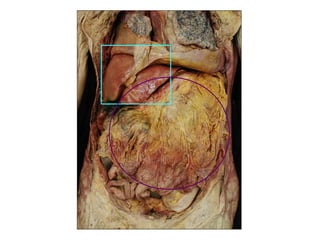
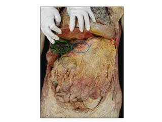
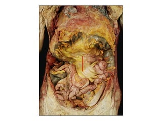
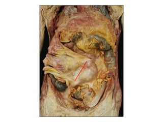
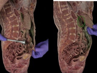
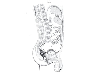
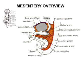
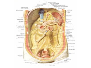
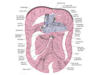
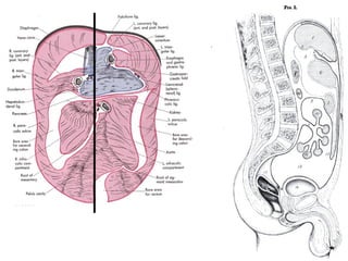
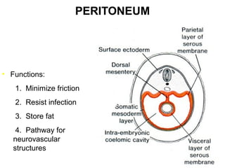
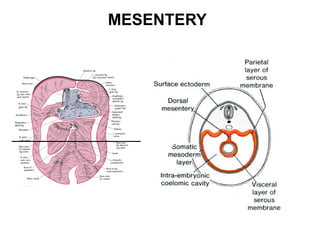
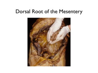
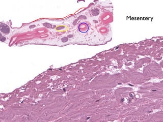
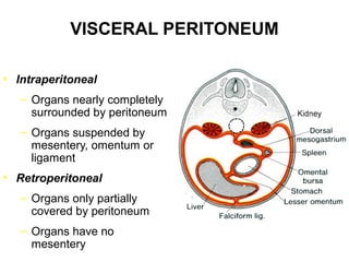
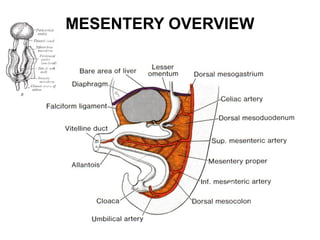
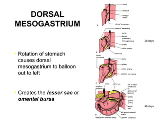
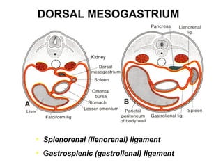
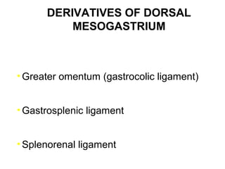
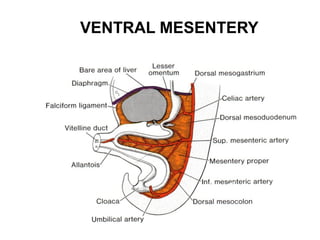
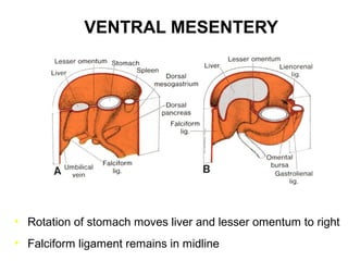
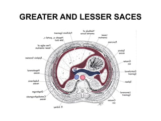
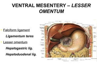
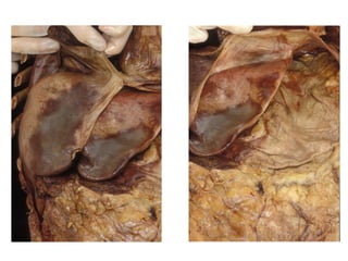
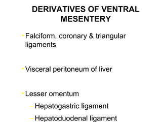
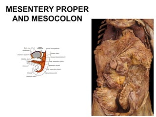
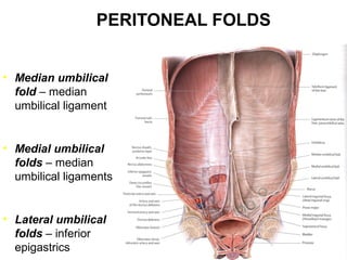
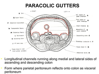
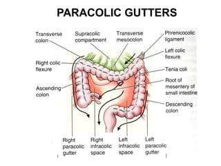
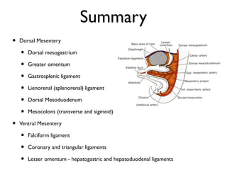

The peritoneum lines the abdominal cavity and surrounds organs. Mesenteries like the dorsal and ventral mesentery suspend and attach organs. The dorsal mesentery includes structures like the greater omentum, gastrosplenic ligament, and splenorenal ligament. The ventral mesentery includes the falciform ligament, coronary and triangular ligaments, and lesser omentum with attachments like the hepatogastric and hepatoduodenal ligaments. Peritoneal folds, recesses, and gutters provide pathways and spaces for structures in the abdomen.


