Histopathological Grossing of Kidney Tumors with the common gross differentials encountered,
reference - TATA memorial grossing techniques , Rosai and ackerman surgical pathology , Fletcher , Springer histopathology Specimen
Leiomyomas are smooth muscle tumors that are common to the uterus. These lesions include a range of presentations and extensions ranging from within the uterus to anywhere in the body, including parasitic leiomyoma, intravenous leiomyomatosis, disseminated peritoneal leiomyomatosis, and benign metastasizing leiomyoma. However, these atypical locations of these tumors present a diagnostic dilemma regarding their nature and benignity. Leiomyomatosis peritonealis disseminata (LPD) is a rare benign disease of unknown etiology of women in reproductive age group. A few reported cases of association with endometriosis have been described, suggesting a possible origin from submesothelial multipotent cells. Here, we present a case of a reproductive age group woman with history of uterine fibroids, now presenting with vague abdominal symptoms and multiple benign leiomyomas scattered throughout the peritoneal cavity. A diagnostic laparoscopy was done and the lesions sampled, which confirmed the imaging diagnosis of disseminated peritoneal leiomyomatosis. We stress the importance of picking up this rare benign pathology and avoiding labeling them as malignant peritoneal disease.
Benign Retroperitoneal Teratoma in young adult--A case report and literature ...iosrjce
IOSR Journal of Dental and Medical Sciences is one of the speciality Journal in Dental Science and Medical Science published by International Organization of Scientific Research (IOSR). The Journal publishes papers of the highest scientific merit and widest possible scope work in all areas related to medical and dental science. The Journal welcome review articles, leading medical and clinical research articles, technical notes, case reports and others.
Malignant Mixed Mullerian Tumor – Case Reports and Review ArticleApollo Hospitals
Malignant mixed mullerian tumors are very rare genital tumors. They are biphasic neoplasms composed of an admixture of malignant epithelial and mesenchymal elements. In descending order of frequency they originate in the uterus, ovaries, fallopian tubes, cervix and vagina. Also they arise denovo from peritoneum. They are highly aggressive and tend to occur in postmenopausal low parity women. Because of rarity, there is as such no treatment guidelines available. Multimodality treatment in the form of radical surgery followed by adjuvant chemotherapy or radiotherapy or combined chemoradiation gives a better prognosis & outcome. Two case reports of such tumors, one from ovary and other from penitoneum are presented along with the review of literature.
Primary Endometrial Stromal Sarcoma arising from Cervixiosrjce
IOSR Journal of Dental and Medical Sciences is one of the speciality Journal in Dental Science and Medical Science published by International Organization of Scientific Research (IOSR). The Journal publishes papers of the highest scientific merit and widest possible scope work in all areas related to medical and dental science. The Journal welcome review articles, leading medical and clinical research articles, technical notes, case reports and others.
Case Report:Massive Ovarian Cyst in a Adolescent GirlTana Kiak
For benign tumours adhesion prevention strategies should be used. Surgical intervention should as much as possible be directed towards preservation of ovarian tissue. There is scarcity of published literature on this subject.
We need bigger studies to address the issue of how much fertility preservation is safely possible.Irrespective of indication for surgery, it is always preferable to attempt conservative, fertility sparing surgery in adolescents.
Liposarcoma of the spermatic cord is a rare entity with only two series and less than 100 cases reported in literature. We report a case of a giant liposarcoma of the spermatic cord.
Liposarcoma of the spermatic cord is a rare entity with only two
series and less than 100 cases reported in literature. We report a case of a giant liposarcoma
of the spermatic cord.
Liposarcoma of the spermatic cord is a rare entity with only two series and less than 100 cases reported in literature. We report a case of a giant liposarcoma of the spermatic cord.
International Journal of Pharmaceutical Science Invention (IJPSI) inventionjournals
International Journal of Pharmaceutical Science Invention (IJPSI) is an international journal intended for professionals and researchers in all fields of Pahrmaceutical Science. IJPSI publishes research articles and reviews within the whole field Pharmacy and Pharmaceutical Science, new teaching methods, assessment, validation and the impact of new technologies and it will continue to provide information on the latest trends and developments in this ever-expanding subject. The publications of papers are selected through double peer reviewed to ensure originality, relevance, and readability. The articles published in our journal can be accessed online
Papillary Hidradenoma is a rare benign tumor of apocrine glands. Very limited number of case reports are available in the literature detailing the cytological features of papillary hidradenoma. Such a rare case specimen came in notice here at Pathology Department of SMS Medical College, Jaipur (Raj.) India. So, one such a rare case of papillary hidradenoma was explored with its cytological findings. A 30 year old female presented with a vulval cyst. Fluid from this cyst was sent for cytology with a clinical diagnosis of Bartholin cyst. The cytology suggested it to be a benign adnexal tumour by the presence of Biphasic pattern of cell arrangement which was further confirmed histologically. So whenever an middle aged female presents with a nodular lesion in the anogenital area, hidradenoma papilliferum should be kept in mind along with other conditions.
PowerPoint presentation on Choledochal Cysts, also known as biliary cyst, uploaded by Dr. Vaskar Humagain, first presented in 31st December, 2013. This presentation contains all the information about Choledochal Cysts, the original and revised Todani classification of choledochal cysts, pathogenesis, other associated congenital anomalies, clinical features in infant and adult, management of choledochal cysts. Comments are highly welcome :)
2. A literature search revealed that rectal duplication cysts6
and anterior sacral meningoceles1
have been associated
with uterus didelphys but only one other case report of a
tailgut cyst associated with uterine didelphys was found. This
was the case of a twelve-year old girl who had mental
retardation, and had hypothyroidism, sacral and coccygeal
agenesis but no chromosomal anomalies.7
Abnormalities of the lower urinary and genital tracts as a
result of Mullerian and Wollfian duct abnormalities have been
reported but none with retrorectal cysts viz. the Mayer
Rokitansky syndrome, which includes uterine didelphys,
imperforate vagina and renal agenesis.8
Double inferior vena cava in itself is a rarity. There are
isolated case reports of its association with renal aplasia9
,
congenital hepatic fibrosis and lung dysgenesis. No report
of double IVC with retrorectal cysts has been published till
date.
Embryologically, tailgut cysts are thought to be the
persistent part of the hind gut in the region of the embryonic
tail that normally involutes.1, 2
Pathologically, tailgut cysts are
usually multicystic or multiloculated and lined by a variety of
epithelia (stratified squammous, transitional, stratified
columnar, mucinous or ciliated columnar, ciliated
pseudostratified columnar or gastric). Well formed but
disorganised smooth muscle fibres are focally present in its
wall unlike the well formed continuous two layer muscle coat
seen in rectal duplication cysts. They are dissimilar to benign
cystic teratomas which contain distinct dermal appendages,
neural elements or mesenchymal derivatives like cartilage
or bone.1
Excision of the tailgut cyst results in its cure. This case
was reported to highlight the congenital anomalies that can
also be associated with this condition.
References
1. Dahan H, Arrivé L, Wendum D, Docou le Pointe H, Djouhri H,
Tubiana JM. Retrorectal developmental cysts in adults: clinical
and radiologic-histopathologic review, differential diagnosis, and
treatment. Radiographics. 2001;21:575–84.
2. Prasad AR, Amin MB, Randolph TL, Lee CS, Ma CK. Retrorectal
cystic hamartoma: report of 5 cases With Malignancy Arising in
2. Arch Pathol Lab Med. 2000;24:725–9.
3. Horenstein MG, Erlandson RA, Gonzalez-Cueto DM, Rosai J.
Presacral carcinoid tumors: a report of 3 cases and review of
literature. Am J Pathol. 1998;22:251–5.
4. Currarino G, Coln D, Votteler T. Triad of anorectal, sacral and
presacral anomlies. AJR Am J Roentgenol. 1981;137:395–8.
5. Kim IS, Oh SY, Choi SJ, Kim JH, Park KH, Park HK, et al. Clinical
and genetic analysis of HLXB9 gene in Korean patients with
Currarino syndrome. J Hum Genet. 2007;52:698–701.
6. Nour S, Kumar D, Dickson JA. Anorectal malformations with sacral
bony abnormalities. Arch Dis Child. 1989;64:1618–20.
7. Galluzzo ML, Bailez MM, Reusmann A, Gonzalez R, Davila MT.
Tailgut cyst (Retrorectal hamartoma): Report of a pediatric case.
Pediatr Dev Pathol 2007;22:1.
8. Stassart JP, Nagel TC, Prem KA, Phipps WR. Uterus didelphys,
obstructed hemivagina and ipsilateral renal agenesis: the
University of Minnesota experience. Fertil Steril.
1992; 57:756–61.
9. Gayer G, Zissin R Strauss S, Hertz M. IVC anomalies and right
renal aplasia on CT: a possible link? Abdom Imaging.
2003;28:395–9.
Figure 2: The figure shows the uterus didelphys displaced anteriorly
by the cyst.
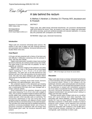
Tropical Gastroenterology 2008.29;2:103–104
Figure 3: The figure shows the retrorectal cyst.