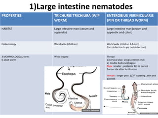
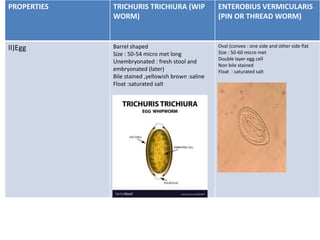
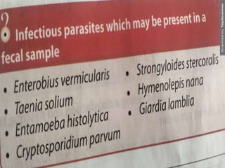
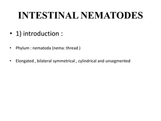
This document summarizes intestinal nematodes (roundworms). It describes their classification, morphology, life cycles, and pathogenic effects. Key points include:
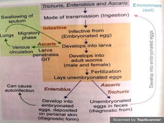
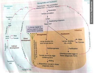
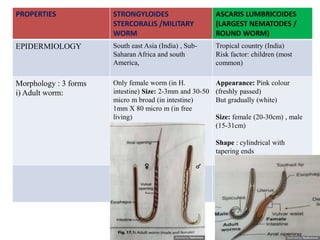
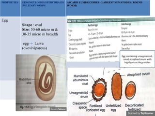
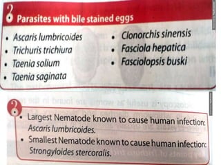
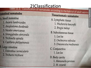
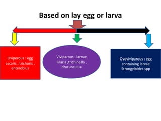
1. Intestinal nematodes are classified based on where they lay eggs or larvae. Major genera discussed are Ascaris, Trichuris, Enterobius, Strongyloides, and Ancylostoma/Necator.
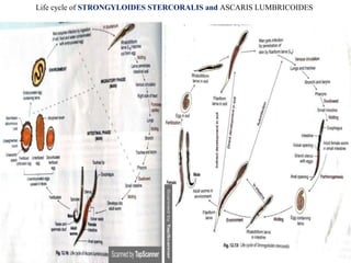
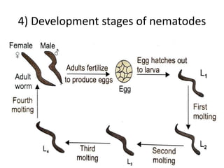
2. The life cycles of these nematodes involve eggs and larvae, with some having direct or indirect life cycles between human and/or insect hosts.
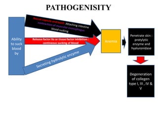
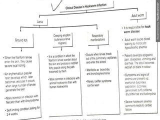
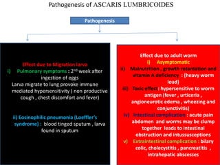
3. Clinical effects range from asymptomatic infection to malnutrition, anemia, intestinal obstruction, and hyperinfection syndromes depending on worm burden and specific nematode.
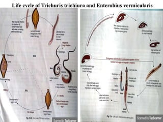
![5) Life cycle
• Life cycle : one host (man) [except –filarial worm-d.h
(man) and i.h(mosquito) and dracunculus- D.H- man
and I.H-cyclops]](https://image.slidesharecdn.com/intestinalnematodes-201128041411/85/Intestinal-nematodes-8-320.jpg)