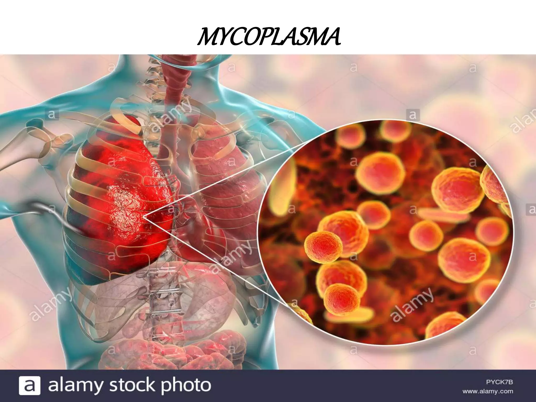
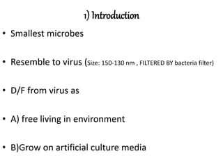
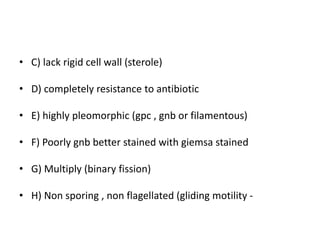
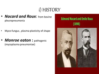
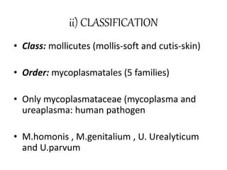
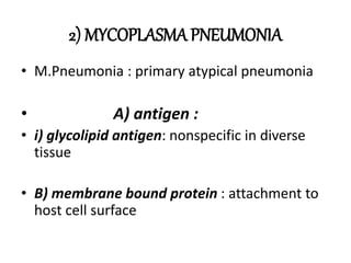
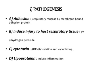
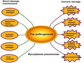
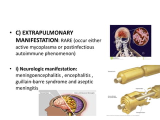
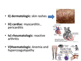
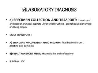
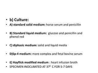
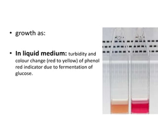
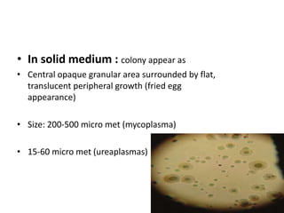
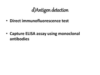
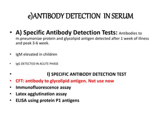
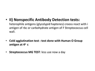
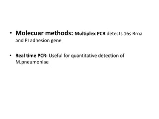
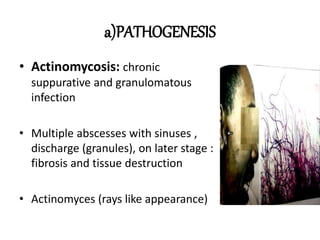
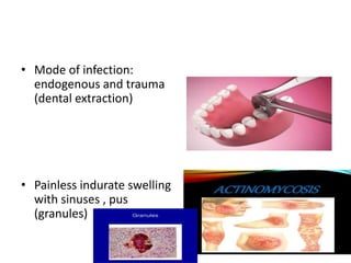
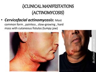
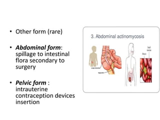
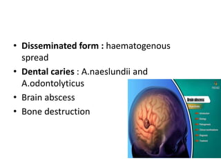
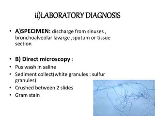
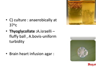
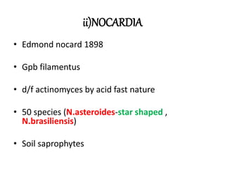
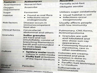
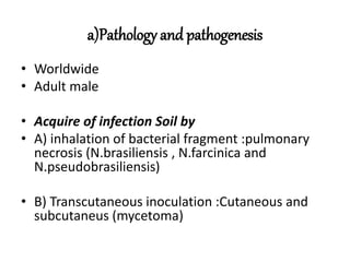
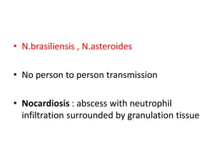
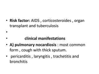
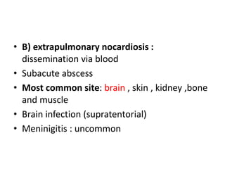
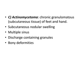
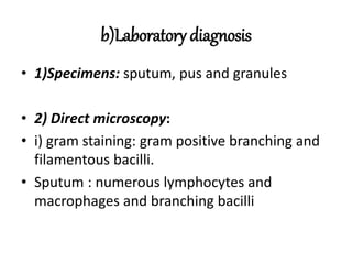
MYCOPLASMA
1) Introduction
• Smallest microbes
• Resemble to virus (Size: 150-130 nm , FILTERED BY bacteria filter)
• D/F from virus as
• A) free living in environment
• B)Grow on artificial culture media
• C) lack rigid cell wall (sterole)
• D) completely resistance to antibiotic
• E) highly pleomorphic (gpc , gnb or filamentous)
• F) Poorly gnb better stained with giemsa stained
• G) Multiply (binary fission)
• H) Non sporing , non flagellated (gliding motility -
i) HISTORY
• Nocard and Roux: from bovine
pleuropneumonia
• Myco-fungus , plasma-plasticity of shape
• Monroe eaton : pathogenic
(mycoplasma pneumoniae)
ii) CLASSIFICATION
• Class: mollicutes (mollis-soft and cutis-skin)
• Order: mycoplasmatales (5 families)
• Only mycoplasmataceae (mycoplasma and
ureaplasma: human pathogen
• M.homonis , M.genitalium , U. Urealyticum
and U.parvum
2) MYCOPLASMA PNEUMONIA
• M.Pneumonia : primary atypical pneumonia
• A) antigen :
• i) glycolipid antigen: nonspecific in diverse
tissue
• B) membrane bound protein : attachment to
host cell surface
i) PATHOGENESIS
• A) Adhesion : respiratory mucosa by membrane bound
adhesion protein
• B) induce injury to host respiratory tissue : by
• i) hydrogen peroxide
• C) cytotoxin : ADP ribosylation and vacuolating
• D) Lipoproteins : induce inflammation
ii) EPIDERMIOLOGY
• Worldwide
• Transmission : respiratory droplets
• i/c: 2-4 weeks
iii)CLINICAL MANIFESTATION
• A) UPPER RESPIRATORY TRACT
INFECTIONS (URTI): pharyngitis ,
tracheobronchitis or otitis media (rarely).
• B)Pneumonia : Atypical community
acquired interstitial pneumonia
(chlamydophila pneumoniae , legionella
pneumophila and viral pneumonia)
• 3-13% of infected person
• Wheeze , dry cough and pribronchial pneumonia with
thickened bronchial marking and streaks of interstitial
infiltration on chest X-ray
• C) EXTRAPULMONARY
MANIFESTATION: RARE (occur either
active mycoplasma or postinfectious
autoimmune phenomenon)
• i) Neurologic manifestation:
meningoencephalitis , encephalitis ,
guillain-barre syndrome and aseptic
meningitis
• Ii) dermatologic: skin rashes
• Iii) cardiac: myocarditis ,
pericarditis
• Iv) rheumatologic: reactive
arthritis
• V)Haematologic: Anemia and
hypercoagulopathy
iv)LABORATORY DIAGNOSIS
• a) SPECIMEN COLLECTION AND TRASPORT: throat swab
and nasopharyngeal aspirate , bronchial brushing , bronchoalveolar lavage
and lung biopsy.
• MUST TRANSPORT :
• A) STANDARD MYCOPLASMA FLUID MEDIUM: fetal bovine serum ,
gelatine and penicillin.
• B)VIRAL TRANSPORT MEDIUM: ampicillin and cefotaxime
• IF DELAY : 40C
• b) Culture:
• A) standard solid medium: horse serum and penicillin
• B) Standard liquid medium: glucose and penicillin and
phenol red
• C) diphasic medium: solid and liquid media
• D)Sp-4 medium: more complex and fetal bovine serum
• E) Hayflick modified medium : heart infusion broth
• SPECIMEN INOCUBATED AT 370 C FOR 5-7 DAYS
• growth as:
• In liquid medium: turbidity and
colour change (red to yellow) of phenol
red indicator due to fermentation of
glucose.
• In solid medium : colony appear as
• Central opaque granular area surrounded by flat,
translucent peripheral growth (fried egg
appearance)
• Size: 200-500 micro met (mycoplasma)
• 15-60 micro met (ureaplasmas)
• Colonies can be examined by:
• i) hand lens
• Ii) dienes staining: plate flooded in alcoholic solution of
methylene blue and examined under low power microscope.
• Mycoplasma retain colour for 2 days
• Ureaplasma : reddish to greenish-blue
c)IDENTIFICATION
• Most mycoplasma: hemolytic colonies
• Hemadsorption test: m.pneumoniae agglutinates guinea
pig red blood cells
• Tetrazolium reduction test : reduce colourless
tetrazolium compound to red coloured formazan
• Growth inhibition test : M.pneumoniae inhibit by adding specific
antisera
d)Antigen detection
• Direct immunofluorescence test
• Capture ELISA assay using monoclonal
antibodies
e)ANTIBODY DETECTION IN SERUM
• A) Specific Antibody Detection Tests: Antibodies to
m.pneumoniae protein and glycolipid antigen detected after 1 week of illness
and peak 3-6 week.
• IgM elevated in children
• IgG DETECTED IN ACUTE PHASE
• I) SPECIFIC ANTIBODY DETECTION TEST
• CFT: antibody to glycolipid antigen. Not use now
• Immunofluorescence assay
• Latex agglutination assay
• ELISA using protein P1 antigens
• II) Nonspecific Antibody Detection tests:
heterophile antigens (glycolypid haptenes) crooss react with i
antigen of rbc or carbohydrate antigen of F Streptococcus cell
wall.
• Cold agglutination test : test done with Human O Group
antigen at 4o c
• Streptococcus MG TEST: less use now a day
• Molecuar methods: Multiplex PCR detects 16s Rrna
and PI adhesion gene
• Real time PCR: Useful for quantitative detection of
M.pneumoniae
e)TREATMENT
• MACROLIDES (AZIHROMYCIN , 500 mg on a
day , then 250 mg on 2-5 days)
• Alternative drugs:
• Doxycycline
• Fluoroquinolones(levofloxacin , moxifloxacin
and gemifloxacin not ciprofloxacin)
3)UROGENITAL MYCOPLASMAS
• M.homonis , M.genitalium , ureaplasma :
urogenital tract disease
• Colonize female lower urogenital tract
• Transmission: sexual contact or mother to fetus during
birth
a)CLINICAL MANIFESTATION
• Non-gonococcal urethritis and epididymitis
(ureaplasma and M.genitalium)
• Pyelonephritis (M.hominis) and urinary calculi
(ureaplasma)
• Pelvic inflammatory disease: (M.hominis)
• M.hominis: brain abscess , wound infection
b)Laboratory Diagnosis
• Culture and pcr
ACTINOMYCETES
1)INTRODUCTION
• GPB
• Non motile
• Non sporing
• Non capsulated
• Branching (fungi)
• Soil saprophytes
2)Genera:
• i)Actinomyces: aerobe , non-acid fast
• Ii)Nocardia: aerobe , acid fast(actinomycetoma and
pulmonary infection)
• Iii) Actinomadura: aerobe , acid fast (actinomycetoma).
• Iv) Streptomyces: aerobe , acid fast(actinomycetoma) ,
source of antibiotoc (streptomycin)
• V) Micropolyspora and thermoactinomyces:
hypersensitivity pneumonitis
i)ACTINOMYCES
• Soil saprophytes
• Oral commensial
• A.israelii: most common infecting man
• A.naeslundii and A.odontolyticus: rare
a)PATHOGENESIS
• Actinomycosis: chronic
suppurative and granulomatous
infection
• Multiple abscesses with sinuses ,
discharge (granules), on later stage :
fibrosis and tissue destruction
• Actinomyces (rays like appearance)
• Mode of infection:
endogenous and trauma
(dental extraction)
• Painless indurate swelling
with sinuses , pus
(granules)
i)CLINICAL MANIFESTATIONS
(ACTINOMYCOSIS)
• Cervicofacial actinomycosis: Most
common form , painless , slow-growing , hard
mass with cutaneous fistulas (lumpy jaw)
• Other form (rare)
• Abdominal form:
spillage to intestinal
flora secondary to
surgery
• Pelvic form :
intrauterine
contraception devices
insertion
• Disseminated form : haematogenous
spread
• Dental caries : A.naeslundii and
A.odontolyticus
• Brain abscess
• Bone destruction
ii)LABORATORY DIAGNOSIS
• A)SPECIMEN: discharge from sinuses ,
bronchoalveolar lavarge ,sputum or tissue
section
• B) Direct microscopy :
• Pus wash in saline
• Sediment collect(white granules : sulfur
granules)
• Crushed between 2 slides
• Gram stain
• C) culture : anaerobically at
37oc
• Thyoglycollate :A.israelli –
fluffy ball , A.bovis-uniform
turbidity
• Brain heart infusion agar :
• Molecular methods : PCR-RFLP
• TREATMENT :
ii)NOCARDIA
• Edmond nocard 1898
• Gpb filamentus
• d/f actinomyces by acid fast nature
• 50 species (N.asteroides-star shaped ,
N.brasiliensis)
• Soil saprophytes
a)Pathology and pathogenesis
• Worldwide
• Adult male
• Acquire of infection Soil by
• A) inhalation of bacterial fragment :pulmonary
necrosis (N.brasiliensis , N.farcinica and
N.pseudobrasiliensis)
• B) Transcutaneous inoculation :Cutaneous and
subcutaneus (mycetoma)
• N.brasiliensis , N.asteroides
• No person to person transmission
• Nocardiosis : abscess with neutrophil
infiltration surrounded by granulation tissue
• Risk factor: AIDS , corticosteroides , organ
transplant and tuberculosis
•
• clinical manifestations
• A) pulmonary nocardiosis : most common
form , cough with thick sputum.
• pericarditis , laryngitis , trachetitis and
bronchitis
• B) extrapulmonary nocardiosis :
dissemination via blood
• Subacute abscess
• Most common site: brain , skin , kidney ,bone
and muscle
• Brain infection (supratentorial)
• Meninigitis : uncommon
• C) Actinomycetoma: chronic granulomatous
(subcutaneous tissue) of feet and hand.
• Subcutaneous nodular swelling
• Multiple sinus
• Discharge containing granules
• Bony deformities
b)Laboratory diagnosis
• 1)Specimens: sputum, pus and granules
• 2) Direct microscopy:
• i) gram staining: gram positive branching and
filamentous bacilli.
• Sputum : numerous lymphocytes and
macrophages and branching bacilli
• Ii)Modified acid fast staining : 1% sulphuric
acid (kinyon method)
• Branching and filamentous red colour bacilli
• 3) culture:
• obligate aerobe
• BHI
• SDA
• AT 37O C for 2 days to 2 weeks
• Cremy , wrinkled , orange or pink pigment
(carotenoid like pigment)
• Some colony :wool ball appearance
• Selective media:
• buffer yeast extract agar (vancomycin ,
polymyxin)
• SDA (chloramphenicol)
• Paraffin bait techniques: carbon source
• LJ medium
• 4)Biochemical:
• Catalase positive
• Non motile
• Decomposition of casein and tyrosine
• Growth in lysozymes
• Acetamide utilization
• Growth at 45oc for 3 days
• Acid from rhamnose
5)Treatment :
• sulphonmide or cotrimoxazole (all form of
nocardiosis)
• Brain abscess or pneumonia: cotrimoxazole
• Duration of treatment:
• For severe disease:(6-12 month – intact immune ,
1 year for immune deficient)
• Lymadenitis and skin abscess:2month
• Actinomycetoma :6-12 month after cure
• Keratitis: 2 month
• ACINOMASURA : SELF STUDY

Mycoplasma and actinomycetes

  • 1.
  • 2.
    1) Introduction • Smallestmicrobes • Resemble to virus (Size: 150-130 nm , FILTERED BY bacteria filter) • D/F from virus as • A) free living in environment • B)Grow on artificial culture media
  • 3.
    • C) lackrigid cell wall (sterole) • D) completely resistance to antibiotic • E) highly pleomorphic (gpc , gnb or filamentous) • F) Poorly gnb better stained with giemsa stained • G) Multiply (binary fission) • H) Non sporing , non flagellated (gliding motility -
  • 6.
    i) HISTORY • Nocardand Roux: from bovine pleuropneumonia • Myco-fungus , plasma-plasticity of shape • Monroe eaton : pathogenic (mycoplasma pneumoniae)
  • 7.
    ii) CLASSIFICATION • Class:mollicutes (mollis-soft and cutis-skin) • Order: mycoplasmatales (5 families) • Only mycoplasmataceae (mycoplasma and ureaplasma: human pathogen • M.homonis , M.genitalium , U. Urealyticum and U.parvum
  • 8.
    2) MYCOPLASMA PNEUMONIA •M.Pneumonia : primary atypical pneumonia • A) antigen : • i) glycolipid antigen: nonspecific in diverse tissue • B) membrane bound protein : attachment to host cell surface
  • 9.
    i) PATHOGENESIS • A)Adhesion : respiratory mucosa by membrane bound adhesion protein • B) induce injury to host respiratory tissue : by • i) hydrogen peroxide • C) cytotoxin : ADP ribosylation and vacuolating • D) Lipoproteins : induce inflammation
  • 11.
    ii) EPIDERMIOLOGY • Worldwide •Transmission : respiratory droplets • i/c: 2-4 weeks
  • 12.
    iii)CLINICAL MANIFESTATION • A)UPPER RESPIRATORY TRACT INFECTIONS (URTI): pharyngitis , tracheobronchitis or otitis media (rarely). • B)Pneumonia : Atypical community acquired interstitial pneumonia (chlamydophila pneumoniae , legionella pneumophila and viral pneumonia)
  • 13.
    • 3-13% ofinfected person • Wheeze , dry cough and pribronchial pneumonia with thickened bronchial marking and streaks of interstitial infiltration on chest X-ray
  • 16.
    • C) EXTRAPULMONARY MANIFESTATION:RARE (occur either active mycoplasma or postinfectious autoimmune phenomenon) • i) Neurologic manifestation: meningoencephalitis , encephalitis , guillain-barre syndrome and aseptic meningitis
  • 17.
    • Ii) dermatologic:skin rashes • Iii) cardiac: myocarditis , pericarditis • Iv) rheumatologic: reactive arthritis • V)Haematologic: Anemia and hypercoagulopathy
  • 18.
    iv)LABORATORY DIAGNOSIS • a)SPECIMEN COLLECTION AND TRASPORT: throat swab and nasopharyngeal aspirate , bronchial brushing , bronchoalveolar lavage and lung biopsy. • MUST TRANSPORT : • A) STANDARD MYCOPLASMA FLUID MEDIUM: fetal bovine serum , gelatine and penicillin. • B)VIRAL TRANSPORT MEDIUM: ampicillin and cefotaxime • IF DELAY : 40C
  • 19.
    • b) Culture: •A) standard solid medium: horse serum and penicillin • B) Standard liquid medium: glucose and penicillin and phenol red • C) diphasic medium: solid and liquid media • D)Sp-4 medium: more complex and fetal bovine serum • E) Hayflick modified medium : heart infusion broth • SPECIMEN INOCUBATED AT 370 C FOR 5-7 DAYS
  • 20.
    • growth as: •In liquid medium: turbidity and colour change (red to yellow) of phenol red indicator due to fermentation of glucose.
  • 21.
    • In solidmedium : colony appear as • Central opaque granular area surrounded by flat, translucent peripheral growth (fried egg appearance) • Size: 200-500 micro met (mycoplasma) • 15-60 micro met (ureaplasmas)
  • 22.
    • Colonies canbe examined by: • i) hand lens • Ii) dienes staining: plate flooded in alcoholic solution of methylene blue and examined under low power microscope. • Mycoplasma retain colour for 2 days • Ureaplasma : reddish to greenish-blue
  • 23.
    c)IDENTIFICATION • Most mycoplasma:hemolytic colonies • Hemadsorption test: m.pneumoniae agglutinates guinea pig red blood cells • Tetrazolium reduction test : reduce colourless tetrazolium compound to red coloured formazan • Growth inhibition test : M.pneumoniae inhibit by adding specific antisera
  • 24.
    d)Antigen detection • Directimmunofluorescence test • Capture ELISA assay using monoclonal antibodies
  • 25.
    e)ANTIBODY DETECTION INSERUM • A) Specific Antibody Detection Tests: Antibodies to m.pneumoniae protein and glycolipid antigen detected after 1 week of illness and peak 3-6 week. • IgM elevated in children • IgG DETECTED IN ACUTE PHASE • I) SPECIFIC ANTIBODY DETECTION TEST • CFT: antibody to glycolipid antigen. Not use now • Immunofluorescence assay • Latex agglutination assay • ELISA using protein P1 antigens
  • 26.
    • II) NonspecificAntibody Detection tests: heterophile antigens (glycolypid haptenes) crooss react with i antigen of rbc or carbohydrate antigen of F Streptococcus cell wall. • Cold agglutination test : test done with Human O Group antigen at 4o c • Streptococcus MG TEST: less use now a day
  • 27.
    • Molecuar methods:Multiplex PCR detects 16s Rrna and PI adhesion gene • Real time PCR: Useful for quantitative detection of M.pneumoniae
  • 28.
    e)TREATMENT • MACROLIDES (AZIHROMYCIN, 500 mg on a day , then 250 mg on 2-5 days) • Alternative drugs: • Doxycycline • Fluoroquinolones(levofloxacin , moxifloxacin and gemifloxacin not ciprofloxacin)
  • 29.
    3)UROGENITAL MYCOPLASMAS • M.homonis, M.genitalium , ureaplasma : urogenital tract disease • Colonize female lower urogenital tract • Transmission: sexual contact or mother to fetus during birth
  • 30.
    a)CLINICAL MANIFESTATION • Non-gonococcalurethritis and epididymitis (ureaplasma and M.genitalium) • Pyelonephritis (M.hominis) and urinary calculi (ureaplasma) • Pelvic inflammatory disease: (M.hominis) • M.hominis: brain abscess , wound infection
  • 31.
  • 32.
  • 33.
    1)INTRODUCTION • GPB • Nonmotile • Non sporing • Non capsulated • Branching (fungi) • Soil saprophytes
  • 34.
    2)Genera: • i)Actinomyces: aerobe, non-acid fast • Ii)Nocardia: aerobe , acid fast(actinomycetoma and pulmonary infection) • Iii) Actinomadura: aerobe , acid fast (actinomycetoma). • Iv) Streptomyces: aerobe , acid fast(actinomycetoma) , source of antibiotoc (streptomycin) • V) Micropolyspora and thermoactinomyces: hypersensitivity pneumonitis
  • 35.
    i)ACTINOMYCES • Soil saprophytes •Oral commensial • A.israelii: most common infecting man • A.naeslundii and A.odontolyticus: rare
  • 36.
    a)PATHOGENESIS • Actinomycosis: chronic suppurativeand granulomatous infection • Multiple abscesses with sinuses , discharge (granules), on later stage : fibrosis and tissue destruction • Actinomyces (rays like appearance)
  • 37.
    • Mode ofinfection: endogenous and trauma (dental extraction) • Painless indurate swelling with sinuses , pus (granules)
  • 38.
    i)CLINICAL MANIFESTATIONS (ACTINOMYCOSIS) • Cervicofacialactinomycosis: Most common form , painless , slow-growing , hard mass with cutaneous fistulas (lumpy jaw)
  • 39.
    • Other form(rare) • Abdominal form: spillage to intestinal flora secondary to surgery • Pelvic form : intrauterine contraception devices insertion
  • 40.
    • Disseminated form: haematogenous spread • Dental caries : A.naeslundii and A.odontolyticus • Brain abscess • Bone destruction
  • 41.
    ii)LABORATORY DIAGNOSIS • A)SPECIMEN:discharge from sinuses , bronchoalveolar lavarge ,sputum or tissue section • B) Direct microscopy : • Pus wash in saline • Sediment collect(white granules : sulfur granules) • Crushed between 2 slides • Gram stain
  • 42.
    • C) culture: anaerobically at 37oc • Thyoglycollate :A.israelli – fluffy ball , A.bovis-uniform turbidity • Brain heart infusion agar :
  • 43.
    • Molecular methods: PCR-RFLP • TREATMENT :
  • 44.
    ii)NOCARDIA • Edmond nocard1898 • Gpb filamentus • d/f actinomyces by acid fast nature • 50 species (N.asteroides-star shaped , N.brasiliensis) • Soil saprophytes
  • 46.
    a)Pathology and pathogenesis •Worldwide • Adult male • Acquire of infection Soil by • A) inhalation of bacterial fragment :pulmonary necrosis (N.brasiliensis , N.farcinica and N.pseudobrasiliensis) • B) Transcutaneous inoculation :Cutaneous and subcutaneus (mycetoma)
  • 47.
    • N.brasiliensis ,N.asteroides • No person to person transmission • Nocardiosis : abscess with neutrophil infiltration surrounded by granulation tissue
  • 48.
    • Risk factor:AIDS , corticosteroides , organ transplant and tuberculosis • • clinical manifestations • A) pulmonary nocardiosis : most common form , cough with thick sputum. • pericarditis , laryngitis , trachetitis and bronchitis
  • 49.
    • B) extrapulmonarynocardiosis : dissemination via blood • Subacute abscess • Most common site: brain , skin , kidney ,bone and muscle • Brain infection (supratentorial) • Meninigitis : uncommon
  • 50.
    • C) Actinomycetoma:chronic granulomatous (subcutaneous tissue) of feet and hand. • Subcutaneous nodular swelling • Multiple sinus • Discharge containing granules • Bony deformities
  • 51.
    b)Laboratory diagnosis • 1)Specimens:sputum, pus and granules • 2) Direct microscopy: • i) gram staining: gram positive branching and filamentous bacilli. • Sputum : numerous lymphocytes and macrophages and branching bacilli
  • 52.
    • Ii)Modified acidfast staining : 1% sulphuric acid (kinyon method) • Branching and filamentous red colour bacilli
  • 53.
    • 3) culture: •obligate aerobe • BHI • SDA • AT 37O C for 2 days to 2 weeks • Cremy , wrinkled , orange or pink pigment (carotenoid like pigment) • Some colony :wool ball appearance
  • 54.
    • Selective media: •buffer yeast extract agar (vancomycin , polymyxin) • SDA (chloramphenicol) • Paraffin bait techniques: carbon source • LJ medium
  • 55.
    • 4)Biochemical: • Catalasepositive • Non motile • Decomposition of casein and tyrosine • Growth in lysozymes • Acetamide utilization • Growth at 45oc for 3 days • Acid from rhamnose
  • 56.
    5)Treatment : • sulphonmideor cotrimoxazole (all form of nocardiosis) • Brain abscess or pneumonia: cotrimoxazole • Duration of treatment: • For severe disease:(6-12 month – intact immune , 1 year for immune deficient) • Lymadenitis and skin abscess:2month • Actinomycetoma :6-12 month after cure • Keratitis: 2 month
  • 57.