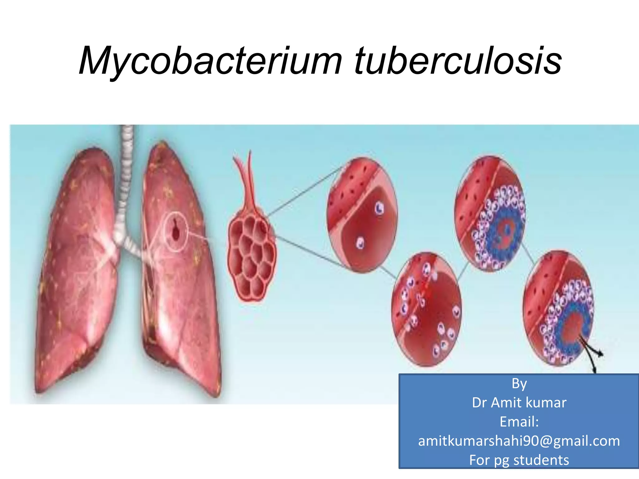
Mycobacterium tuberculosis
By
Dr Amit kumar
Email:
amitkumarshahi90@gmail.com
For pg students
1) GENERAL
CHARACTERISTICS
• Definition: The genus Mycobacterium belong to
genus in the family Mycobacteriaceae, order:
Actinomycetales, containing mycolic acid.
• acid-alcohol fastness (i.e. resist decolorization by
acidified alcohol
• Mycolic acids containing 60–90 carbon atoms,
which are cleaved to C22–C26 fatty acid methyl esters
by pyrolysis.
• G + C content of the DNA 61–71 mol% with the
only exception being M. leprae (>57 percent)
• Mycobacteria are nonmotile, nonspore-forming,
weakly gram-positive, aerobic or
microaerophilic, straight or slightly curved rod-
shaped bacteria (0.2– 0.6 X 1.0–10 micro met) .
Nutritional requirements and metabolic
activities:
M. genavense, M. paratuberculosis, and M.
haemophilum are not fastidious. Nutritions include
carbon, nitrogen, oxygen, phosphorus, sulfur, iron,
sodium, potassium, magnesium, and trace elements
(primarily zinc and manganese).
1) CARBON AND NITROGEN SOURCES: Important
carbon sources are carbohydrates (e.g. glucose,
fructose, sucrose, mannose, trehalose, inositol,
mannitol), organic acids, including short-chain
(pyruvic, acetic, and citric) and long-chain (oleic
and palmitic) acids, hydrocarbons (polycyclic
aromatic compounds), and even CO2, which is
essential for optimal growth of many species
Gas phase (5–10 percent): in media as
NaHCO3/Na2CO3 act CO2 in the gas phase.
• Laboratory culture medium contains glycerol
as carbon source (utilized by all members of
the genus), pyruvate:( C-source for M. bovis).
Tween 80 can also be used as sources of
carbon which can be hydrolyzed to oleic acid.
In growth media asparagine or glutamine act
as a source of carbon.NH4+ salts and amides,
amines, amino acids, nucleosides can also be
used as sources of nitrogen .
• 2) ACQUISITION OF IRON:
• Iron is essential for the growth of all bacteria
(mycobacteria) except lactobacilli and borrelia
burgdorferi. Mycobacteria aquire iron by small
water-soluble iron binding siderophores called
exochelins
• Mycobacteria produce two type of exochelins
The MS-type of exochelin (e.g. produced by M.
smegmatis) is water-soluble and MB-type
exochelin (choloroform soluble) , produced by
pathogenic micobacteria ( M. tuberculosis ).
• Exochelins has very high affinity for ferric ions
which is used as a growth supplement for
fastidious mycobacteria such as M. genavense.
• Transfer of iron in mycobacteria is mediated by
ferriexochelin and ferrimycobactin
• Release of the iron from either ferriexochelin or
ferrimycobactin is then converted from ferric ion
to ferrous ion by an NADH-dependent
reductase.
Metabolic pathways in mycobacteria
• Metabolic activities of mycobacteria depends on
presence of mycolic acids or the phenolic glycolipid
I of M. leprae and sets of enzymatic activities.
• Metabolic pathway of mycobacteria studied in two
environment.
a) Invivo: nutritional features of the intracellular
environment and persistant of mycobacteria in the
human host for long periods as metabolically
dormant state.
• An in vitro : tubercle bacilli are settle down to
the bottom of a liquid culture and these bacilli
cease to replicate, reduce their oxidative
metabolism, and survive in anaerobic
conditions much longer than actively
metabolizing cells. Conversion of lipids into
carbohydrates through the glyoxylate-shunt
pathway allowing M. tuberculosis cells long-
time survival in the host
Cell-wall structure and components
• Cell wall of Mycobacteria is made up of outer
envelope, The inner most layer is the plasma
membrane made up of proteins,
phosphatidylinositol mannosides,
lipoarabinomannan and peptidoglyca layer
contain repeating disaccharide units of N-
acetylglucosamine(b1–4)-N-glycolylmuramic
acid cross-linked via L-alanylD-isoglutaminyl-
meso diaminopimelyl-D-alanine tetrapeptides,
except in M. leprae, where the L-alanine is
replaced by glycine
• the outer surface of the mycobacterium is
formed by the intercalation of medium-chain
(e.g. mycocerosates) and short-chain (e.g.
acylglycerols) lipids, glycolipids, and
peptidoglycolipids converted into mycolic
acids.
• LIPIDS :lipids are long-chain fatty acids (e.g.
CH3(CH2)nCOOH), which are often modified by the
presence of unsaturated bonds, cyclopropane rings,
or side groups such as methyl, methoxy, hydroxy,
and keto groups.
• Outer surface of mycobacteria is high content of
lipid and contributes to several biological features,
including the hydrophobicity of mycobacteria, their
tendency to form clumps and cords, their resistance
to common lysis procedures, and their ability to
survive for a long period of time.
• Lipoarabinomannan: also called
LAM, is a glycolipid, and
associated with virulence factor
Mycobacterium tuberculosis.
• Its primary function is to
inactivate macrophages, inhibition
of T-cell proliferation(via
diminished IFN-γ response) ,and
destruction of oxidative radicals.
• The inactivation of macrophages
allows mycobacteria to
disseminate other part of body.
The destruction of oxidative
radicals allows for the survival of
the bacteria, as oxidative free
radicals are an important
mechanism by which our bodies
try to rid ourselves of infection.
• Many proposed mechanisms behind ManLAM , ManLAM is able to activate the
serine/threonine kinase Akt via phosphorylation and converted to Dephosphorylated
Bad which is serves as a pro-apoptotic protein and its activation allows for cell survival
and bacteria are able to up-regulate signaling pathways to control host cell apoptosis.
• ManLAM may also directly activate SHP-1 (Tyrosine-protein phosphatase non-
receptor type 6 or Src homology region 2 domain-containing phosphatase-1 ), a
phosphotyrosine phosphatase known to be involved in terminating activation signals.
SHP-1 related to the actions of IFN-γ and insulin.Once activated, SHP-1 translocates
from the cytosol to the membrane. LAM can inhibit LPS and IFN-γ deactivate IL-12. IL-
12 is important for innate resistance to M. tuberculosis infections. It activates natural
killer cells which produce IFN-γ to activate macrophages. By impairing the function of
these two molecules by SHP-1 activation, ManLAM may promote intracellular survival.
• Other models suggest that ManLAM acts as mediate immunosuppressive effects
through suppression of LPS-induced IL-12 p40 protein production. ManLAM is thought
to inhibit the IL-1 receptor-associated kinase (IRAK)-TRAF6 interaction, IκB-α
phosphorylation, and nuclear translocation of c-Rel and p50 which causes reduced IL-
12 p40 production.
• Mycolic acids: Mycolic acids are a defining
characteristic of members of the genus
Mycobacterium.
• ACETYLATED TREHALOSES : according to Bloch In
1950, trehalose dimycolate (TDM) is the cell-surface
component responsible for the ability of virulent
tubercle bacilli to form serpentine cords and to absorb
the cationic phenazine dye neutral red.
• Other trehalose-based lipids found in M. tuberculosis
are sulfolipids, which are thought to play a role in the
intracellular survival of tubercle bacilli by inhibiting
phagosome activation
• MYCOSIDES: categorized into two major groups:
• 1. peptidoglycolipids, which contain mycoserosic acid,
sugars and amino acids
• 2. phenol-phthiocerol glycosides.
• Peptidoglycolipids often contain unusual sugar
residues such as O-methyl rhamnose, fucose, or
deoxytalose and play roles in determining serotype,
colony morphology, and virulence.
• phenol-phthiocerol (donot contain amino acid)
.Phenolic glycolipid I (PGL-I) is found only in M.
leprae . PGL-I is used in immunodiagnostic tests to
detect people infected with M. leprae. In addition
it play a role in the ability of M. leprae to survive
within macrophages .PGL-I is also used as playing a
role in the specific immunosuppression seen in
lepromatous leprosy patients.
Laboratory diagnosis
1) Collection of specimens and preparation for primary culture :
Specimens should always be collected and submitted in leakproof,
disposable, appropriately labeled laboratoryapproved containers without
any fixatives.
Sputum: samples should not be saliva , three consecutive sample should be
examned
Urine : three consecutive early morning sample should be examned
Pus : should be collected in universal container or in syring , if pus is very
small then two separate swab should for microscopic and culture.
Aspirate fluids (pleural fluid , ascitic fluid) is liable to clot that why it should
be collected in containers containing anticoagulant (tri-sodium citrate).
CSF: should always be collected and submitted in leakproof, plastic disposable
without anticoagulant.
• 2) Acid-fast smear microscopy: Sensitivity of smear (range from 22 to 80
percent) depends upon the number of tubercular bacilli must be between 5 x 10³
and 5 x 10⁴ AFB in 1 ml of sputum. While 106 AFB/ml of specimen usually results
in a positive smear, only 60 percent of the smears are positive if 104 AFB/ml are
present.
• Ziehl–Neelsen acid-fast stain (hot staining procedure), Kinyoun acid-fast stain
(cold staining procedure), or auramine O fluorescence acidfast stain are used for
detection of mycobacterium by microscopically. It should be noted that heat
fixing of smear in bunsen flame donot kill micobacteria.
• A volume of approximately 0.01 ml of specimen is smeared over a 2-cm2 area of
the slide and heat-fixed.
• Fuchsin-staining shows Mycobacteria appear as red to pink rods on a blue
background. If there is any delay between smear preparation and staining slide
must be treated with 10% formal saline
• All fluorochrome-positive smears should be confirmed by a carbol-fuchsin
staining method.
• 2) CULTURE: For detection of mycobacteria in clinical specimens,
the current ‘gold standard’ consists of a combination of solid and liquid
media.
• culture of mycobacteria require following steps
• HOMOGINIZATION , DECONTAMINATION , CENTRIFUGATION ,
NEUTRILIZATION , INOCULATION IN CULTUTE MEDIA
• HOMOGINIZATION: it is essential to release the mycobacteria from body
fluid or tissue. Tissue require mechanical homoginization before
decontamination. For other samples homoginization and decontamination
can be done in one operation.
• DECONTAMINATION: it is used to isolate mycobacterium from heavily
contaminated samples with commensal flora. 4% sodium hydroxide to be
useful decontamination agent for sputa , biopsies and body fluids etc ,
followed by neutralization with 14% potassium dihydrogen
orthophosphate buffer with phenol indicator.
• Solid media: divided into two categories
• A) egg with buffer salt either with glycerol or sodium pyruvate (use for isolation
of mycobacteriun) : glycerol containing media (L J media used for
mycobacterium tuberculosis ) and pyruvate containing media (used for some
M.bovis)
• B) Agar based media : Middlebrook’s 7H11 7H10
• Liquid media : Middlebrook’s 7H9 (for sensitivity testing ) , kirchener or dubos
containing selective agent (PANTA : POLYMIXIN , AMPHOTERICIN , NALIDIXIC
ACID, TRIMETHOPRIM AND AZLOCILLIN) in order to prevent overgrowth of
contaminants. Cultures are normally normally incubate at 37 oc for 12 weeks.
• semiautomated BACTEC 460TB System is the most efficient and rapid technique
to culture mycobacteria . By this method, 14C-labeled palmitic acid act as a
carbon source in the medium is metabolized by microorganisms to 14CO2, which
is monitored by the instrument. The amount of 14CO2 and the rate at which the
gas is produced are directly proportional to the growth rate of the organisms.
For M. tuberculosis, an average detection time of 8 days . For smear-negative
specimens, an average recovery time of M. tuberculosis between 14 days in the
BACTEC 460TB .
• Other techniques are based on
nonradiometric liquid media eg. Mycobacteria
Growth Indicator Tube (MGIT), Becton to fully
automated systems (BACTEC MGIT 960,
Becton Dickinson; MB Bac/T, ESP Culture
System II.

Mycobacteriun tuberculosis

  • 1.
    Mycobacterium tuberculosis By Dr Amitkumar Email: amitkumarshahi90@gmail.com For pg students
  • 2.
    1) GENERAL CHARACTERISTICS • Definition:The genus Mycobacterium belong to genus in the family Mycobacteriaceae, order: Actinomycetales, containing mycolic acid. • acid-alcohol fastness (i.e. resist decolorization by acidified alcohol • Mycolic acids containing 60–90 carbon atoms, which are cleaved to C22–C26 fatty acid methyl esters by pyrolysis.
  • 3.
    • G +C content of the DNA 61–71 mol% with the only exception being M. leprae (>57 percent) • Mycobacteria are nonmotile, nonspore-forming, weakly gram-positive, aerobic or microaerophilic, straight or slightly curved rod- shaped bacteria (0.2– 0.6 X 1.0–10 micro met) .
  • 4.
    Nutritional requirements andmetabolic activities: M. genavense, M. paratuberculosis, and M. haemophilum are not fastidious. Nutritions include carbon, nitrogen, oxygen, phosphorus, sulfur, iron, sodium, potassium, magnesium, and trace elements (primarily zinc and manganese).
  • 5.
    1) CARBON ANDNITROGEN SOURCES: Important carbon sources are carbohydrates (e.g. glucose, fructose, sucrose, mannose, trehalose, inositol, mannitol), organic acids, including short-chain (pyruvic, acetic, and citric) and long-chain (oleic and palmitic) acids, hydrocarbons (polycyclic aromatic compounds), and even CO2, which is essential for optimal growth of many species
  • 6.
    Gas phase (5–10percent): in media as NaHCO3/Na2CO3 act CO2 in the gas phase. • Laboratory culture medium contains glycerol as carbon source (utilized by all members of the genus), pyruvate:( C-source for M. bovis). Tween 80 can also be used as sources of carbon which can be hydrolyzed to oleic acid. In growth media asparagine or glutamine act as a source of carbon.NH4+ salts and amides, amines, amino acids, nucleosides can also be used as sources of nitrogen .
  • 7.
    • 2) ACQUISITIONOF IRON: • Iron is essential for the growth of all bacteria (mycobacteria) except lactobacilli and borrelia burgdorferi. Mycobacteria aquire iron by small water-soluble iron binding siderophores called exochelins • Mycobacteria produce two type of exochelins The MS-type of exochelin (e.g. produced by M. smegmatis) is water-soluble and MB-type exochelin (choloroform soluble) , produced by pathogenic micobacteria ( M. tuberculosis ). • Exochelins has very high affinity for ferric ions which is used as a growth supplement for fastidious mycobacteria such as M. genavense. • Transfer of iron in mycobacteria is mediated by ferriexochelin and ferrimycobactin • Release of the iron from either ferriexochelin or ferrimycobactin is then converted from ferric ion to ferrous ion by an NADH-dependent reductase.
  • 8.
    Metabolic pathways inmycobacteria • Metabolic activities of mycobacteria depends on presence of mycolic acids or the phenolic glycolipid I of M. leprae and sets of enzymatic activities. • Metabolic pathway of mycobacteria studied in two environment. a) Invivo: nutritional features of the intracellular environment and persistant of mycobacteria in the human host for long periods as metabolically dormant state.
  • 9.
    • An invitro : tubercle bacilli are settle down to the bottom of a liquid culture and these bacilli cease to replicate, reduce their oxidative metabolism, and survive in anaerobic conditions much longer than actively metabolizing cells. Conversion of lipids into carbohydrates through the glyoxylate-shunt pathway allowing M. tuberculosis cells long- time survival in the host
  • 10.
    Cell-wall structure andcomponents • Cell wall of Mycobacteria is made up of outer envelope, The inner most layer is the plasma membrane made up of proteins, phosphatidylinositol mannosides, lipoarabinomannan and peptidoglyca layer contain repeating disaccharide units of N- acetylglucosamine(b1–4)-N-glycolylmuramic acid cross-linked via L-alanylD-isoglutaminyl- meso diaminopimelyl-D-alanine tetrapeptides, except in M. leprae, where the L-alanine is replaced by glycine
  • 11.
    • the outersurface of the mycobacterium is formed by the intercalation of medium-chain (e.g. mycocerosates) and short-chain (e.g. acylglycerols) lipids, glycolipids, and peptidoglycolipids converted into mycolic acids.
  • 12.
    • LIPIDS :lipidsare long-chain fatty acids (e.g. CH3(CH2)nCOOH), which are often modified by the presence of unsaturated bonds, cyclopropane rings, or side groups such as methyl, methoxy, hydroxy, and keto groups. • Outer surface of mycobacteria is high content of lipid and contributes to several biological features, including the hydrophobicity of mycobacteria, their tendency to form clumps and cords, their resistance to common lysis procedures, and their ability to survive for a long period of time.
  • 13.
    • Lipoarabinomannan: alsocalled LAM, is a glycolipid, and associated with virulence factor Mycobacterium tuberculosis. • Its primary function is to inactivate macrophages, inhibition of T-cell proliferation(via diminished IFN-γ response) ,and destruction of oxidative radicals. • The inactivation of macrophages allows mycobacteria to disseminate other part of body. The destruction of oxidative radicals allows for the survival of the bacteria, as oxidative free radicals are an important mechanism by which our bodies try to rid ourselves of infection. • Many proposed mechanisms behind ManLAM , ManLAM is able to activate the serine/threonine kinase Akt via phosphorylation and converted to Dephosphorylated Bad which is serves as a pro-apoptotic protein and its activation allows for cell survival and bacteria are able to up-regulate signaling pathways to control host cell apoptosis. • ManLAM may also directly activate SHP-1 (Tyrosine-protein phosphatase non- receptor type 6 or Src homology region 2 domain-containing phosphatase-1 ), a phosphotyrosine phosphatase known to be involved in terminating activation signals. SHP-1 related to the actions of IFN-γ and insulin.Once activated, SHP-1 translocates from the cytosol to the membrane. LAM can inhibit LPS and IFN-γ deactivate IL-12. IL- 12 is important for innate resistance to M. tuberculosis infections. It activates natural killer cells which produce IFN-γ to activate macrophages. By impairing the function of these two molecules by SHP-1 activation, ManLAM may promote intracellular survival. • Other models suggest that ManLAM acts as mediate immunosuppressive effects through suppression of LPS-induced IL-12 p40 protein production. ManLAM is thought to inhibit the IL-1 receptor-associated kinase (IRAK)-TRAF6 interaction, IκB-α phosphorylation, and nuclear translocation of c-Rel and p50 which causes reduced IL- 12 p40 production.
  • 14.
    • Mycolic acids:Mycolic acids are a defining characteristic of members of the genus Mycobacterium. • ACETYLATED TREHALOSES : according to Bloch In 1950, trehalose dimycolate (TDM) is the cell-surface component responsible for the ability of virulent tubercle bacilli to form serpentine cords and to absorb the cationic phenazine dye neutral red. • Other trehalose-based lipids found in M. tuberculosis are sulfolipids, which are thought to play a role in the intracellular survival of tubercle bacilli by inhibiting phagosome activation • MYCOSIDES: categorized into two major groups: • 1. peptidoglycolipids, which contain mycoserosic acid, sugars and amino acids
  • 15.
    • 2. phenol-phthiocerolglycosides. • Peptidoglycolipids often contain unusual sugar residues such as O-methyl rhamnose, fucose, or deoxytalose and play roles in determining serotype, colony morphology, and virulence. • phenol-phthiocerol (donot contain amino acid) .Phenolic glycolipid I (PGL-I) is found only in M. leprae . PGL-I is used in immunodiagnostic tests to detect people infected with M. leprae. In addition it play a role in the ability of M. leprae to survive within macrophages .PGL-I is also used as playing a role in the specific immunosuppression seen in lepromatous leprosy patients.
  • 16.
    Laboratory diagnosis 1) Collectionof specimens and preparation for primary culture : Specimens should always be collected and submitted in leakproof, disposable, appropriately labeled laboratoryapproved containers without any fixatives. Sputum: samples should not be saliva , three consecutive sample should be examned Urine : three consecutive early morning sample should be examned Pus : should be collected in universal container or in syring , if pus is very small then two separate swab should for microscopic and culture. Aspirate fluids (pleural fluid , ascitic fluid) is liable to clot that why it should be collected in containers containing anticoagulant (tri-sodium citrate). CSF: should always be collected and submitted in leakproof, plastic disposable without anticoagulant.
  • 17.
    • 2) Acid-fastsmear microscopy: Sensitivity of smear (range from 22 to 80 percent) depends upon the number of tubercular bacilli must be between 5 x 10³ and 5 x 10⁴ AFB in 1 ml of sputum. While 106 AFB/ml of specimen usually results in a positive smear, only 60 percent of the smears are positive if 104 AFB/ml are present. • Ziehl–Neelsen acid-fast stain (hot staining procedure), Kinyoun acid-fast stain (cold staining procedure), or auramine O fluorescence acidfast stain are used for detection of mycobacterium by microscopically. It should be noted that heat fixing of smear in bunsen flame donot kill micobacteria. • A volume of approximately 0.01 ml of specimen is smeared over a 2-cm2 area of the slide and heat-fixed. • Fuchsin-staining shows Mycobacteria appear as red to pink rods on a blue background. If there is any delay between smear preparation and staining slide must be treated with 10% formal saline • All fluorochrome-positive smears should be confirmed by a carbol-fuchsin staining method.
  • 18.
    • 2) CULTURE:For detection of mycobacteria in clinical specimens, the current ‘gold standard’ consists of a combination of solid and liquid media. • culture of mycobacteria require following steps • HOMOGINIZATION , DECONTAMINATION , CENTRIFUGATION , NEUTRILIZATION , INOCULATION IN CULTUTE MEDIA • HOMOGINIZATION: it is essential to release the mycobacteria from body fluid or tissue. Tissue require mechanical homoginization before decontamination. For other samples homoginization and decontamination can be done in one operation. • DECONTAMINATION: it is used to isolate mycobacterium from heavily contaminated samples with commensal flora. 4% sodium hydroxide to be useful decontamination agent for sputa , biopsies and body fluids etc , followed by neutralization with 14% potassium dihydrogen orthophosphate buffer with phenol indicator.
  • 19.
    • Solid media:divided into two categories • A) egg with buffer salt either with glycerol or sodium pyruvate (use for isolation of mycobacteriun) : glycerol containing media (L J media used for mycobacterium tuberculosis ) and pyruvate containing media (used for some M.bovis) • B) Agar based media : Middlebrook’s 7H11 7H10 • Liquid media : Middlebrook’s 7H9 (for sensitivity testing ) , kirchener or dubos containing selective agent (PANTA : POLYMIXIN , AMPHOTERICIN , NALIDIXIC ACID, TRIMETHOPRIM AND AZLOCILLIN) in order to prevent overgrowth of contaminants. Cultures are normally normally incubate at 37 oc for 12 weeks. • semiautomated BACTEC 460TB System is the most efficient and rapid technique to culture mycobacteria . By this method, 14C-labeled palmitic acid act as a carbon source in the medium is metabolized by microorganisms to 14CO2, which is monitored by the instrument. The amount of 14CO2 and the rate at which the gas is produced are directly proportional to the growth rate of the organisms. For M. tuberculosis, an average detection time of 8 days . For smear-negative specimens, an average recovery time of M. tuberculosis between 14 days in the BACTEC 460TB .
  • 20.
    • Other techniquesare based on nonradiometric liquid media eg. Mycobacteria Growth Indicator Tube (MGIT), Becton to fully automated systems (BACTEC MGIT 960, Becton Dickinson; MB Bac/T, ESP Culture System II.