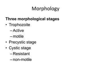
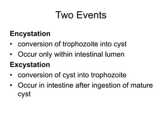
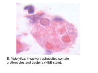
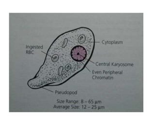
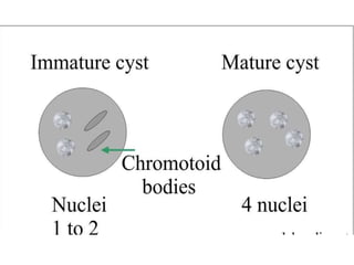
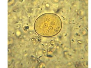
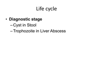
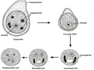
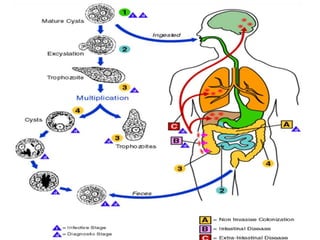
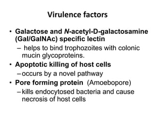
Entamoeba
Dr. Md. Abdullah Yusuf
Associate Professor of Microbiology
Email: ayusuf75@yahoo.com
Objectives
• Protozoa
• Basic meaning
• Members of the genus
• Morphology
• Life cycle
• Virulence factors and related events
• Mode of transmission
• Clinical features
• Sequelae and complications
• Diagnosis
• Treatment
Protozoa
• Definition
–A single cell like unit which is
morphologically & functionally complete
• Differences from metazoa
–Unicellular
–Single cell perform all function
• Morphology
–Cytoplasm: Ectoplasm & Endoplasm
–Nucleus
Classification: Protozoa
According to organ of Locomotion
• Motile
–Pseudopodia
–Flagella
–Cilia
• Non-Motile
–Plasmodium species
Organ of Locomotion
Motility with Flagella
• Urogenital Flagellates
–Trichomonas vaginalis
• Intestinal Flagellates
–Giardia intestinalis
• Haemo- & Tissue flagellates (Blood &
Tissue)
–Leishmania species
–Trypanosoma Species
Organ of Locomotion
• Motility with Pseudopodia
–Entamoeba histolytica
–Naegleria fowleri
–Acanthamoeba castellanii
• Motility with Cilia
–Balantidium coli
Classification: Protozoa
According to Host Requirement
• Second Host not Required
–Entamoeba histolytica
–Giardia intestinalis
• Second Host not Required
–Trypanosoma cruzi
–Plasmodium vivax
–Leishmania donovani
Classification: Protozoa
According to Location/Habitate
• Large Intestine
–Entamoeba histolytica
• Small intestine (Intestinal Flagellates)
–Giardia intestinalis
• Urogenital Flagellates (Vagina in female
& Urethra in Male)
–Trichomonas vaginalis
Classification (Conti..)
• Blood
–Plasmodium species (RBC)
–Leishmania species (Monocyte)
–Trypanosoma species (Plasma)
Classification
• Intestinal protozoa
–Giardia lamblia (flagellate)
–Entamoeba histolytica (ameba)
–Cryptosporidium hominis (sporozoa)
–Cyclospora cayetanensis (sporozoa)
• Sexually transmitted protozoan
infection
–Trichomonas vaginalis (flagellate)
Classification
Blood and tissue protozoan infections
• Flagellates
–Trypanosoma
–Leishmania
• Amebae
–Entamoeba histolytica
–Naegleria fowleri
–Acanthamoeba castellanii
Classification
• Sporozoa
–Plasmodium Species
–Babesia microti
–Toxoplasma gondii
• Microsporidia
Important Protozoa
• Entamoeba histolytica
• Giardia lamblia
• Trichomonas vaginalis
• Leishmania Species
• Plasmodium Species
• Toxoplasma gondii
Entamoeba histolytica
Basic meaning and members
• Basic meaning of Entamoeba
–Enta means intestine
–amoeba means protozoa with
pseudopods
Member of the Genus
• Pathogenic
• Non-Pathogenic
Member of the genus
• Pathogenic
–Entamoeba histolytica (histo-tissue,
lytica-that lyze)
Member of the genus
Non-Pathogenic
Entamoeba coli
–coli-colon
–reside in colon
Entamoeba dispar
–dispar-disparity with E. histolytica
Non-Pathogenic Entamoeba
Entamoeba hartmanni
–Hartman is the discoverer)
Entamoeba gingivalis
–Gingiva means gum
–reside in mouth
Morphology
Three morphological stages
• Trophozoite
–Active
–motile
• Precystic stage
• Cystic stage
–Resistant
–non-motile
Two Events
Encystation
• conversion of trophozoite into cyst
• Occur only within intestinal lumen
Excystation
• conversion of cyst into trophozoite
• Occur in intestine after ingestion of mature
cyst
Methods of Reproduction
• Maturation of cyst
–from one nucleus stage to 4 nuclei stage
• Trophozoites multiply
–by simple binary fission
• Cyst not multiply
• Encystation not a reproductive process
Morphology of Trophozoite
• Growing or feeding stage
• Actively motile by pseudopodia
• Shape changes due to pseudopodial
movement
• Cytoplasm divided into
–clear outer ectoplasm
–inner granular endoplasm
Morphology of Trophozoite
Endoplasm contains
• Nucleus
• Food vacuoles
Food vacuoles contain
–bacteria or red blood cells
• Can’t get in stool examination
E.histolytica: trophozoite of E.histolytica with ingested erythrocytes of
Gomori‘s trichrome
E. histolytica: invasive trophozoites contain
erythrocytes and bacteria (H&E stain).
Morphology of Cyst
• Spherical in shape
• Thin transparent wall
• Contain 4 nuclei in mature cysts
• Fine uniform granular Peripheral chromatin
–with small discrete central karyosome
–Differentiates it from other nonpathogenic
species
Morphology of Cyst
• Can find in stool examination
• Can’t found in Liver abscess
• Cyst not develop
–in Intestine wall
–Inside tissues of human
–In liver abscess
–In lungs abscess
–In metastatic Invasion of other organs
Life cycle
• Infective stage/Form
–Mature Quadrinucleated Cyst
• Pathogenic stage
–Trophozoite
• Route of infection
–Feco-oral
• Site of lesion
–Large intestine
Life cycle
• Diagnostic stage
–Cyst in Stool
–Trophozoite in Liver Abscess
Spread of Infection
Mature cyst in stool
• House fly carrying the cyst from faeces to
food
• Infected faeces used as manure for
growing vegetables
• Carrier handling the food without hand
washing
Why cyst infective?
• Ingested qudrinucleated cysts
–resists the action of gastric juice
• Ingested Trophozoites
–Destroyed by gastric juice
Life cycle
• excystation occurs in Lumen of Caecum
–young trophozites come out
• Cysts can survive in environment for 12 to
30 days
–resistant to chemicals
Life Cycle
• Trophozoites live and multiply in
–Crypts of mucosa of large intestine
–Feed starches & mucosal secretions
–Interacting with normal gut flora (bacteria)
–Hydrolyze mucosal cells
–Causing amoebic colitis
–Reach submucosa & blood vessels
• Travel in blood to other organs
–liver, lungs, skin
Virulence factors
• Galactose and N-acetyl-D-galactosamine
(Gal/GalNAc) specific lectin
– helps to bind trophozoites with colonic
mucin glycoproteins.
• Apoptotic killing of host cells
–occurs by a novel pathway
• Pore forming protein (Amoebopore)
–kills endocytosed bacteria and cause
necrosis of host cells
Virulence factors (Conti..)
• Anti complement activity
–resist destruction by lectin mediated
complement activation
• Motility
–probably promote invasion by the
cytoskeleton-induced motility
• Enzymes
–like Proteases, Collagenases, elastase,
hyaluronidase that degrade the
extracellular matrix
Virulence Events
Invasion is facilitated by many factors like
– pore forming proteins
– Proteases
– Motility
– phagocytosis
Diseases Caused by E. histolytica
• Primary or Intestinal Lesion
–Acute amoebic dysentery (extensive
ulcers)
–Amoebic colitis/Chronic Intestinal
Amoebiasis
–Amoebomas
Diseases Caused by E. histolytica
• Secondary or Metastatic or Extra-
intestinal Lesion
–Amobic liver abscess/Hepatic amoebiasis
–Pulmonary amoebiasis: Primary &
Secondary
–Cerebral Amoebiasis/Brain abscess
–Cutaneous Amoebiasis
–Splenic abscess
Amoebomas
• These are tumour-like lesions of the
colonic wall
• can measure up to several cm
Hall mark of amoebic dysentery
Amoebic ulcer
• Trophozoites invade tissues
• cause extensive destruction of the colonic
mucosa
• Initially produces small, discrete erosions
• then extend through the mucosa into the
submucosa
• expand laterally to produce flask-shaped
lesions
Ulcer Characteristics
• Flask shaped ulcer
• Ragged margins & bases
–due to digesting effect of proteases
• Floor remains full of altered blood from
oozing of the side walls
• Base of the ulcer usually remains limited to
muscularis mucosa
–rarely reach to submucosa and up to
muscularis externa
Clinical Features of Intestinal
Amoebiasis
• Passage of stool mixed with Mucus ±Blood
• Intermittent diarrhoea
–usually 4-6 times per day
• Tormina
–Severe abdominal cramps
• Tenesmus
–Feeling of incomplete Evacuation
• General malaise
• weight loss in chronic cases
Clinical Features of Hepatic
Amoebiasis
• Pain in right hypochondrium or
generalized abdominal pain
• Low grade irregular prolonged fever
• Enlarged tender liver
• Anorexia and weight loss
• Jaundice
• Raised right dome of the diaphragm
Characteristic of Pus of Amoebic
Liver Abscess
• Chocolate brown colour
–Anchovy-sauce pus
• Thick consistency
• Bacteriological sterile
• Mixed of slough liver tissue & blood
• Cyst not found
• Trophozoites found
• Usually single large, abscess in
Ultrasonography
Anchovy Fish/
Stolephorus commersonii
Differences between Amoebic & Bacillary
Liver Abscess
Points Amoebic Abscess Bacillary
Abscess
Size Large Small
Number Single Multiple
Pus colour Chocolate brown Pale colour
Fever Low grade High grade
Susceptible
person
Chronic amebiasis IV drug abusers
Culture No bacteria Bacteria found
Pus cell Occasionally pus Plenty of pus cell
Complications of Intestinal
Amoebiasis
• Haemorrhage from ulcer
• Perforation of ulcer
• Ameboma growth into intestinal lumen
–Causes Intestinal obstruction &
intussuception
• Abscess formation
• Toxic Megacolon
–dilation of colon causes 40% mortality
Metastatic Amoebiasis
• Cerebral abscess
• Lungs abscess
• Liver abscess
• Splenic Abscess
• Amoebiasis cutis
Laboratory Diagnosis
Principle
• Specimen
• Microscopic Examination
• Culture
• Serology
• Molecular test
Basic Principles
• Microscopic demonstration of
–trophozoite or cyst of E. histolytica
• Antigen detection from stool
• Nucleic acids techniques
• Hepatic amoebiasis diagnosed by
–Antigen detection from blood and pus
from liver
–Antibody detection from serum
Microscopic Examination
• Freshly passed stool
–Cyst
Diagnosis of Hepatic Amoebiasis
• Diagnostic aspiration
• Liver biopsy
• Exam of blood
• Exam of stool
• Serological tests
• Radiological exam
• Ultrasonography of Hepatobiliary system
Treatment
• Drug of choice for symptomatic intestinal
amoebiasis
–Metronidazole 400mg tds fro 5 days
–Secnidazole 1000mg 2 tab stat
–Tinidazole
–Ornidazole
Treatment
Asymptomatic cyst passers
• Diloxanide furoate
• Iodoquinol (Diiodohydroxyquin)
• Paromomycin
• Nitazoxanide
• Secnidazole
• Chloroquin
Entamoeba dispar
• E. dispar is non invasive
• Morphologically similar to E. histolytica
• First identified in 1925
• In 1978, isoenzyme analysis separate the
two species
Free Living Amoeba
• Name
–Acanthemoeba
–Negleria
• Disease
–Primary meningoencephalitis
Clinical & Lab Differences between
Amoebic & Bacillary Dysentery
Points Amoebic Bacillary
Number of
Motion
6 to 8 >10
Odour Offensive odourless
Colour Dark red Bright red
Composition Blood & mucus
mixed faeces
Blood & mucus
No faeces
RBC clumps Rouleaux
Pus cell Scanty Plenty
Culture E. histolytica Bacteria
Thank You

Entamoeba histolytica 2020

  • 1.
    Entamoeba Dr. Md. AbdullahYusuf Associate Professor of Microbiology Email: ayusuf75@yahoo.com
  • 2.
    Objectives • Protozoa • Basicmeaning • Members of the genus • Morphology • Life cycle • Virulence factors and related events • Mode of transmission • Clinical features • Sequelae and complications • Diagnosis • Treatment
  • 3.
    Protozoa • Definition –A singlecell like unit which is morphologically & functionally complete • Differences from metazoa –Unicellular –Single cell perform all function • Morphology –Cytoplasm: Ectoplasm & Endoplasm –Nucleus
  • 4.
    Classification: Protozoa According toorgan of Locomotion • Motile –Pseudopodia –Flagella –Cilia • Non-Motile –Plasmodium species
  • 5.
    Organ of Locomotion Motilitywith Flagella • Urogenital Flagellates –Trichomonas vaginalis • Intestinal Flagellates –Giardia intestinalis • Haemo- & Tissue flagellates (Blood & Tissue) –Leishmania species –Trypanosoma Species
  • 6.
    Organ of Locomotion •Motility with Pseudopodia –Entamoeba histolytica –Naegleria fowleri –Acanthamoeba castellanii • Motility with Cilia –Balantidium coli
  • 7.
    Classification: Protozoa According toHost Requirement • Second Host not Required –Entamoeba histolytica –Giardia intestinalis • Second Host not Required –Trypanosoma cruzi –Plasmodium vivax –Leishmania donovani
  • 8.
    Classification: Protozoa According toLocation/Habitate • Large Intestine –Entamoeba histolytica • Small intestine (Intestinal Flagellates) –Giardia intestinalis • Urogenital Flagellates (Vagina in female & Urethra in Male) –Trichomonas vaginalis
  • 9.
    Classification (Conti..) • Blood –Plasmodiumspecies (RBC) –Leishmania species (Monocyte) –Trypanosoma species (Plasma)
  • 10.
    Classification • Intestinal protozoa –Giardialamblia (flagellate) –Entamoeba histolytica (ameba) –Cryptosporidium hominis (sporozoa) –Cyclospora cayetanensis (sporozoa) • Sexually transmitted protozoan infection –Trichomonas vaginalis (flagellate)
  • 11.
    Classification Blood and tissueprotozoan infections • Flagellates –Trypanosoma –Leishmania • Amebae –Entamoeba histolytica –Naegleria fowleri –Acanthamoeba castellanii
  • 12.
    Classification • Sporozoa –Plasmodium Species –Babesiamicroti –Toxoplasma gondii • Microsporidia
  • 13.
    Important Protozoa • Entamoebahistolytica • Giardia lamblia • Trichomonas vaginalis • Leishmania Species • Plasmodium Species • Toxoplasma gondii
  • 14.
  • 15.
    Basic meaning andmembers • Basic meaning of Entamoeba –Enta means intestine –amoeba means protozoa with pseudopods
  • 16.
    Member of theGenus • Pathogenic • Non-Pathogenic
  • 17.
    Member of thegenus • Pathogenic –Entamoeba histolytica (histo-tissue, lytica-that lyze)
  • 18.
    Member of thegenus Non-Pathogenic Entamoeba coli –coli-colon –reside in colon Entamoeba dispar –dispar-disparity with E. histolytica
  • 19.
    Non-Pathogenic Entamoeba Entamoeba hartmanni –Hartmanis the discoverer) Entamoeba gingivalis –Gingiva means gum –reside in mouth
  • 20.
    Morphology Three morphological stages •Trophozoite –Active –motile • Precystic stage • Cystic stage –Resistant –non-motile
  • 21.
    Two Events Encystation • conversionof trophozoite into cyst • Occur only within intestinal lumen Excystation • conversion of cyst into trophozoite • Occur in intestine after ingestion of mature cyst
  • 22.
    Methods of Reproduction •Maturation of cyst –from one nucleus stage to 4 nuclei stage • Trophozoites multiply –by simple binary fission • Cyst not multiply • Encystation not a reproductive process
  • 23.
    Morphology of Trophozoite •Growing or feeding stage • Actively motile by pseudopodia • Shape changes due to pseudopodial movement • Cytoplasm divided into –clear outer ectoplasm –inner granular endoplasm
  • 24.
    Morphology of Trophozoite Endoplasmcontains • Nucleus • Food vacuoles Food vacuoles contain –bacteria or red blood cells • Can’t get in stool examination
  • 25.
    E.histolytica: trophozoite ofE.histolytica with ingested erythrocytes of Gomori‘s trichrome
  • 26.
    E. histolytica: invasivetrophozoites contain erythrocytes and bacteria (H&E stain).
  • 28.
    Morphology of Cyst •Spherical in shape • Thin transparent wall • Contain 4 nuclei in mature cysts • Fine uniform granular Peripheral chromatin –with small discrete central karyosome –Differentiates it from other nonpathogenic species
  • 29.
    Morphology of Cyst •Can find in stool examination • Can’t found in Liver abscess • Cyst not develop –in Intestine wall –Inside tissues of human –In liver abscess –In lungs abscess –In metastatic Invasion of other organs
  • 32.
    Life cycle • Infectivestage/Form –Mature Quadrinucleated Cyst • Pathogenic stage –Trophozoite • Route of infection –Feco-oral • Site of lesion –Large intestine
  • 33.
    Life cycle • Diagnosticstage –Cyst in Stool –Trophozoite in Liver Abscess
  • 35.
    Spread of Infection Maturecyst in stool • House fly carrying the cyst from faeces to food • Infected faeces used as manure for growing vegetables • Carrier handling the food without hand washing
  • 36.
    Why cyst infective? •Ingested qudrinucleated cysts –resists the action of gastric juice • Ingested Trophozoites –Destroyed by gastric juice
  • 37.
    Life cycle • excystationoccurs in Lumen of Caecum –young trophozites come out • Cysts can survive in environment for 12 to 30 days –resistant to chemicals
  • 38.
    Life Cycle • Trophozoiteslive and multiply in –Crypts of mucosa of large intestine –Feed starches & mucosal secretions –Interacting with normal gut flora (bacteria) –Hydrolyze mucosal cells –Causing amoebic colitis –Reach submucosa & blood vessels • Travel in blood to other organs –liver, lungs, skin
  • 40.
    Virulence factors • Galactoseand N-acetyl-D-galactosamine (Gal/GalNAc) specific lectin – helps to bind trophozoites with colonic mucin glycoproteins. • Apoptotic killing of host cells –occurs by a novel pathway • Pore forming protein (Amoebopore) –kills endocytosed bacteria and cause necrosis of host cells
  • 41.
    Virulence factors (Conti..) •Anti complement activity –resist destruction by lectin mediated complement activation • Motility –probably promote invasion by the cytoskeleton-induced motility • Enzymes –like Proteases, Collagenases, elastase, hyaluronidase that degrade the extracellular matrix
  • 42.
    Virulence Events Invasion isfacilitated by many factors like – pore forming proteins – Proteases – Motility – phagocytosis
  • 43.
    Diseases Caused byE. histolytica • Primary or Intestinal Lesion –Acute amoebic dysentery (extensive ulcers) –Amoebic colitis/Chronic Intestinal Amoebiasis –Amoebomas
  • 44.
    Diseases Caused byE. histolytica • Secondary or Metastatic or Extra- intestinal Lesion –Amobic liver abscess/Hepatic amoebiasis –Pulmonary amoebiasis: Primary & Secondary –Cerebral Amoebiasis/Brain abscess –Cutaneous Amoebiasis –Splenic abscess
  • 45.
    Amoebomas • These aretumour-like lesions of the colonic wall • can measure up to several cm
  • 46.
    Hall mark ofamoebic dysentery Amoebic ulcer • Trophozoites invade tissues • cause extensive destruction of the colonic mucosa • Initially produces small, discrete erosions • then extend through the mucosa into the submucosa • expand laterally to produce flask-shaped lesions
  • 47.
    Ulcer Characteristics • Flaskshaped ulcer • Ragged margins & bases –due to digesting effect of proteases • Floor remains full of altered blood from oozing of the side walls • Base of the ulcer usually remains limited to muscularis mucosa –rarely reach to submucosa and up to muscularis externa
  • 48.
    Clinical Features ofIntestinal Amoebiasis • Passage of stool mixed with Mucus ±Blood • Intermittent diarrhoea –usually 4-6 times per day • Tormina –Severe abdominal cramps • Tenesmus –Feeling of incomplete Evacuation • General malaise • weight loss in chronic cases
  • 49.
    Clinical Features ofHepatic Amoebiasis • Pain in right hypochondrium or generalized abdominal pain • Low grade irregular prolonged fever • Enlarged tender liver • Anorexia and weight loss • Jaundice • Raised right dome of the diaphragm
  • 51.
    Characteristic of Pusof Amoebic Liver Abscess • Chocolate brown colour –Anchovy-sauce pus • Thick consistency • Bacteriological sterile • Mixed of slough liver tissue & blood • Cyst not found • Trophozoites found • Usually single large, abscess in Ultrasonography
  • 52.
  • 55.
    Differences between Amoebic& Bacillary Liver Abscess Points Amoebic Abscess Bacillary Abscess Size Large Small Number Single Multiple Pus colour Chocolate brown Pale colour Fever Low grade High grade Susceptible person Chronic amebiasis IV drug abusers Culture No bacteria Bacteria found Pus cell Occasionally pus Plenty of pus cell
  • 56.
    Complications of Intestinal Amoebiasis •Haemorrhage from ulcer • Perforation of ulcer • Ameboma growth into intestinal lumen –Causes Intestinal obstruction & intussuception • Abscess formation • Toxic Megacolon –dilation of colon causes 40% mortality
  • 57.
    Metastatic Amoebiasis • Cerebralabscess • Lungs abscess • Liver abscess • Splenic Abscess • Amoebiasis cutis
  • 58.
    Laboratory Diagnosis Principle • Specimen •Microscopic Examination • Culture • Serology • Molecular test
  • 59.
    Basic Principles • Microscopicdemonstration of –trophozoite or cyst of E. histolytica • Antigen detection from stool • Nucleic acids techniques • Hepatic amoebiasis diagnosed by –Antigen detection from blood and pus from liver –Antibody detection from serum
  • 60.
  • 61.
    Diagnosis of HepaticAmoebiasis • Diagnostic aspiration • Liver biopsy • Exam of blood • Exam of stool • Serological tests • Radiological exam • Ultrasonography of Hepatobiliary system
  • 62.
    Treatment • Drug ofchoice for symptomatic intestinal amoebiasis –Metronidazole 400mg tds fro 5 days –Secnidazole 1000mg 2 tab stat –Tinidazole –Ornidazole
  • 63.
    Treatment Asymptomatic cyst passers •Diloxanide furoate • Iodoquinol (Diiodohydroxyquin) • Paromomycin • Nitazoxanide • Secnidazole • Chloroquin
  • 64.
    Entamoeba dispar • E.dispar is non invasive • Morphologically similar to E. histolytica • First identified in 1925 • In 1978, isoenzyme analysis separate the two species
  • 65.
    Free Living Amoeba •Name –Acanthemoeba –Negleria • Disease –Primary meningoencephalitis
  • 66.
    Clinical & LabDifferences between Amoebic & Bacillary Dysentery Points Amoebic Bacillary Number of Motion 6 to 8 >10 Odour Offensive odourless Colour Dark red Bright red Composition Blood & mucus mixed faeces Blood & mucus No faeces RBC clumps Rouleaux Pus cell Scanty Plenty Culture E. histolytica Bacteria
  • 67.