This document provides guidance on evaluating and managing dermatological conditions. It discusses the functions of skin, taking a thorough history, conducting a full examination of the skin and appendages, selecting appropriate investigations like skin scrapings or biopsies, and developing a treatment plan. Topical therapies are often first-line and include creams, ointments, or gels applied correctly based on the skin condition and amount of surface area involved. Close monitoring of the patient's response to treatment is important.
• In recent years, the usefulness of trichoscopy (scalp dermoscopy) (videodermatoscopy) has been reported for diagnosing hair loss diseases. This method allows viewing of the hair and scalp at X20 to X160 magnifications. Characteristic trichoscopy features of alopecia areata are black dots, tapering hairs (exclamation mark hairs), broken hairs, yellow dots, and short vellus hairs. In androgenetic alopecia (AGA), hair diameter diversity (HDD), perifollicular pigmentation/peripilar sign, and yellow dots are trichoscopically observed. In all cases of AGA and female AGA, HDD, more than 20%, which corresponds to vellus transformation, can be seen. In cicatricial alopecia (CA), the loss of orifices, a hallmark of CA, and the associated changes including perifollicular erythema or scale and hair tufting were observed. Different hair shafts variation such as vellus, terminal, micro-exclamation mark type, monilethrix, Netherton type, and pili annulati hairs can be seen . The number of hairs in one pilosebaceous unit can be assessed. Healthy Hair follicles variation healthy, empty, fibrotic ("white dots"), filled with hyperkeratotic plugs ("yellow dots"), or containing dead hair ("black dots"). Abnormalities of scalp skin color or structure include honeycomb-type hyperpigmentation, perifollicular discoloration (hyperpigmentation), and scaling are also seen with the help of trichoscopy.
Subspecialty of dermatology and pathology focused on performing and interpreting tests on human tissue samples to provide scientific data and consultative opinions to referring clinicians
Androgenetic alopecia (AGA) is a nonscarring progressive miniaturization of the hair follicle in genetically predisposed men and women, usually in a specific pattern distribution.
Multifactorial and polygenetic etiology.
Clinical features:
-History of hair loss is -
long standing
slowly progressing reduction of hair density, diameter
Miniaturization of hair
Diminished anagen hair and increased telogen hair
-Pattern of hair loss in male:
Hamilton- Norwood type: recession of frontal hair line, latter followed by a vertex thinning with progression until top of the scalp is completely bald.
-Pattern of hair loss in female:
Centrofrontal hair loss with preservation of frontal hair line
(Ludwig type) {figure - left}
Christmas tree pattern {figure- right}
-Family history of AGA often positive
In female
signs of hyperandrogenism should be evaluated
gynecological history
progesterone containing pills
-To exclude other causes history should be taken regarding-
Thyroid disease,
Surgery, infection in last 6months to 1 year
Drug history
Iron deficiency
Smoking
UV exposure
Hair color, cosmetics use.
Allergic contact dermatitis
Treatment:
Androgenic alopecia is naturally progressive , so main strategy is to prevent progression and increase hair density.
1.Topical minoxidil:
2% for female and 5% spray for male 1 ml twice daily or half cup foam once daily.
There is transitory telogen shedding within first 8 weeks observed.
Response should be assessed after 6 months.
If response occurs, will be continued as main stay of treatment.
2.Finasteride oral ad Dutasteride oral
1 mg finasteride per day prevents progression of AGA .
0.5 mg daily dutasteride is alternative.
Combination of topical minoxidil and finasteride is good option
Response evaluated after 6 months . not indicated in women. Contraindicated in pregnant and child bearing female.
3.Antiandrogen and estrogenic drugs:
Given in hyperandrogenism in female. Not indicated in male.
Spironolactone 100-200 mg daily
Cyproterone acetate can be used
4.Hair transplantation
5.Low-level laser therapy
6.Miscellaneous: low level of evidence.
Platelet rich plasma therapy and microneedling
Herbal preparations
Topical melatonin
Nutritional supplement of- biotin, copper, zinc, aminoacids, micronutrients
The contents :
Skin over view
Types of skin lesions
Hypersensitivity reactions and the skin
Eczema over view
Approach to a Skin Rash
Atopic dermatitis
MCQ Questions
Psoriasis is a chronic autoimmune skin disorder characterized by the rapid and excessive growth of skin cells. It affects around 2-3% of the global population and can manifest in various forms, from mild to severe. This condition is often associated with genetic factors and involves the immune system mistakenly attacking healthy skin cells.
Lung Cancer: Artificial Intelligence, Synergetics, Complex System Analysis, S...Oleg Kshivets
RESULTS: Overall life span (LS) was 2252.1±1742.5 days and cumulative 5-year survival (5YS) reached 73.2%, 10 years – 64.8%, 20 years – 42.5%. 513 LCP lived more than 5 years (LS=3124.6±1525.6 days), 148 LCP – more than 10 years (LS=5054.4±1504.1 days).199 LCP died because of LC (LS=562.7±374.5 days). 5YS of LCP after bi/lobectomies was significantly superior in comparison with LCP after pneumonectomies (78.1% vs.63.7%, P=0.00001 by log-rank test). AT significantly improved 5YS (66.3% vs. 34.8%) (P=0.00000 by log-rank test) only for LCP with N1-2. Cox modeling displayed that 5YS of LCP significantly depended on: phase transition (PT) early-invasive LC in terms of synergetics, PT N0—N12, cell ratio factors (ratio between cancer cells- CC and blood cells subpopulations), G1-3, histology, glucose, AT, blood cell circuit, prothrombin index, heparin tolerance, recalcification time (P=0.000-0.038). Neural networks, genetic algorithm selection and bootstrap simulation revealed relationships between 5YS and PT early-invasive LC (rank=1), PT N0—N12 (rank=2), thrombocytes/CC (3), erythrocytes/CC (4), eosinophils/CC (5), healthy cells/CC (6), lymphocytes/CC (7), segmented neutrophils/CC (8), stick neutrophils/CC (9), monocytes/CC (10); leucocytes/CC (11). Correct prediction of 5YS was 100% by neural networks computing (area under ROC curve=1.0; error=0.0).
CONCLUSIONS: 5YS of LCP after radical procedures significantly depended on: 1) PT early-invasive cancer; 2) PT N0--N12; 3) cell ratio factors; 4) blood cell circuit; 5) biochemical factors; 6) hemostasis system; 7) AT; 8) LC characteristics; 9) LC cell dynamics; 10) surgery type: lobectomy/pneumonectomy; 11) anthropometric data. Optimal diagnosis and treatment strategies for LC are: 1) screening and early detection of LC; 2) availability of experienced thoracic surgeons because of complexity of radical procedures; 3) aggressive en block surgery and adequate lymph node dissection for completeness; 4) precise prediction; 5) adjuvant chemoimmunoradiotherapy for LCP with unfavorable prognosis.
MANAGEMENT OF ATRIOVENTRICULAR CONDUCTION BLOCK.pdfJim Jacob Roy
Cardiac conduction defects can occur due to various causes.
Atrioventricular conduction blocks ( AV blocks ) are classified into 3 types.
This document describes the acute management of AV block.
Title: Sense of Smell
Presenter: Dr. Faiza, Assistant Professor of Physiology
Qualifications:
MBBS (Best Graduate, AIMC Lahore)
FCPS Physiology
ICMT, CHPE, DHPE (STMU)
MPH (GC University, Faisalabad)
MBA (Virtual University of Pakistan)
Learning Objectives:
Describe the primary categories of smells and the concept of odor blindness.
Explain the structure and location of the olfactory membrane and mucosa, including the types and roles of cells involved in olfaction.
Describe the pathway and mechanisms of olfactory signal transmission from the olfactory receptors to the brain.
Illustrate the biochemical cascade triggered by odorant binding to olfactory receptors, including the role of G-proteins and second messengers in generating an action potential.
Identify different types of olfactory disorders such as anosmia, hyposmia, hyperosmia, and dysosmia, including their potential causes.
Key Topics:
Olfactory Genes:
3% of the human genome accounts for olfactory genes.
400 genes for odorant receptors.
Olfactory Membrane:
Located in the superior part of the nasal cavity.
Medially: Folds downward along the superior septum.
Laterally: Folds over the superior turbinate and upper surface of the middle turbinate.
Total surface area: 5-10 square centimeters.
Olfactory Mucosa:
Olfactory Cells: Bipolar nerve cells derived from the CNS (100 million), with 4-25 olfactory cilia per cell.
Sustentacular Cells: Produce mucus and maintain ionic and molecular environment.
Basal Cells: Replace worn-out olfactory cells with an average lifespan of 1-2 months.
Bowman’s Gland: Secretes mucus.
Stimulation of Olfactory Cells:
Odorant dissolves in mucus and attaches to receptors on olfactory cilia.
Involves a cascade effect through G-proteins and second messengers, leading to depolarization and action potential generation in the olfactory nerve.
Quality of a Good Odorant:
Small (3-20 Carbon atoms), volatile, water-soluble, and lipid-soluble.
Facilitated by odorant-binding proteins in mucus.
Membrane Potential and Action Potential:
Resting membrane potential: -55mV.
Action potential frequency in the olfactory nerve increases with odorant strength.
Adaptation Towards the Sense of Smell:
Rapid adaptation within the first second, with further slow adaptation.
Psychological adaptation greater than receptor adaptation, involving feedback inhibition from the central nervous system.
Primary Sensations of Smell:
Camphoraceous, Musky, Floral, Pepperminty, Ethereal, Pungent, Putrid.
Odor Detection Threshold:
Examples: Hydrogen sulfide (0.0005 ppm), Methyl-mercaptan (0.002 ppm).
Some toxic substances are odorless at lethal concentrations.
Characteristics of Smell:
Odor blindness for single substances due to lack of appropriate receptor protein.
Behavioral and emotional influences of smell.
Transmission of Olfactory Signals:
From olfactory cells to glomeruli in the olfactory bulb, involving lateral inhibition.
Primitive, less old, and new olfactory systems with different path
Pulmonary Thromboembolism - etilogy, types, medical- Surgical and nursing man...VarunMahajani
Disruption of blood supply to lung alveoli due to blockage of one or more pulmonary blood vessels is called as Pulmonary thromboembolism. In this presentation we will discuss its causes, types and its management in depth.
Couples presenting to the infertility clinic- Do they really have infertility...Sujoy Dasgupta
Dr Sujoy Dasgupta presented the study on "Couples presenting to the infertility clinic- Do they really have infertility? – The unexplored stories of non-consummation" in the 13th Congress of the Asia Pacific Initiative on Reproduction (ASPIRE 2024) at Manila on 24 May, 2024.
These lecture slides, by Dr Sidra Arshad, offer a quick overview of physiological basis of a normal electrocardiogram.
Learning objectives:
1. Define an electrocardiogram (ECG) and electrocardiography
2. Describe how dipoles generated by the heart produce the waveforms of the ECG
3. Describe the components of a normal electrocardiogram of a typical bipolar leads (limb II)
4. Differentiate between intervals and segments
5. Enlist some common indications for obtaining an ECG
Study Resources:
1. Chapter 11, Guyton and Hall Textbook of Medical Physiology, 14th edition
2. Chapter 9, Human Physiology - From Cells to Systems, Lauralee Sherwood, 9th edition
3. Chapter 29, Ganong’s Review of Medical Physiology, 26th edition
4. Electrocardiogram, StatPearls - https://www.ncbi.nlm.nih.gov/books/NBK549803/
5. ECG in Medical Practice by ABM Abdullah, 4th edition
6. ECG Basics, http://www.nataliescasebook.com/tag/e-c-g-basics
These simplified slides by Dr. Sidra Arshad present an overview of the non-respiratory functions of the respiratory tract.
Learning objectives:
1. Enlist the non-respiratory functions of the respiratory tract
2. Briefly explain how these functions are carried out
3. Discuss the significance of dead space
4. Differentiate between minute ventilation and alveolar ventilation
5. Describe the cough and sneeze reflexes
Study Resources:
1. Chapter 39, Guyton and Hall Textbook of Medical Physiology, 14th edition
2. Chapter 34, Ganong’s Review of Medical Physiology, 26th edition
3. Chapter 17, Human Physiology by Lauralee Sherwood, 9th edition
4. Non-respiratory functions of the lungs https://academic.oup.com/bjaed/article/13/3/98/278874
- Video recording of this lecture in English language: https://youtu.be/lK81BzxMqdo
- Video recording of this lecture in Arabic language: https://youtu.be/Ve4P0COk9OI
- Link to download the book free: https://nephrotube.blogspot.com/p/nephrotube-nephrology-books.html
- Link to NephroTube website: www.NephroTube.com
- Link to NephroTube social media accounts: https://nephrotube.blogspot.com/p/join-nephrotube-on-social-media.html
Tom Selleck Health: A Comprehensive Look at the Iconic Actor’s Wellness Journeygreendigital
Tom Selleck, an enduring figure in Hollywood. has captivated audiences for decades with his rugged charm, iconic moustache. and memorable roles in television and film. From his breakout role as Thomas Magnum in Magnum P.I. to his current portrayal of Frank Reagan in Blue Bloods. Selleck's career has spanned over 50 years. But beyond his professional achievements. fans have often been curious about Tom Selleck Health. especially as he has aged in the public eye.
Follow us on: Pinterest
Introduction
Many have been interested in Tom Selleck health. not only because of his enduring presence on screen but also because of the challenges. and lifestyle choices he has faced and made over the years. This article delves into the various aspects of Tom Selleck health. exploring his fitness regimen, diet, mental health. and the challenges he has encountered as he ages. We'll look at how he maintains his well-being. the health issues he has faced, and his approach to ageing .
Early Life and Career
Childhood and Athletic Beginnings
Tom Selleck was born on January 29, 1945, in Detroit, Michigan, and grew up in Sherman Oaks, California. From an early age, he was involved in sports, particularly basketball. which played a significant role in his physical development. His athletic pursuits continued into college. where he attended the University of Southern California (USC) on a basketball scholarship. This early involvement in sports laid a strong foundation for his physical health and disciplined lifestyle.
Transition to Acting
Selleck's transition from an athlete to an actor came with its physical demands. His first significant role in "Magnum P.I." required him to perform various stunts and maintain a fit appearance. This role, which he played from 1980 to 1988. necessitated a rigorous fitness routine to meet the show's demands. setting the stage for his long-term commitment to health and wellness.
Fitness Regimen
Workout Routine
Tom Selleck health and fitness regimen has evolved. adapting to his changing roles and age. During his "Magnum, P.I." days. Selleck's workouts were intense and focused on building and maintaining muscle mass. His routine included weightlifting, cardiovascular exercises. and specific training for the stunts he performed on the show.
Selleck adjusted his fitness routine as he aged to suit his body's needs. Today, his workouts focus on maintaining flexibility, strength, and cardiovascular health. He incorporates low-impact exercises such as swimming, walking, and light weightlifting. This balanced approach helps him stay fit without putting undue strain on his joints and muscles.
Importance of Flexibility and Mobility
In recent years, Selleck has emphasized the importance of flexibility and mobility in his fitness regimen. Understanding the natural decline in muscle mass and joint flexibility with age. he includes stretching and yoga in his routine. These practices help prevent injuries, improve posture, and maintain mobilit
Ozempic: Preoperative Management of Patients on GLP-1 Receptor Agonists Saeid Safari
Preoperative Management of Patients on GLP-1 Receptor Agonists like Ozempic and Semiglutide
ASA GUIDELINE
NYSORA Guideline
2 Case Reports of Gastric Ultrasound
Ozempic: Preoperative Management of Patients on GLP-1 Receptor Agonists
Initial skin lesion
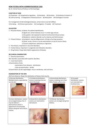
1. HOW TO DEAL WITH A DERMATOLOGICAL CASE.
By: Dr. Ashraf Hamza (Professor of Dermatology)
FUNCTION OF SKIN
(1) Protection (2) Temperature regulation (3) Sensation (4) Excretion (5) Synthesis of vitamin D
(6) UVR screening (7) Regulation of blood pressure (8) Absorption (9) Psychogenic function
For management of dermatological disease, certain items must be fulfilled;
1) Hx taking (2) Clinical examination (3) Investigation -if needed (4) Treatment
(A) HISTORY
1 – Personal History: a) Name: for patient identification.
b) Age & sex: certain diseases occur in certain age and sex.
c) Occupation: skin exposed to external environment (house wives).
d) Residence: endemic disorder such as leprosy and leishmaniasis.
2 – Present History: a) Complaint: may be disfigurement, itching or burning sensation.
b) Onset: acute, chronic or acute exacerbation on top of chronic illness.
c) Course: progressive, stationary or regressive.
3 – Past History: Important in recurrent disorders.
4 – Family History: Important in congenital and infestation disorders.
5 – Drug history: Drugs taken before appearance of disease.
(B) CLINICAL EXAMINATION
1 – General examination
Skin disorders associated with systemic disorders.
2 – Local examination
a) Examination of skin:
- examination from distance - distribution
- close-up examination - border
b) Examination od skin appendages: mucus membrane, nails and hairs.
EXAMINATION OF THE SKIN
(A) From distance: Shows the distribution of lesions that may be:
1) Discrete distribution:
Multiple lesions separated by
normal skin.
2) Unilateral distribution:
Lesions involving only one
side of the body.
3) Generalized distribution:
Lesions involving more than
50% of body surface area.
4) Grouped
distribution: Lesions
are restricted to a
localized area.
5) Linear distribution: Lesions
are arranged along a line. It
may be Kobner’s
phenomenon which is
appearance of isomorphic
lesions along the site of blunt
trauma.
6) Zosteriform distribution:
Lesions are restricted to
dermatome.
7) Follicular distribution:
Lesions are arranged along hair
follicles.
2. (B) Close-up examination: Shows the border of lesions that may be:
1) Well-defined border: Marked
separation between the edge of the
lesion and normal skin.
2) Ill-defined border: Difficult to
identify the separation line between
the lesion and normal skin.
3) Circinate border: The lesion
increases in size by peripheral
extension and healing at the centre.
TYPES OF SKIN LESIONS
Skin lesions may be: initial lesions, secondary lesions or specific lesions.
(A) INITIAL LESIONS
1) Macule: Discoloration of skin <1cm diameter. Patch: if >1cm.
2) Papule: Solid elevation of the skin <1cm diameter. Plaque: if >1cm
(a) Dome shaped: papule with a smooth convex surface.
(b) Flat topped: papule with a flat surface. It is describe as lichenoid papule.
(c) Umbilicated: dome shaped papule with central notch.
(d) Verrucous: papule with fine mammilated surface.
3) Nodule: Elevated solid skin lesion with dermal extension.
4) Vesicle: Fluid-containing lesion <1cm diameter. Bulla: if >1cm
(a) Intraepidermal separation: flaccid bulla
(b) Subepidermal separation: Tense bulla
(B) SECONDARY LESIONS
1) Pustule: Elevated lesion containing pus.
2) Scales: Dry surface dt abnormal keratinization.
(a) Fine branny: Pytriasis versicolor
(b) Greasy: Seborrheic dermatitis
(c) Lamellar: Psoriasis
(d) Fish scales: Ichthyosis
(e) Collarette: Pityriasis rosea
(f) Horny (keratotic): Discoid LE
3) Crust: Dried exudates, either pus or blood.
4) Erosion: Superficial epidermal loss.
5) Ulcer: Deep dermal loss, thus it has characteristic edge.
6) Fissure: Longitudinal discontinuity off the skin.
7) Atrophy: Thinning of skin dt thinning of epidermis or dermis or both.
8) Scar: Replacement of the skin by FT
9) Lichenification: Descriptive term of 3 criteria:
(a) Thickening of skin. (b) Hyperpigmentation. (c) Increased skin markings.
(C) SPECIFIC INITIAL LESIONS
1) Wheal: Specific to urticaria. It is edematous erythematous lesion which is migratory.
2) Scutulum: Specific for favus. It is concavo-convex golden yellow cup stuck to scalp.
3. 3) Comedone: Specific to acne. Either: - Black head: papule with central black spot.
- White head: small pale papule.
4) Tunnel (burrow): Specific for scabies. It is a curved line dt burrowing of female mite to skin.
5) Target lesion: Specific to erythema multiforme. Consist of 3 zones:
(a) Central zone: cyanotic. (b) Intermediate zone: pale (c) Outer zone: erythematous
6) Herald patch: specific for P.rosea. It has 3 concentric zones:
(a) Central cafe au lait. (b) Intermediate collaretic scales. (c) Peripheral erythematous.
EXAMINATION OF SKIN APPENDAGES
(A) Examination of mucus membranes:
Check for any erosion, ulceration, plaque, pigmentation, white streaks.
(B) Examination of nails:
Nail pitting: in cases of ................
Nail discoloration: ........................
Nail fold swelling: ........................
Nail dystrophy: ..............................
(C) Examination of hair.
1) Hair loss: either
Diffuse hair loss.
Patchy or alopecia Cicatrical or non cicatrical (Differentiate by ........................)
2) Hair growth in abnormal sites. (In woman: hirsutism)
INVESTIGATION
There are certain investigations specific to the skin that can help in the dx of some skin diseases.
1) Wood’s light: It is a special UV light which if thrown to:
Normal skin – reflects deep violet colour.
Pityriasis versicolor – reflects golden yellow colour.
Erythrasma – reflects deep red colour
Tinea capitis – reflects brilliant green colour
2) Skin scraping: Used for dx of fungal infection of skin.
Procedure:
Skin is scratched by scalpel. The resulted scales are places on glass slide, then 10% KOH is added and examined under
microscope. (eg: hyphae of dermatophytes)
Hair sample: endothrix
ectothrix
3) Patch testing: Used for dx of contact dermatitis.
Procedure: aluminium strip with multiple holes is fixed on the back. The antigens are placed each in one hole. Then,
another aluminium strip is placed over the previous one and left for 48 hours. On removal of the strip, we examine the
sites o different allergenfor erythema and vesicles. If present, the test is positive.
4) Immunoflourescent tests: Used for dx of autoimmune disorders. Either:
Direct test – Detect antibody in the skin. Skin biopsy is taken and flourescent anti-antibody is placed on it.
Indirect test – Detect antibody in serum of patient. Flourescent anti-antibody is added to the patient serum.
5) Skin biopsy and histopathology:
Demonstrate the pathological
changes in the diseased area.
Usually diagnostic.
Histology of skin (epidermal layer):
1) Horny layer (stratum cornium)
2) Granular cell layer
(stratum granulosum)
3) Prickle cell layer
(stratum spinosum)
4. 4) Basal cell layer
(stratum basale)
Pathological terms:
1) Hyperkeratosis: increased thickness of horny layer (stratum cornium).
2) Parakeratosis: retention of nuclei in horny layer (stratum cornium).
3) Hypergranulosis: increased thickness of granular cell layer (stratum granulosum).
4) Acanthosis: increased thickness of prickle cell layer. Either uniform or saw tooth acanthosis.
5) Spongiosis: edema of prickle cell layer (stratum spinosum).
6) Acantholysis: Loss of coherence between cells of prickle cell layer (stratum spinosum)/
Hyperkeratosis Parakeratosis Acanthosis
Spongiosis Acantholysis
THERAPY IN DERMATOLOGY
Principles of topical therapy:
1) Type of skin lesion: Wet lesions need creams, dry lesions need ointments.
2) Never do any harm: don’t use irritant and sensitizers.
3) Never overtreat: This usually occurs when a patient ask a friend and use many medications.
4) Instruct the patient adequately: It is not important to tell a patient how to swallow a pill but it is essential to tell
patients how to apply local medications.
5) Prescribe the correct amount of medication for the area and dermatoses to be treated.
6) Change the therapy as the response indicates.
7) If the prescription is expensive, explain this fact to the patient.
8) Therapy plus is usually indicated, advise the patient to continue to apply the medications for a specific period after
the dermatoses apparently cleared. This is to prevent recurrence.
9) Ask the patient to cantact you if there is any question or if the medicine appeared to irritate the dermatoses.
EFFECTS OF LOCALLY APPLIED DRUGS
5. 1) Antipruritic Agents: Relieving itching in various ways. Eg: Calamine lotion.
2) Keratoplastic Agents: Increased the thickness of horny layer. Eg: Tar
3) Keratolytic Agents: Remove or soften horny layer. Eg: Salicylic acid 4%-6%, urea 20%-40%, lactic acid 20%
4) Antieczematous: Stop the secretions by various actions. Eg: Corticosteroids.
5) Antiparasitic Agents: Destroy or inhibit living infestations. Eg: Permethrim 5%
6) Antiseptics: Destroy or inhibit bacteria, viruses or fungi.
7) Antibacterial topical medications: destroy or inhibit bacteria. Eg: Gentamycin, fusidic acid, erythromycin,
tetracycline, neomycin, and chloramphenicol.
8) Antiviral topical agents: destroy or inhibit viruses. Eg: Acyclovir.
9) Emollient Agents: Soften skin surface. Eg: Cold cream and Vaseline.
TYPES OF TOPICAL MEDICATIONS
1) Compresses: Remove the crust. Eg: Potassium permanganate 1/8000 and saline.
2) Drying agents: Dry oozing skin. Eg: Gentian violet 1% and microchrome 1%
3) Creams: They are semisolid emulsion systems containing both water and oil. They are water miscible, cooling and
soothing, and are well absorbed into the skin.
Used in acute oozing skin disorders.
4) Ointments: They have oil or grease. They are semisolid and anhydrous substances.
Used in chronic, dry skin disorders. Eg: Vaseline petroleum jelly
5) Gels: They are semisolid preparations gelled with high molecular weight polymers, such as methylcellulose.
Non-greasy, water miscible, easy to apply and wash off. Especially suitable for treating hairy parts of body.
6) Paints: Liquid preparations, either aqueous, or alcoholic (tinctures), which are usually applied with a brush to the
skin. They are evaporate, and are therefore cooling as well astringent and antiseptic. They may also be used as
protectives to seal abrasions.
7) Lotions: Combination of powder and water. Able to cover wide surface area of skin dt increased evaporating surface.
Not suitable for xerosis pruritus. Eg: Calamine lotion.
QUANTITY OF CREAMS TO PRESCRIBE
FACTOR AFFECTING THE QUANTITY
1) Types of dermatoses: acute or chronic.
2) Base of topical medication: Ointments spread over skin more than creams.
3) Intelligence of patients: educated patients usually consume smaller amounts.
HOW TO ASSESS THE QUANTITY OF TOPICAL MEDICATIONS?
1) Use the fingertip unit to assess the quantity of cream needed to cover a certain body surface area.
2) One fingertip unit is approximately 500mg.
Amount needed Duration of application Frequency of application Surface area
15gm 14 days b.i.d Hand
30gm 14 days b.i.d Arm
60gm 14 days b.i.d Leg
480-960gm 14 days b.i.d Entire body