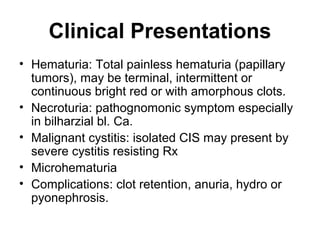
Bladder Tumor
Mohamed Adel Atta
Classification
• Epithelial: papilloma, carcinoma..
• Mesenchymal: leiomyoma, sarcoma.
Epidemiology
• Commonest cancer in males in Egypt.
• Male:female ratio 3
• Age of peak incidence 50-70, in Egypt 30-
50
Bilharzial bladder carcinoma
• Bilharziasis results in younger age,
• Higher male:female ratio,
• More squamous cell carcinoma,
• Presents in advanced stage because bilh.
cystitis masks symptoms of the tumor
Pathogenesis
• Bilharziasis
• Smoking
• Aniline dyes
• Balkan residency
• Bladder extrophy (adenocarcinoma), all
have strong etiologic relationship to
bladder carcinoma.
Spread
Bladder carcinoma spreads equally by all
routes local, lymphatic and vascular.
Bladder carcinoma spreads mainly locally in
bilharzial bladder because of intense
fibrosis that limits lymph and vascular
spread.
Staging
T0: Intraepithelial, Tis: High
grade intraepithelial
neoplasia (CIS).
T1: Lamnia propria invasion
T2: invasion of muscle
layer:
T2a: superficial muscle
layer
T2b: deep muscle layer.
T3: invasion of perivesical
fat
T3a: microscopic
invasion
T3b: macroscopic
invasion.
T4: invasion of pelvic wall
or nearby organs.
Staging
• Superficial bladder tumor (non muscle
invasive bladder carcinoma NMIBC ):
Ta, T1, CIS
• Invasive Bladder carcinoma (MIBC):
T2-4
Natural History
• Superficial bladder tumor: Ta&T1 and CIS,
usually recur in other site of the bladder(70%),
but rarely invades bladder wall (10%), solitary or
multiple with mobile fronds long stalk and narrow
base. CIS carcinoma in situ: high grade
intraepithelial carcinoma, appear as velvety
hyperemic areas.
• Invasive deep tumor:T2-4 cauliflower, nodular or
ulcerative with necrotic surface.
Gross Types
• Papillary: sea-weed appearance
• Cauliflower mass with stunt fronds, some
necrotic.
• Nodular
• Ulcerative.
Invasive Cauliflower Multiple
Tumors
GROSS TYPES
Superficial Bladder Tumor
Clinical Presentations
• Hematuria: Total painless hematuria (papillary
tumors), may be terminal, intermittent or
continuous bright red or with amorphous clots.
• Necroturia: pathognomonic symptom especially
in bilharzial bl. Ca.
• Malignant cystitis: isolated CIS may present by
severe cystitis resisting Rx
• Microhematuria
• Complications: clot retention, anuria, hydro or
pyonephrosis.
Diagnosis
• Ultrasonography: echogenic
intravesical mass
• Plain&IVU: bladder filling defect
• Pelvic and abdominal CT: confirm
and stage bladder carcinoma
CT bladder tumors
Diagnosis
• Urine cytology
• Cystoscopy and biopsy: tumor and
tumor bed for proper staging,
bimanual examination under
anathesia to asses the degree of
pelvic spread of the tumor
• Metastatic workup X-ray chest and
bone scan.
Urine cytology
Cytology is the detection of cells in fluid,
cells should be viable to take up the stain
Malignant cells have ameboid movement
due to loss of intercellular attachments
(nexi) and according can be seen in urine
frequently especially in CIS and grade 2
carcinoma
Voided urine cytology high grade uroth ca: nuclear
hyperchromatism and irregular nucl memb.
Voided urine cytology high grade uroth ca: nuclear
hyperchromatism and irregular nucl memb.
Bladder wash cytology clump malignant cells
nuclear hyperchromatisia, vacuolated cytoplasm
Bladder wash cytology low grade car, thick nuclear
memb, hypochromatasia, homogenous cytoplasm
Voided urine cytology low grade
carcinoma
Transurethral Biopsy: The definitive
Diagnosis
• 1- Tumor Tissue
• 2- Tumor bed biopsy to properly stage
muscle infiltration
• 3- Bimanual examination under
anasthesia to asses clinically infiltration of
nearby organs and pelvic wall.
Histopathology
• 1- Transitional cell carcinoma: the commonest
type
• 2- Squamous cell carcinoma: develops on top of
squamous metaplasia due to bilharziasis
• 3- Verrucous Ca: subtype of sq. c. ca.,
hyperkerratotic low grade squamous ca., locally
malignant with no vascular spread.
• 4- Adenocarcinoma: bladder dome on top of
allantoic remnant, or bladder base on top cloacal
remnants.
The papilloma is composed of a delicate
fibrovascular core covered by normal urothelium
Low Grade urothelial
carcinoma
The low-grade
papillary urothelial
carcinoma group
includes all former
grade 1 (WHO
1973) cases and
some former grade
2 cases (if a
variation of
architectural and
cytological features
exist at high
magnification).
High grade Urothelial
carcinoma
High grade urothelial
carcinoma showing
atypical urothelial
cells that vary in size
and shape. The
nuclei are enlarged,
with coarsely
granular chromatin,
hyperchromasia,
abnormal nuclear
contours and
prominent nucleoli
High grade Urothelial
carcinoma
High grade urothelial
carcinoma showing
atypical urothelial
cells that vary in size
and shape. The
nuclei are enlarged,
with coarsely
granular chromatin,
hyperchromasia,
abnormal nuclear
contours and
prominent nucleoli
CIS
High grade
urothelial
carcinoma limitted
to the urothelium.
No invasion of the
underlying
basement.. Lamnia
propria underneath
shows
angiogenesis
Lymphovascular invasion
Muscularis propria invasion
Squmous cell carcinoma
Bilharzial egg S Hemmatobiu
Treatment
Superficial Bladder Tumor
• 1- Transurethral resection (TURT):
• 2- In multiple, big,T1, and recurrent
tumors: Intravesical chemotherapy
(thiotepa, mitomycin, adriamycin) or better
immunotherapy (BCG Vaccine) is advised
to reduce tumor recurrence and avoid
tumor progression 6 weekly instillations
followed by maintenance 3 weekly inst.
every 6 months.
Treatment Superficial Bladder
Tumor
• 3- Followup Protocol: including US, urine
cytology, cystoscopy and biopsy
• 4- Radical cystectomy in high grade
tumors resisting treatment and rapidly
recurrent.
Treatment Of Invasive Tumors
• Radical cystectomy is the gold standard excision
of bladder, lower ureters, as well as prostate,
seminal vesicles in males and uterus upper
vagina and ovaries in females together with
pelvic lymph nodes.
• Radical radiotherapy: less efficient
• Bladder saving protocol using initial
chemotherapy followed by radiotherapy in
responding tumors or salvage cystectomy in
non-responding tumors.
Post-Cystectomy Urinary
Reconstruction
1- Orthotopic bladder substitutes
2- Ectopic bladder substitutes:
A- Cutaneous:
a- Wet stoma: ileal conduit
b- Continent stoma: cont.reservoir
B- Anal:
a- Ureterosigmoidostomy & its variants
b- Rectal bladder with left terminal colostomy
Post-Cystectomy Urinary
Reconstruction
Any part of GI tract can be used: ileum,
colon or stomach.
Detubularization and refashioning in the
form of a sphere results in bigger (3 times
the volume of the tubular intestine) and
less intraluminal pressure (la Place law).
Effect of detubularization
Orthotopic Neobladder
• Detubularized intestinal segment
fashioned in the form of sphere is
anastomosed to the urethra and both
ureters are anastomosed to the
pouch with an antireflux mechanism.
• Is the first option unless tumor
invades the proximal urethra.
Serous-lined W-shape
neobladder
Sigmoid neobladder
Ileal Conduit
• Both ureters are anastomosed to 15
cm ileal segment , one end is closed
and the other end is anastomosed to
the skin.
• Urine bag is applied to the stoma
Ileal Conduit
Ileal conduit
Continent Reservoir
• Detubularized intestinal segment is
fashioned in the form of sphere, both
ureters are anastomosed to the pouch
with antireflux mechanism.
• The pouch is anastomosed to the
umblicus with continent mechanism to
prevent urine leakage
• Patient uses plastic catheter to evacuate
the pouch every 6-8 hours
Kock Reservoir
Indiana & Florida pouches
Ureterosigmoidostomy
• First known continent diversion, both
ureters are anastomosed to the sigmoid
colon with proper antireflux technique
• Sequelae: electrolyte imbalance
hyperchloremic hypokalemic acidosis,
ascending infection, colonic
carcinogenesis.
• New variants are introduced to avoid such
sequelae with better outcome.
Ureterosigmoidostomy
UERETEROSIGMOIDOSTOM
Y
1- Drawbacks: electrolyte imbalance
hypokalemic hyperchloremic
acidosis, repeated UTI, colonic
cancer, inconvenient evacuation
2- Less with the new modifications
3- Not accepted in all centers
Detubularized Isolated
Ureterosigmoidostomy (DIUS)
Detubularized Isolated
Ureterosigmoidostomy (DIUS)

8 bladder tumor

  • 1.
  • 2.
    Classification • Epithelial: papilloma,carcinoma.. • Mesenchymal: leiomyoma, sarcoma.
  • 3.
    Epidemiology • Commonest cancerin males in Egypt. • Male:female ratio 3 • Age of peak incidence 50-70, in Egypt 30- 50
  • 4.
    Bilharzial bladder carcinoma •Bilharziasis results in younger age, • Higher male:female ratio, • More squamous cell carcinoma, • Presents in advanced stage because bilh. cystitis masks symptoms of the tumor
  • 5.
    Pathogenesis • Bilharziasis • Smoking •Aniline dyes • Balkan residency • Bladder extrophy (adenocarcinoma), all have strong etiologic relationship to bladder carcinoma.
  • 6.
    Spread Bladder carcinoma spreadsequally by all routes local, lymphatic and vascular. Bladder carcinoma spreads mainly locally in bilharzial bladder because of intense fibrosis that limits lymph and vascular spread.
  • 7.
    Staging T0: Intraepithelial, Tis:High grade intraepithelial neoplasia (CIS). T1: Lamnia propria invasion T2: invasion of muscle layer: T2a: superficial muscle layer T2b: deep muscle layer. T3: invasion of perivesical fat T3a: microscopic invasion T3b: macroscopic invasion. T4: invasion of pelvic wall or nearby organs.
  • 8.
    Staging • Superficial bladdertumor (non muscle invasive bladder carcinoma NMIBC ): Ta, T1, CIS • Invasive Bladder carcinoma (MIBC): T2-4
  • 9.
    Natural History • Superficialbladder tumor: Ta&T1 and CIS, usually recur in other site of the bladder(70%), but rarely invades bladder wall (10%), solitary or multiple with mobile fronds long stalk and narrow base. CIS carcinoma in situ: high grade intraepithelial carcinoma, appear as velvety hyperemic areas. • Invasive deep tumor:T2-4 cauliflower, nodular or ulcerative with necrotic surface.
  • 10.
    Gross Types • Papillary:sea-weed appearance • Cauliflower mass with stunt fronds, some necrotic. • Nodular • Ulcerative.
  • 11.
  • 12.
  • 13.
  • 15.
    Clinical Presentations • Hematuria:Total painless hematuria (papillary tumors), may be terminal, intermittent or continuous bright red or with amorphous clots. • Necroturia: pathognomonic symptom especially in bilharzial bl. Ca. • Malignant cystitis: isolated CIS may present by severe cystitis resisting Rx • Microhematuria • Complications: clot retention, anuria, hydro or pyonephrosis.
  • 16.
    Diagnosis • Ultrasonography: echogenic intravesicalmass • Plain&IVU: bladder filling defect • Pelvic and abdominal CT: confirm and stage bladder carcinoma
  • 19.
  • 20.
    Diagnosis • Urine cytology •Cystoscopy and biopsy: tumor and tumor bed for proper staging, bimanual examination under anathesia to asses the degree of pelvic spread of the tumor • Metastatic workup X-ray chest and bone scan.
  • 21.
    Urine cytology Cytology isthe detection of cells in fluid, cells should be viable to take up the stain Malignant cells have ameboid movement due to loss of intercellular attachments (nexi) and according can be seen in urine frequently especially in CIS and grade 2 carcinoma
  • 22.
    Voided urine cytologyhigh grade uroth ca: nuclear hyperchromatism and irregular nucl memb.
  • 23.
    Voided urine cytologyhigh grade uroth ca: nuclear hyperchromatism and irregular nucl memb.
  • 24.
    Bladder wash cytologyclump malignant cells nuclear hyperchromatisia, vacuolated cytoplasm
  • 25.
    Bladder wash cytologylow grade car, thick nuclear memb, hypochromatasia, homogenous cytoplasm
  • 26.
    Voided urine cytologylow grade carcinoma
  • 27.
    Transurethral Biopsy: Thedefinitive Diagnosis • 1- Tumor Tissue • 2- Tumor bed biopsy to properly stage muscle infiltration • 3- Bimanual examination under anasthesia to asses clinically infiltration of nearby organs and pelvic wall.
  • 28.
    Histopathology • 1- Transitionalcell carcinoma: the commonest type • 2- Squamous cell carcinoma: develops on top of squamous metaplasia due to bilharziasis • 3- Verrucous Ca: subtype of sq. c. ca., hyperkerratotic low grade squamous ca., locally malignant with no vascular spread. • 4- Adenocarcinoma: bladder dome on top of allantoic remnant, or bladder base on top cloacal remnants.
  • 29.
    The papilloma iscomposed of a delicate fibrovascular core covered by normal urothelium
  • 30.
    Low Grade urothelial carcinoma Thelow-grade papillary urothelial carcinoma group includes all former grade 1 (WHO 1973) cases and some former grade 2 cases (if a variation of architectural and cytological features exist at high magnification).
  • 31.
    High grade Urothelial carcinoma Highgrade urothelial carcinoma showing atypical urothelial cells that vary in size and shape. The nuclei are enlarged, with coarsely granular chromatin, hyperchromasia, abnormal nuclear contours and prominent nucleoli
  • 32.
    High grade Urothelial carcinoma Highgrade urothelial carcinoma showing atypical urothelial cells that vary in size and shape. The nuclei are enlarged, with coarsely granular chromatin, hyperchromasia, abnormal nuclear contours and prominent nucleoli
  • 33.
    CIS High grade urothelial carcinoma limitted tothe urothelium. No invasion of the underlying basement.. Lamnia propria underneath shows angiogenesis
  • 34.
  • 35.
  • 36.
  • 37.
    Bilharzial egg SHemmatobiu
  • 38.
    Treatment Superficial Bladder Tumor •1- Transurethral resection (TURT): • 2- In multiple, big,T1, and recurrent tumors: Intravesical chemotherapy (thiotepa, mitomycin, adriamycin) or better immunotherapy (BCG Vaccine) is advised to reduce tumor recurrence and avoid tumor progression 6 weekly instillations followed by maintenance 3 weekly inst. every 6 months.
  • 39.
    Treatment Superficial Bladder Tumor •3- Followup Protocol: including US, urine cytology, cystoscopy and biopsy • 4- Radical cystectomy in high grade tumors resisting treatment and rapidly recurrent.
  • 40.
    Treatment Of InvasiveTumors • Radical cystectomy is the gold standard excision of bladder, lower ureters, as well as prostate, seminal vesicles in males and uterus upper vagina and ovaries in females together with pelvic lymph nodes. • Radical radiotherapy: less efficient • Bladder saving protocol using initial chemotherapy followed by radiotherapy in responding tumors or salvage cystectomy in non-responding tumors.
  • 41.
    Post-Cystectomy Urinary Reconstruction 1- Orthotopicbladder substitutes 2- Ectopic bladder substitutes: A- Cutaneous: a- Wet stoma: ileal conduit b- Continent stoma: cont.reservoir B- Anal: a- Ureterosigmoidostomy & its variants b- Rectal bladder with left terminal colostomy
  • 42.
    Post-Cystectomy Urinary Reconstruction Any partof GI tract can be used: ileum, colon or stomach. Detubularization and refashioning in the form of a sphere results in bigger (3 times the volume of the tubular intestine) and less intraluminal pressure (la Place law).
  • 43.
  • 44.
    Orthotopic Neobladder • Detubularizedintestinal segment fashioned in the form of sphere is anastomosed to the urethra and both ureters are anastomosed to the pouch with an antireflux mechanism. • Is the first option unless tumor invades the proximal urethra.
  • 45.
  • 46.
  • 47.
    Ileal Conduit • Bothureters are anastomosed to 15 cm ileal segment , one end is closed and the other end is anastomosed to the skin. • Urine bag is applied to the stoma
  • 48.
  • 49.
  • 50.
    Continent Reservoir • Detubularizedintestinal segment is fashioned in the form of sphere, both ureters are anastomosed to the pouch with antireflux mechanism. • The pouch is anastomosed to the umblicus with continent mechanism to prevent urine leakage • Patient uses plastic catheter to evacuate the pouch every 6-8 hours
  • 51.
  • 52.
  • 53.
    Ureterosigmoidostomy • First knowncontinent diversion, both ureters are anastomosed to the sigmoid colon with proper antireflux technique • Sequelae: electrolyte imbalance hyperchloremic hypokalemic acidosis, ascending infection, colonic carcinogenesis. • New variants are introduced to avoid such sequelae with better outcome.
  • 54.
  • 55.
  • 56.
    UERETEROSIGMOIDOSTOM Y 1- Drawbacks: electrolyteimbalance hypokalemic hyperchloremic acidosis, repeated UTI, colonic cancer, inconvenient evacuation 2- Less with the new modifications 3- Not accepted in all centers
  • 57.
  • 58.