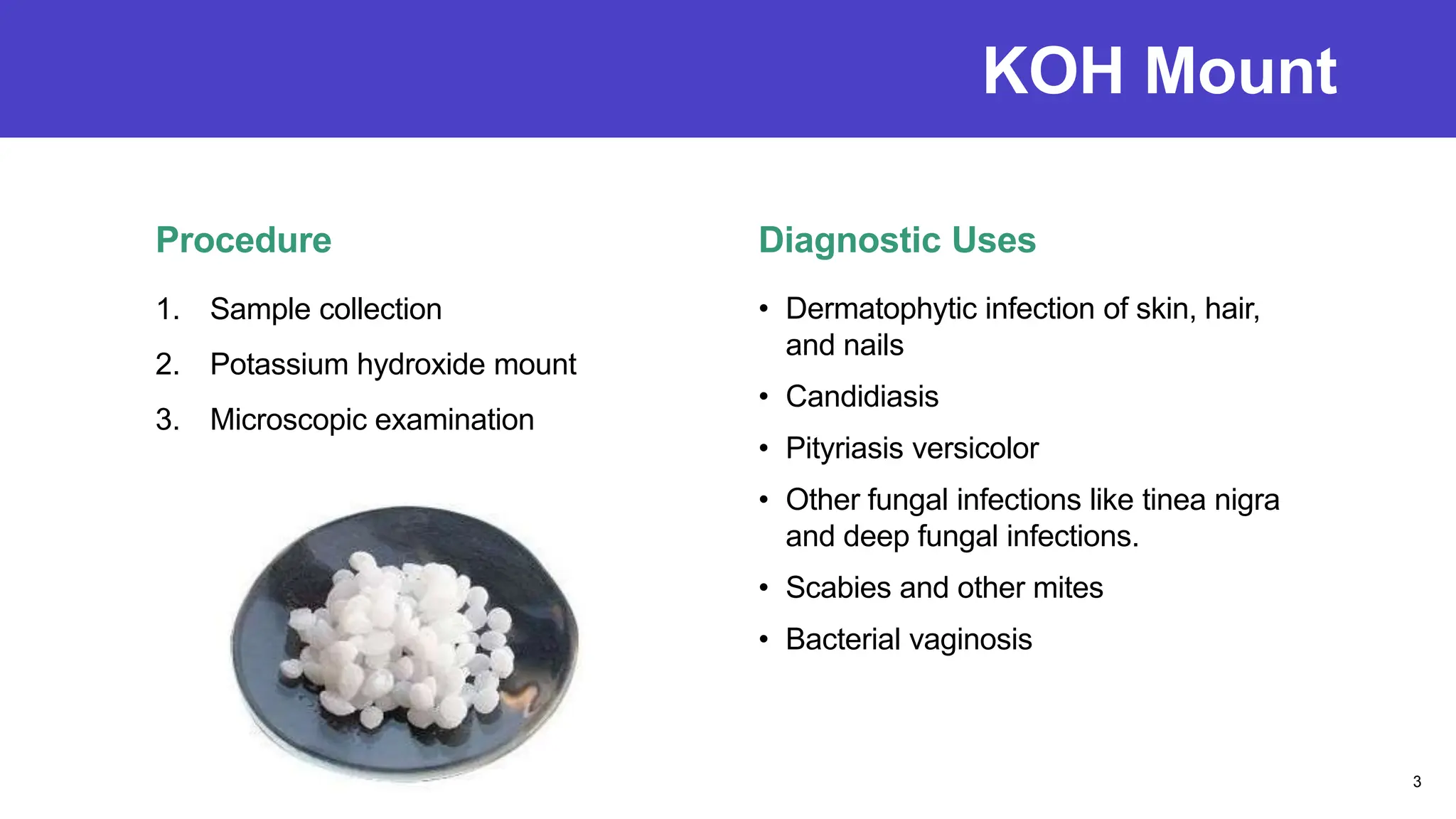
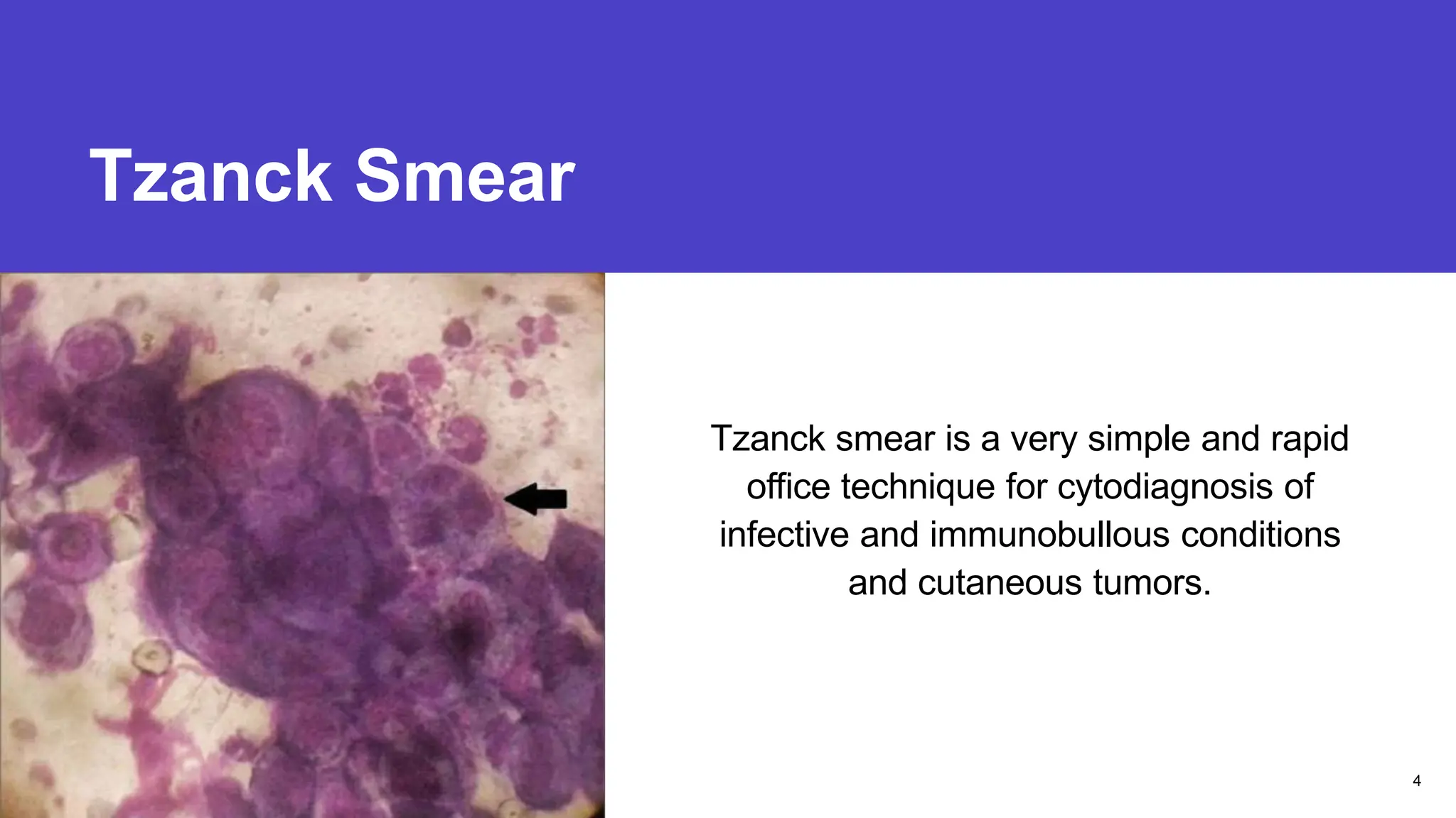
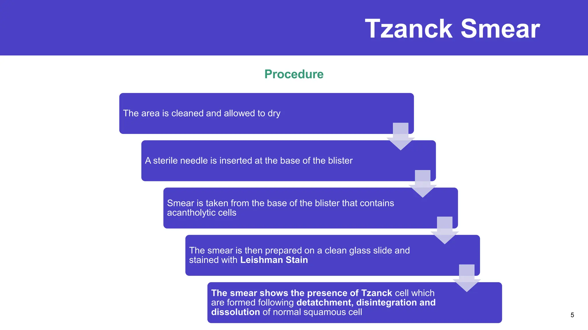
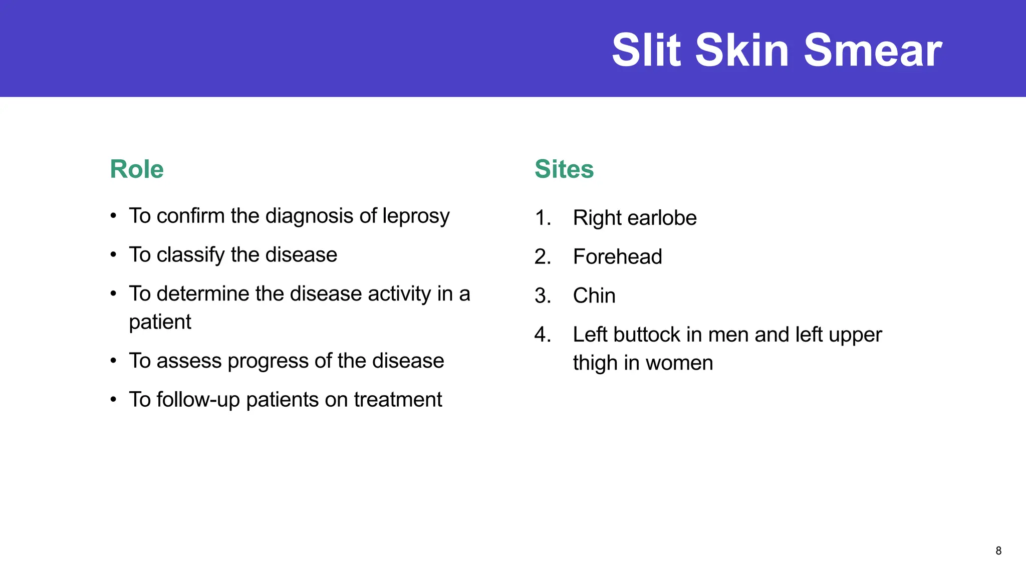
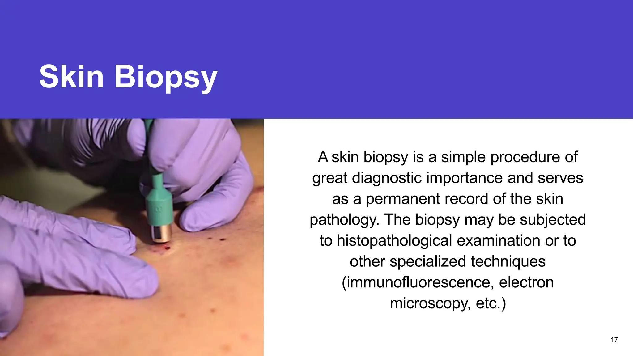
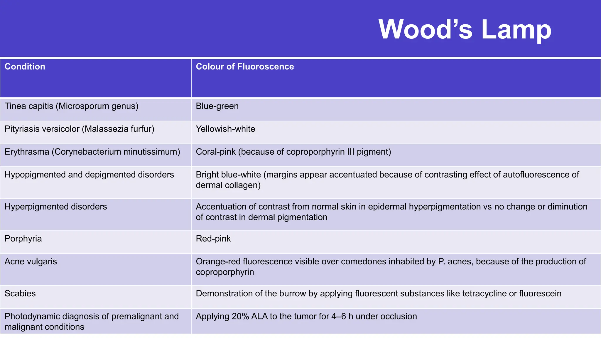
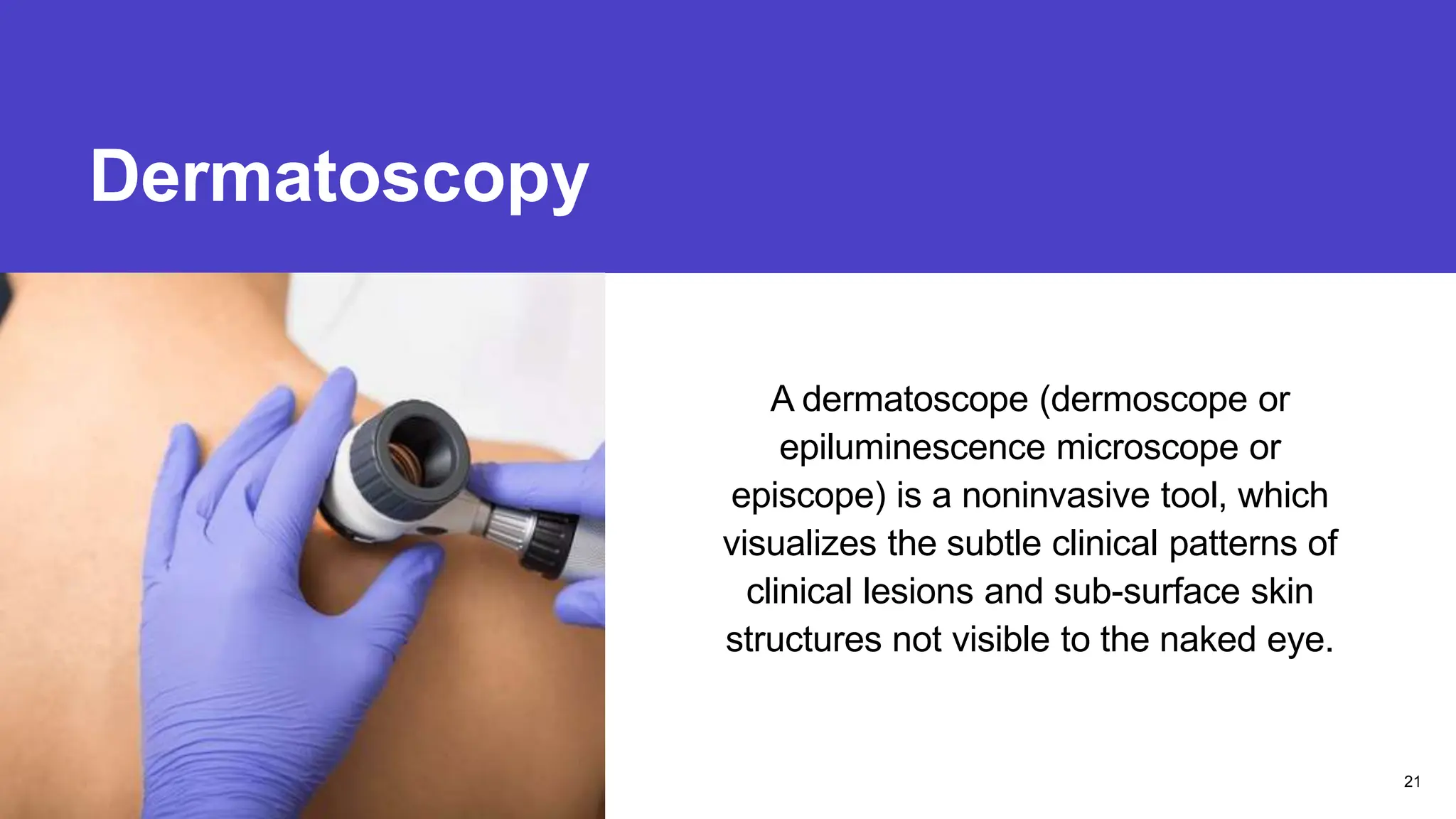
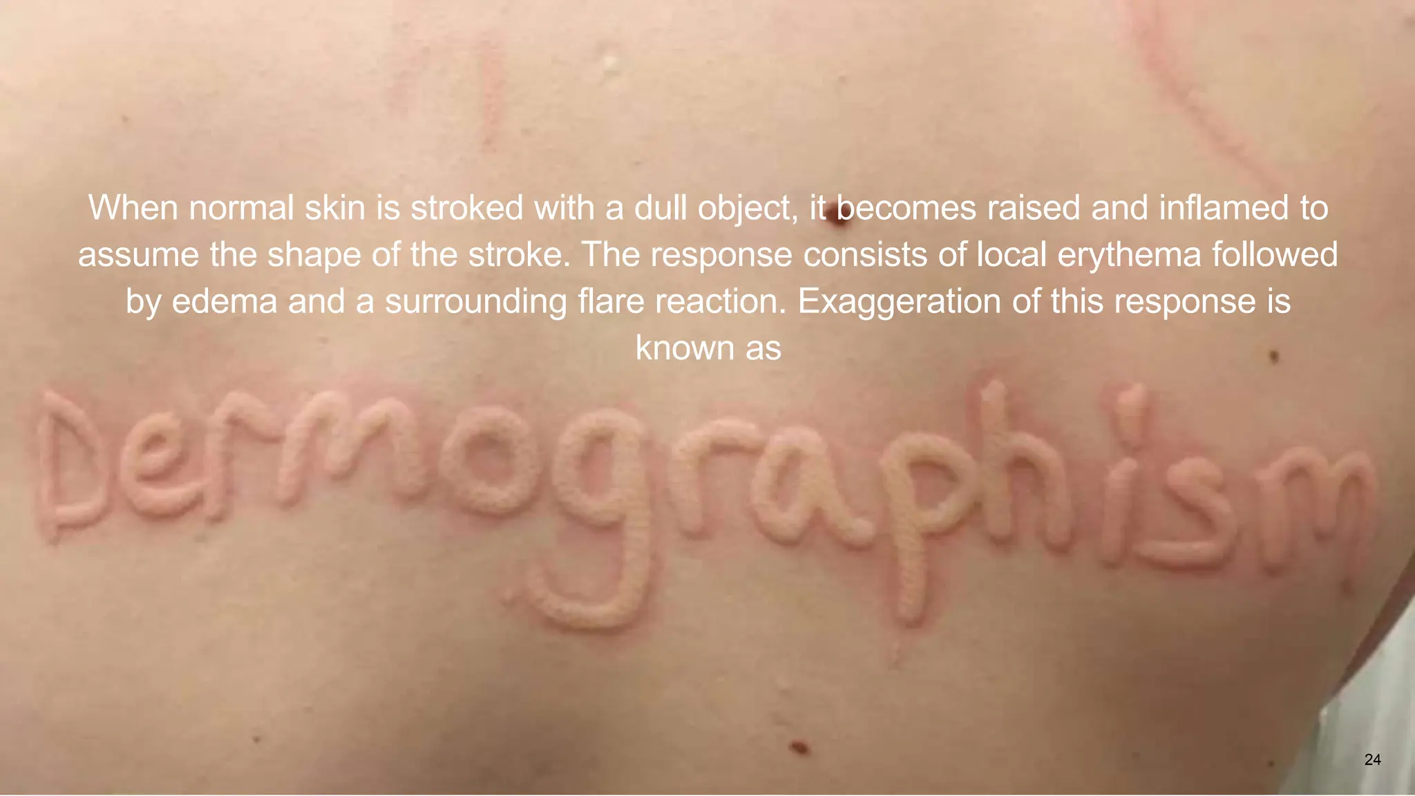
Bedside Tests
in
Dermatology
Dr. Nishkarsh Chugh
PG Resident, Dept. of Dermatology
Potassium Hydroxide Mount
Presentation title
A potassium hydroxide (KOH) mount of
a skin scraping is a commonly
performed procedure to demonstrate the
evidence of fungal infection in the skin,
hairs, and nails.
2
KOH Mount
Procedure
1. Sample collection
2. Potassium hydroxide mount
3. Microscopic examination
Diagnostic Uses
• Dermatophytic infection of skin, hair,
and nails
• Candidiasis
• Pityriasis versicolor
• Other fungal infections like tinea nigra
and deep fungal infections.
• Scabies and other mites
• Bacterial vaginosis
3
Tzanck Smear
Presentation title
Tzanck smear is a very simple and rapid
office technique for cytodiagnosis of
infective and immunobullous conditions
and cutaneous tumors.
4
Tzanck Smear
Procedure
5
The area is cleaned and allowed to dry
A sterile needle is inserted at the base of the blister
Smear is taken from the base of the blister that contains
acantholytic cells
The smear is then prepared on a clean glass slide and
stained with Leishman Stain
The smear shows the presence of Tzanck cell which
are formed following detatchment, disintegration and
dissolution of normal squamous cell
Tzanck Smear
6
Diagnostic Uses
Disease Finding
Pemphigus Acantholytic cells (rounded cells with a relatively large nucleus and a condensed
cytoplasm) with hazy nucleoli
Bullous pemphigoid Non-specific findings. Scarcity of epithelial cells and an abundance of leukocytes,
particularly eosinophils with leukocyte adherence, is seen
Stevens-Johnson syndrome No acantholytic cells, but plenty of leukocytes
Toxic epidermal necrolysis Necrotic basal cells and leukocytes
Staphylococcal scalded skin syndrome Minimal or no inflammation, dyskeratotic Acantholytic cells
Herpes simplex, varicella, herpes zoster Ballooning multinucleate giant cells
Molluscum contagiosum Henderson-Patterson bodies
Leishmaniasis Leishman-Donovan bodies
Hailey-Hailey disease Acantholytic cells with normal nucleoli
Darier’s disease Corps, ronds and grains
BCC Basaloid cells
Paget’s disease of breast Paget cells
Mastocytoma Mast cells
Histiocytosis Atypical Langerhan cells
Slit Skin Smear
Presentation title
It is the most important lab test to detect
lepra bacilli in suspected Hansen’s
patches
7
Slit Skin Smear
Role
• To confirm the diagnosis of leprosy
• To classify the disease
• To determine the disease activity in a
patient
• To assess progress of the disease
• To follow-up patients on treatment
Sites
1. Right earlobe
2. Forehead
3. Chin
4. Left buttock in men and left upper
thigh in women
8
Slit Skin Smear
Procedure
9
Lesion is cleaned with spirit.
After pinching the skin between thumb and index
finger, a 5mm long and 2mm deep cut is made
with sterile blade (Bard Parker No. 15)
Base is scraped and the material is smeared (8-
10mm) on a glass slide.
After drying and heat fixing the smear, Ziehl-
Neelsen staining is done.
Slit Skin Smear
Bacterial Index Morphological Index
Percentage of solid stained bacilli,
calculated after examining 200 bacilli
lying singly
Bacilli are considered solid staining if:
1. Entire organism is uniformly stained
2. Longitudinal sides are parallel
3. Both ends are rounded
4. Length is five times its width
10
0 0 bacilli in 100 OIFs
1+ 1-10 bacilli/100 fields
2+ 1-10 bacilli/10 fields
3+ 1-10 bacilli/field
4+ 10-100 bacilli/field
5+ 100-1000 bacilli/field
6+ >1000 bacilli/field
Dark Field Microscopy
Presentation title
It is the most specific and sensitive
technique to diagnose syphilis when an
active chancre or condyloma lata is
present
11
Dark Field Microscopy
Principle
The dark ground microscope creates a
contrast between the object and the
surrounding field, such that, the
background is dark and the object is
bright.
Interpretation
Treponema pallidum appear as brightly
illuminated objects against a dark background.
0.25-0.3µm wide and 6-16µm long
organism with 8-14 regular, tightly wound, deep
spirals.
It exhibits quick and abrupt movements.
The organism rotates slowly along the
longitudinal axis (corkscrew motion)
accompanied by bending and twisting in the
middle. 12
Diascopy
Presentation title
Pressing a glass slide or a clear,
colourless piece of plastic onto the skin
compresses blood out of small vessels,
to allow evaluation of other colours.
13
Diascopy
Typical Uses
1. Granulomatous lesions: Yellowish green color is seen in plaques of lupus vulgaris
known as apple jelly nodules and grains of sand appearance in sarcoidosis.
2. To distinguish between nevus depigmentosus and nevus anemicus – in nevus
anemicus, there is a localised area of vasoconstriction, other pigments are unaltered
– diascopy of adjacent skin therefore reveals an identical colour to that of the
‘depigmented’ area.
3. Application of medium pressure to a spider naevus can compress radiating
arterioles and allow visualisation of pulsatile flow in the feeding vessel.
14
Intradermal Tests
Presentation title
Intradermal tests are widely used in
dermatology to support the diagnosis of
infectious diseases as well as for
detection of immediate (Type I) and
delayed (Type IV) hypersensitivity
towards exogenous or endogenous
antigens.
15
Intradermal Tests
Technique
The antigen is injected into the superficial
layer of the dermis through a fine-bore
(26 or 27-G) needle with its bevel
pointing upward.
The usual quantity injected is 0.1 mL.
The proximal part of the flexor aspect of
the forearm is conventionally used.
Interpretation
Tests for the detection of DTH are
typically read at 48–72 hours. The size of
the induration is more important than
erythema while interpreting Type IV
hypersensitivity.
16
Skin Biopsy
Presentation title
A skin biopsy is a simple procedure of
great diagnostic importance and serves
as a permanent record of the skin
pathology. The biopsy may be subjected
to histopathological examination or to
other specialized techniques
(immunofluorescence, electron
microscopy, etc.)
17
Skin Biopsy
Types
1. Incisional
2. Superficial shave biopsy
3. Punch biopsy
4. Excisional
Choice of Lesion
• Fully evolved, untreated lesion
• In blistering disorders, the smallest vesicle should
be biopsied to keep the roof intact.
• Excoriated or ulcerated lesions or lesions modified
because of steroid application should be avoided.
• Advancing edge with maximal elevation should be
biopsied for annular lesions such as granuloma
annulare or porokeratosis.
• Sites to be avoided: legs, bony prominences, and
pressure bearing areas
18
Wood’s Lamp
Presentation title
A Wood’s lamp is a mercury vapor lamp that
emits longwave UV radiation generated by a
high-pressure mercury arc fitted with the “Wood’s
filter” made of barium silicate with 9% nickel
oxide. Wood’s filter is opaque to all light rays,
except a band between 320 and 400 nm with a
peak at 365 nm.
After absorption by a substance or tissue, the
emission of a longer wavelength light results in
visible fluorescence.
19
Wood’s Lamp
20
Condition Colour of Fluoroscence
Tinea capitis (Microsporum genus) Blue-green
Pityriasis versicolor (Malassezia furfur) Yellowish-white
Erythrasma (Corynebacterium minutissimum) Coral-pink (because of coproporphyrin III pigment)
Hypopigmented and depigmented disorders Bright blue-white (margins appear accentuated because of contrasting effect of autofluorescence of
dermal collagen)
Hyperpigmented disorders Accentuation of contrast from normal skin in epidermal hyperpigmentation vs no change or diminution
of contrast in dermal pigmentation
Porphyria Red-pink
Acne vulgaris Orange-red fluorescence visible over comedones inhabited by P. acnes, because of the production of
coproporphyrin
Scabies Demonstration of the burrow by applying fluorescent substances like tetracycline or fluorescein
Photodynamic diagnosis of premalignant and
malignant conditions
Applying 20% ALA to the tumor for 4–6 h under occlusion
Dermatoscopy
Presentation title
A dermatoscope (dermoscope or
epiluminescence microscope or
episcope) is a noninvasive tool, which
visualizes the subtle clinical patterns of
clinical lesions and sub-surface skin
structures not visible to the naked eye.
21
Dermatoscopy
Principle
Physical properties of the skin influence
the outcome of light incident over it
(reflection, refraction, diffraction, and
absorption).
While the dry scaly skin reflects most
light, smooth oily skin allows penetration
till deeper dermis.
22
Dermatoscopy
Normal Dermoscopic Appearance
23
Site Colour/Pattern
Normal skin over trunk Fine reticulate pigmentary network
Normal skin over extremities Fine reticulate pigmentary network interrupted by
crisscross skin markings
Normal skin over the face Pseudoreticular Pattern
Upper Epidermis – S. Corneum – Spinosum Black
DEJ Light to Dark Brown
Papillary Dermis Slate Blue
Reticular Dermis Steel Blue
24
When normal skin is stroked with a dull object, it becomes raised and inflamed to
assume the shape of the stroke. The response consists of local erythema followed
by edema and a surrounding flare reaction. Exaggeration of this response is
known as
Dermatographism
Triple Response of Lewis
1. Initial red line (capillary dilatation)
2. Axon-reflex flare with broadening
erythema (arteriolar dilatation)
3. Formation of a linear wheal
(transudation of fluid/edema)
Dermatographism usually develops
within five minutes of stroking the skin
and persists for 15- 30 min in contrast to
the normal triple response of Lewis that
subsides in less than 5-10 min
Time
The exact mechanism of
dermatographism remains uncertain but
it is likely to be caused by
‘mechanicoimmunological’ stimulation of
mast cells that release histamine.
25
Nikolsky Sign
Presentation title
The sign is best elicited by applying lateral
pressure with the thumb or fingerpad on skin
over a bony prominence. This results in a
shearing force that dislodges the upper layers of
epidermis from the lower epidermis.
A positive Nikolskiy sign indicates intraepidermal
cleavage and differentiates intraepidermal
blisters from subepidermal blisters.
It is pathognomonic of pemphigus and
staphylococcal scalded skin syndrome.
26
Bulla Spread Sign
Presentation title
Slow, careful and unidirectional pressure
applied by a finger to the bulla causes
peripheral extension of the bulla beyond the
marked margin.
The bulla thus extended has an irregular
angulated border in pemphigus vulgaris, while
a regular rounded border is observed in
bullous pemphigoid or other subepidermal
blistering disorders
27
Auspitz Sign
Presentation title 28
In Psoriasis, when the scales are completely
scraped off, the basement membrane is
exposed and is seen as a moist red surface
(membrane of Bulkeley) through which
dilated capillaries at the tip of elongated
dermal papillae are torn, leading to multiple
bleeding points
Carpet Tack/Cat’s Tongue Sign
Presentation title 29
In DLE, characteristic lesions are well
defined erythematous plaques with
partially adherent scales entering a
patulous follicle. When the scale is
removed, its undersurface shows horny
plugs that had occupied follicles
Thank You
Dr. Nishkarsh Chugh
PG Resident, Department of
Dermatology

Bedside Tests in Dermatology

  • 1.
    Bedside Tests in Dermatology Dr. NishkarshChugh PG Resident, Dept. of Dermatology
  • 2.
    Potassium Hydroxide Mount Presentationtitle A potassium hydroxide (KOH) mount of a skin scraping is a commonly performed procedure to demonstrate the evidence of fungal infection in the skin, hairs, and nails. 2
  • 3.
    KOH Mount Procedure 1. Samplecollection 2. Potassium hydroxide mount 3. Microscopic examination Diagnostic Uses • Dermatophytic infection of skin, hair, and nails • Candidiasis • Pityriasis versicolor • Other fungal infections like tinea nigra and deep fungal infections. • Scabies and other mites • Bacterial vaginosis 3
  • 4.
    Tzanck Smear Presentation title Tzancksmear is a very simple and rapid office technique for cytodiagnosis of infective and immunobullous conditions and cutaneous tumors. 4
  • 5.
    Tzanck Smear Procedure 5 The areais cleaned and allowed to dry A sterile needle is inserted at the base of the blister Smear is taken from the base of the blister that contains acantholytic cells The smear is then prepared on a clean glass slide and stained with Leishman Stain The smear shows the presence of Tzanck cell which are formed following detatchment, disintegration and dissolution of normal squamous cell
  • 6.
    Tzanck Smear 6 Diagnostic Uses DiseaseFinding Pemphigus Acantholytic cells (rounded cells with a relatively large nucleus and a condensed cytoplasm) with hazy nucleoli Bullous pemphigoid Non-specific findings. Scarcity of epithelial cells and an abundance of leukocytes, particularly eosinophils with leukocyte adherence, is seen Stevens-Johnson syndrome No acantholytic cells, but plenty of leukocytes Toxic epidermal necrolysis Necrotic basal cells and leukocytes Staphylococcal scalded skin syndrome Minimal or no inflammation, dyskeratotic Acantholytic cells Herpes simplex, varicella, herpes zoster Ballooning multinucleate giant cells Molluscum contagiosum Henderson-Patterson bodies Leishmaniasis Leishman-Donovan bodies Hailey-Hailey disease Acantholytic cells with normal nucleoli Darier’s disease Corps, ronds and grains BCC Basaloid cells Paget’s disease of breast Paget cells Mastocytoma Mast cells Histiocytosis Atypical Langerhan cells
  • 7.
    Slit Skin Smear Presentationtitle It is the most important lab test to detect lepra bacilli in suspected Hansen’s patches 7
  • 8.
    Slit Skin Smear Role •To confirm the diagnosis of leprosy • To classify the disease • To determine the disease activity in a patient • To assess progress of the disease • To follow-up patients on treatment Sites 1. Right earlobe 2. Forehead 3. Chin 4. Left buttock in men and left upper thigh in women 8
  • 9.
    Slit Skin Smear Procedure 9 Lesionis cleaned with spirit. After pinching the skin between thumb and index finger, a 5mm long and 2mm deep cut is made with sterile blade (Bard Parker No. 15) Base is scraped and the material is smeared (8- 10mm) on a glass slide. After drying and heat fixing the smear, Ziehl- Neelsen staining is done.
  • 10.
    Slit Skin Smear BacterialIndex Morphological Index Percentage of solid stained bacilli, calculated after examining 200 bacilli lying singly Bacilli are considered solid staining if: 1. Entire organism is uniformly stained 2. Longitudinal sides are parallel 3. Both ends are rounded 4. Length is five times its width 10 0 0 bacilli in 100 OIFs 1+ 1-10 bacilli/100 fields 2+ 1-10 bacilli/10 fields 3+ 1-10 bacilli/field 4+ 10-100 bacilli/field 5+ 100-1000 bacilli/field 6+ >1000 bacilli/field
  • 11.
    Dark Field Microscopy Presentationtitle It is the most specific and sensitive technique to diagnose syphilis when an active chancre or condyloma lata is present 11
  • 12.
    Dark Field Microscopy Principle Thedark ground microscope creates a contrast between the object and the surrounding field, such that, the background is dark and the object is bright. Interpretation Treponema pallidum appear as brightly illuminated objects against a dark background. 0.25-0.3µm wide and 6-16µm long organism with 8-14 regular, tightly wound, deep spirals. It exhibits quick and abrupt movements. The organism rotates slowly along the longitudinal axis (corkscrew motion) accompanied by bending and twisting in the middle. 12
  • 13.
    Diascopy Presentation title Pressing aglass slide or a clear, colourless piece of plastic onto the skin compresses blood out of small vessels, to allow evaluation of other colours. 13
  • 14.
    Diascopy Typical Uses 1. Granulomatouslesions: Yellowish green color is seen in plaques of lupus vulgaris known as apple jelly nodules and grains of sand appearance in sarcoidosis. 2. To distinguish between nevus depigmentosus and nevus anemicus – in nevus anemicus, there is a localised area of vasoconstriction, other pigments are unaltered – diascopy of adjacent skin therefore reveals an identical colour to that of the ‘depigmented’ area. 3. Application of medium pressure to a spider naevus can compress radiating arterioles and allow visualisation of pulsatile flow in the feeding vessel. 14
  • 15.
    Intradermal Tests Presentation title Intradermaltests are widely used in dermatology to support the diagnosis of infectious diseases as well as for detection of immediate (Type I) and delayed (Type IV) hypersensitivity towards exogenous or endogenous antigens. 15
  • 16.
    Intradermal Tests Technique The antigenis injected into the superficial layer of the dermis through a fine-bore (26 or 27-G) needle with its bevel pointing upward. The usual quantity injected is 0.1 mL. The proximal part of the flexor aspect of the forearm is conventionally used. Interpretation Tests for the detection of DTH are typically read at 48–72 hours. The size of the induration is more important than erythema while interpreting Type IV hypersensitivity. 16
  • 17.
    Skin Biopsy Presentation title Askin biopsy is a simple procedure of great diagnostic importance and serves as a permanent record of the skin pathology. The biopsy may be subjected to histopathological examination or to other specialized techniques (immunofluorescence, electron microscopy, etc.) 17
  • 18.
    Skin Biopsy Types 1. Incisional 2.Superficial shave biopsy 3. Punch biopsy 4. Excisional Choice of Lesion • Fully evolved, untreated lesion • In blistering disorders, the smallest vesicle should be biopsied to keep the roof intact. • Excoriated or ulcerated lesions or lesions modified because of steroid application should be avoided. • Advancing edge with maximal elevation should be biopsied for annular lesions such as granuloma annulare or porokeratosis. • Sites to be avoided: legs, bony prominences, and pressure bearing areas 18
  • 19.
    Wood’s Lamp Presentation title AWood’s lamp is a mercury vapor lamp that emits longwave UV radiation generated by a high-pressure mercury arc fitted with the “Wood’s filter” made of barium silicate with 9% nickel oxide. Wood’s filter is opaque to all light rays, except a band between 320 and 400 nm with a peak at 365 nm. After absorption by a substance or tissue, the emission of a longer wavelength light results in visible fluorescence. 19
  • 20.
    Wood’s Lamp 20 Condition Colourof Fluoroscence Tinea capitis (Microsporum genus) Blue-green Pityriasis versicolor (Malassezia furfur) Yellowish-white Erythrasma (Corynebacterium minutissimum) Coral-pink (because of coproporphyrin III pigment) Hypopigmented and depigmented disorders Bright blue-white (margins appear accentuated because of contrasting effect of autofluorescence of dermal collagen) Hyperpigmented disorders Accentuation of contrast from normal skin in epidermal hyperpigmentation vs no change or diminution of contrast in dermal pigmentation Porphyria Red-pink Acne vulgaris Orange-red fluorescence visible over comedones inhabited by P. acnes, because of the production of coproporphyrin Scabies Demonstration of the burrow by applying fluorescent substances like tetracycline or fluorescein Photodynamic diagnosis of premalignant and malignant conditions Applying 20% ALA to the tumor for 4–6 h under occlusion
  • 21.
    Dermatoscopy Presentation title A dermatoscope(dermoscope or epiluminescence microscope or episcope) is a noninvasive tool, which visualizes the subtle clinical patterns of clinical lesions and sub-surface skin structures not visible to the naked eye. 21
  • 22.
    Dermatoscopy Principle Physical properties ofthe skin influence the outcome of light incident over it (reflection, refraction, diffraction, and absorption). While the dry scaly skin reflects most light, smooth oily skin allows penetration till deeper dermis. 22
  • 23.
    Dermatoscopy Normal Dermoscopic Appearance 23 SiteColour/Pattern Normal skin over trunk Fine reticulate pigmentary network Normal skin over extremities Fine reticulate pigmentary network interrupted by crisscross skin markings Normal skin over the face Pseudoreticular Pattern Upper Epidermis – S. Corneum – Spinosum Black DEJ Light to Dark Brown Papillary Dermis Slate Blue Reticular Dermis Steel Blue
  • 24.
    24 When normal skinis stroked with a dull object, it becomes raised and inflamed to assume the shape of the stroke. The response consists of local erythema followed by edema and a surrounding flare reaction. Exaggeration of this response is known as
  • 25.
    Dermatographism Triple Response ofLewis 1. Initial red line (capillary dilatation) 2. Axon-reflex flare with broadening erythema (arteriolar dilatation) 3. Formation of a linear wheal (transudation of fluid/edema) Dermatographism usually develops within five minutes of stroking the skin and persists for 15- 30 min in contrast to the normal triple response of Lewis that subsides in less than 5-10 min Time The exact mechanism of dermatographism remains uncertain but it is likely to be caused by ‘mechanicoimmunological’ stimulation of mast cells that release histamine. 25
  • 26.
    Nikolsky Sign Presentation title Thesign is best elicited by applying lateral pressure with the thumb or fingerpad on skin over a bony prominence. This results in a shearing force that dislodges the upper layers of epidermis from the lower epidermis. A positive Nikolskiy sign indicates intraepidermal cleavage and differentiates intraepidermal blisters from subepidermal blisters. It is pathognomonic of pemphigus and staphylococcal scalded skin syndrome. 26
  • 27.
    Bulla Spread Sign Presentationtitle Slow, careful and unidirectional pressure applied by a finger to the bulla causes peripheral extension of the bulla beyond the marked margin. The bulla thus extended has an irregular angulated border in pemphigus vulgaris, while a regular rounded border is observed in bullous pemphigoid or other subepidermal blistering disorders 27
  • 28.
    Auspitz Sign Presentation title28 In Psoriasis, when the scales are completely scraped off, the basement membrane is exposed and is seen as a moist red surface (membrane of Bulkeley) through which dilated capillaries at the tip of elongated dermal papillae are torn, leading to multiple bleeding points
  • 29.
    Carpet Tack/Cat’s TongueSign Presentation title 29 In DLE, characteristic lesions are well defined erythematous plaques with partially adherent scales entering a patulous follicle. When the scale is removed, its undersurface shows horny plugs that had occupied follicles
  • 30.
    Thank You Dr. NishkarshChugh PG Resident, Department of Dermatology

Editor's Notes

  • #4 Sample collection: The lesion should be cleaned with alcohol, skin is gently scraped with the scalpel edge, and scrapings collected in a black paper or directly onto the glass slide. Hair should be plucked with forceps. A nail sample is obtained by scraping the undersurface of the nail plate and may include subungual debris. Potassium hydroxide mount: Potassium hydroxide 10% (20% for nails) is added to the collected material, covered by a cover slip, and gently heated. For hyperkeratotic specimens, KOH preparation may be left for digestion of excess keratin for 0.5–2 h. The clearing time for nails may extend up to 24–48 h. Microscopic examination: Fungal spores or hyphae are detected by direct microscopic examination Dermatophytic infection of skin, hair, and nails: Refractile, long, branching, and septate hyphae are seen with or without arthroconidiospores. Candidiasis: Budding yeast cells and pseudohyphae are seen. Pityriasis versicolor: Thick-walled clustered spherical yeast cells, often with short filaments giving a “spaghetti and meatballs” or “banana and grapes” appearance. Other fungal infections like tinea nigra and deep fungal infections. Scabies and other mites: Sarcoptes scabiei and Demodex folliculorum (in rosacea-like lesions) can be demonstrated. Bacterial vaginosis: Fishy odor is detected when a few drops of 10% KOH are added to the vaginal discharge.
  • #6 Stains used for Tzanck smears include Giemsa, hematoxylin and eosin, Wright, methylene blue, Papanicolaou, and toluidine blue
  • #11 OIF – Oil immersion field
  • #13 Special condenser is used, which prevents the transmitted light from directly illuminating the specimen. Only oblique scattered light reaches the specimen and passes onto the lens system causing the object to appear bright against a dark background
  • #14 The purpose of this procedure is to empty blood from the superficial vessels to determine if skin redness is due to blood within vessels (erythema) or extravasated into the skin (petechiae, purpura). The former will blanch with pressure, the latter will not.
  • #17 Avoidance of systemic steroids or immunosuppressive agents for at least 3 days and anti-histaminic drugs for at least 7 days before the procedure is advised to prevent inhibition of DTH reaction.
  • #19 It is the technique preferred by most dermatologists as it is convenient, easy, and quick. Punches are tubular stainless steel instruments with sharpened edges and are available in various sizes. After local anesthesia has been obtained with 1% lignocaine, the instrument is pressed onto the stretched skin with unidirectional rotatory movements until it enters the subcutis. The base is then snipped off with a pair of scissors and a single stitch is usually sufficient. legs (because of stasis changes), bony prominences, and pressure bearing areas (because of the presence of mild epidermal hyperplasia and non-specific lymphocytic infiltrate and delayed healing).
  • #21 Detection of drug deposits by demonstration of yellow fluorescence because of tetracycline in teeth and mepacrine in nails. Wood’s light has a sterilizing effect on S. aureus and mycobacteria and may be used to sterilize culture media.
  • #29 It is attributed to parakeratosis, suprapapillary thinning of the stratum malphighii, elongation of dermal papillae and dilatation and tortuosity of the papillary capillaries.