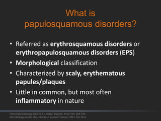
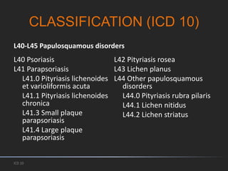
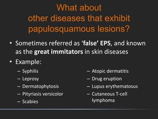
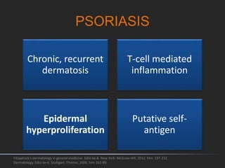
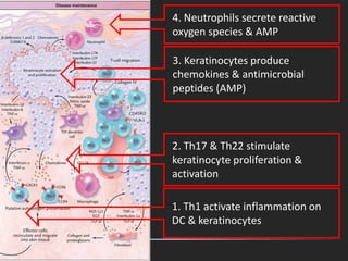
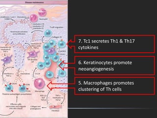
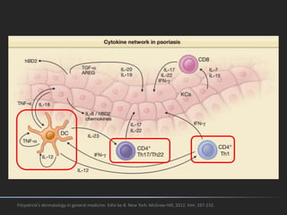
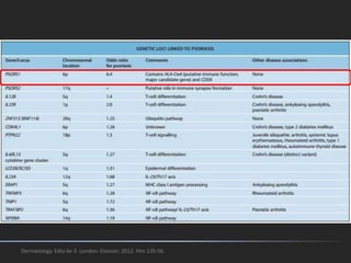
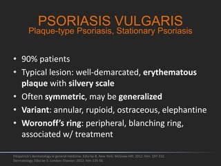
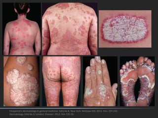
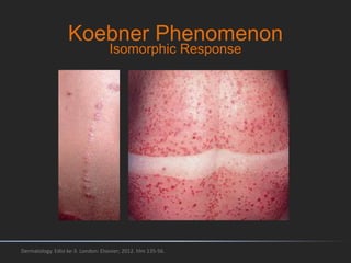
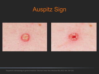
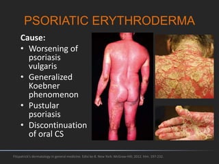
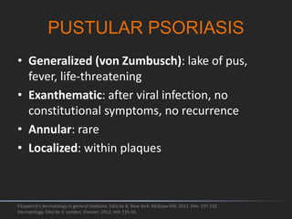
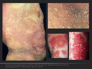
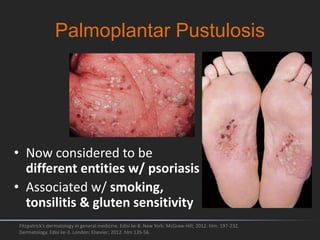
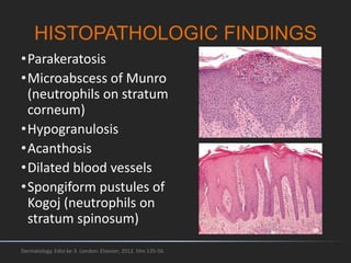
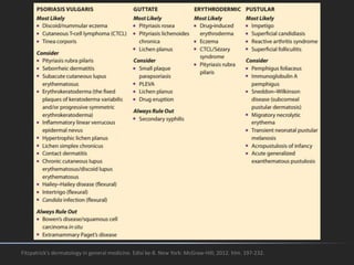
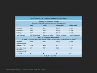
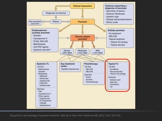
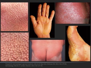
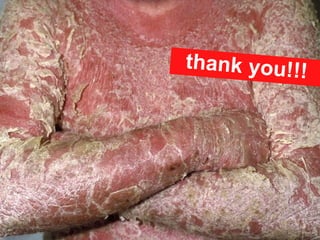
This document discusses papulosquamous disorders, which are characterized by scaly, erythematous papules and plaques. It begins by defining papulosquamous disorders and providing a morphological classification. It then discusses specific disorders like psoriasis, parapsoriasis, and pityriasis rosea. For each disorder, it describes clinical features, pathogenesis, histopathology, and management approaches. The document aims to comprehensively cover papulosquamous disorders through classification, descriptions of individual disorders, and inclusion of relevant images and diagrams.
2. What is
papulosquamous disorders?
• Referred as erythrosquamous disorders or
erythropapulosquamous disorders (EPS)
• Morphological classification
• Characterized by scaly, erythematous
papules/plaques
• Little in common, but most often
inflammatory in nature
Clinical dermatology. Edisi ke-5. London: Elsevier; 2010. hlm. 264-334.
Dermatology secrets plus. Edisi ke-4. London: Elsevier; 2011. hlm 50-6.
4. What about
other diseases that exhibit
papulosquamous lesions?
• Sometimes referred as ‘false’ EPS, and known
as the great immitators in skin diseases
• Example:
– Syphilis
– Leprosy
– Dermatophytosis
– Pityriasis versicolor
– Scabies
– Atopic dermatitis
– Drug eruption
– Lupus erythematosus
– Cutaneous T-cell
lymphoma
9. Fitzpatrick's dermatology in general medicine. Edisi ke-8. New York: McGraw-Hill; 2012. hlm. 197-232.
Normal skin (healthy
individual): Langerhans cells
(L), immature dendritic cells
(D), memory T cells (T)
Normal-appearing psoriatic
skin: slight capillary dilatation
& curvature, mononuclear
cells & mast cells (M),
epidermal thickness ↑
10. Developing lesion:
Dermis: capillary dilatation &
tortuosity, macrophages (MP), T &
M degranulation (small arrows) ↑↑↑
Epidermis: thickness ↑, prominent
rete pegs, spotty loss of the
granular layer & parakeratosis. L
begin to exit the epidermis,
inflammatory dendritic epidermal
cells (I) & CD8+ T cells (8) begin to
enter the epidermis
Fitzpatrick's dermatology in general medicine. Edisi ke-8. New York: McGraw-Hill; 2012. hlm. 197-232.
11. Fully developed lesion:
Dermis: 10-fold increase in blood
flow, M under the basement
membrane & dermal T (mainly
CD4+) making contact with
maturing D ↑
Epidermis: 10-fold keratinocyte
hyperproliferation, loss of the
granular layer, overlying
parakeratosis, CD8+ T cells ↑,
Munro’s microabscesses
Fitzpatrick's dermatology in general medicine. Edisi ke-8. New York: McGraw-Hill; 2012. hlm. 197-232.
12. Dermatology. Edisi ke-3. London: Elsevier; 2012. hlm 135-56.
Psoriasis. Nestle FO, Kaplan DH, Barker J. N Engl J Med 2009; 361:496-509July 30, 2009. DOI: 10.1056/NEJMra0804595
13. 1. Self-DNA & RNA from
stressed keratinocytes form
complexes w/ cathelicidin LL-37
2. Plasmacytoid dendritic cell
(pDC) produce IFNα to activate
dermal DC
3. dDC promote differentiation
of naïve T cells to Th1, Th17 &
Th22 in lymph nodes
14. 1. Th1 activate inflammation on
DC & keratinocytes
2. Th17 & Th22 stimulate
keratinocyte proliferation &
activation
4. Neutrophils secrete reactive
oxygen species & AMP
3. Keratinocytes produce
chemokines & antimicrobial
peptides (AMP)
24. • Generalized, small papules/plaques (3-15
mm) with adherent scales
• Children > adults
• Often preceded by streptococcal pharyngitis
(antibiotics are not beneficial)
• May have spontaneous remission in children
Fitzpatrick's dermatology in general medicine. Edisi ke-8. New York: McGraw-Hill; 2012. hlm. 197-232.
Dermatology. Edisi ke-3. London: Elsevier; 2012. hlm 135-56.
GUTTATE PSORIASIS
Eruptive Psoriasis
26. INVERSE PSORIASIS
Fitzpatrick's dermatology in general medicine. Edisi ke-8. New York: McGraw-Hill; 2012. hlm. 197-232.
Dermatology. Edisi ke-3. London: Elsevier; 2012. hlm 135-56.
Flexural Psoriasis
• Located on major skin folds
• Scaling: minimal/absent!!!
27. SEBOPSORIASIS
Fitzpatrick's dermatology in general medicine. Edisi ke-8. New York: McGraw-Hill; 2012. hlm. 197-232.
Dermatology. Edisi ke-3. London: Elsevier; 2012. hlm 135-56.
• Common
• Located on seborrheic areas
28. PSORIATIC ERYTHRODERMA
Fitzpatrick's dermatology in general medicine. Edisi ke-8. New York: McGraw-Hill; 2012. hlm. 197-232.
Cause:
• Worsening of
psoriasis
vulgaris
• Generalized
Koebner
phenomenon
• Pustular
psoriasis
• Discontinuation
of oral CS
29. • Generalized (von Zumbusch): lake of pus,
fever, life-threatening
• Exanthematic: after viral infection, no
constitutional symptoms, no recurrence
• Annular: rare
• Localized: within plaques
Fitzpatrick's dermatology in general medicine. Edisi ke-8. New York: McGraw-Hill; 2012. hlm. 197-232.
Dermatology. Edisi ke-3. London: Elsevier; 2012. hlm 135-56.
PUSTULAR PSORIASIS
30. Fitzpatrick's dermatology in general medicine. Edisi ke-8. New York: McGraw-Hill; 2012. hlm. 197-232.
Dermatology. Edisi ke-3. London: Elsevier; 2012. hlm 135-56.
31. Palmoplantar Pustulosis
Fitzpatrick's dermatology in general medicine. Edisi ke-8. New York: McGraw-Hill; 2012. hlm. 197-232.
Dermatology. Edisi ke-3. London: Elsevier; 2012. hlm 135-56.
• Now considered to be
different entities w/ psoriasis
• Associated w/ smoking,
tonsilitis & gluten sensitivity
32. Acrodermatitis Continua
Fitzpatrick's dermatology in general medicine. Edisi ke-8. New York: McGraw-Hill; 2012. hlm. 197-232.
Dermatology. Edisi ke-3. London: Elsevier; 2012. hlm 135-56.
• Rare
• Pustular eruptions of the tips of the fingers &
toes, slowly extends proximally
• Nail destruction & distal phalanx atrophy
of Hallopeau
33. NAIL PSORIASIS
Fitzpatrick's dermatology in general medicine. Edisi ke-8. New York: McGraw-Hill; 2012. hlm. 197-232.
Dermatology. Edisi ke-3. London: Elsevier; 2012. hlm 135-56.
• 40% patients
• Pitting nail, oil spot, salmon patch, splinter
hemorrhage, subungual hyperkeratosis,
onycholysis
34. GEOGRAPHIC TONGUE
Fitzpatrick's dermatology in general medicine. Edisi ke-8. New York: McGraw-Hill; 2012. hlm. 197-232.
• Relatively common in normal individuals
• Local loss of filiform papillae
Benign Migratory Glossitis
40. TOPICAL TREATMENTS
• Absorption: ointment > cream
• Corticosteroids (CS): 1st line, long-term use may
cause tachyphylaxis & many side effects
• Vitamin D analogs: no tachyphylaxis, often used
alternately/combined w/ CS
• Coal tar: often combined with salicylic acid,
irritating, stains clothing, carcinogenic
• Salicylic acid: keratolytic agent, enhances
absorption
• Emollients
Fitzpatrick's dermatology in general medicine. Edisi ke-8. New York: McGraw-Hill; 2012. hlm. 197-232.