7. Fungal infections
•Download as PPTX, PDF•
3 likes•408 views

Dermatology Power point slides Useful, Important & Easy Way To Understand The Chapter

Recommended
More Related Content
What's hot
What's hot (20)
Similar to 7. Fungal infections
Similar to 7. Fungal infections (20)
More from Dr.Bijay Yadav
More from Dr.Bijay Yadav (20)
Recently uploaded
Recently uploaded (20)
7. Fungal infections
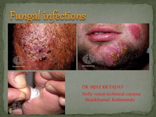
- 1. DR. BIJAY KR.YADAV Holly vision technical campus Shankhamul, Kathmandu
- 2. Dermatophytes are a group of fungi invading the dead keratin of skin, hair, nails. The infection can be Anthropophilic (spread from person-to-person), Zoophilic ( animal to person), Geophilic(soil to person). • Fungal infections are also called Mycosis. • 4th most common skin disease in the world. • Causative organism :- A) Epidermophyton B) Trichophyton C) Microsporum
- 3. Depending on the site: Tinea capitis : Head Tinea faceii : Face Tinea barbae : Beard area Tinea corporis : Body Tinea cruris : Groin area Tinea manuum : Hand Tinea pediis : Leg Tinea unguium ( onychomycosis) : Nails Tinea incognito : Fungal infection that can’t be recognize by area naked eye, which should be diagnosed by KOH preparation.
- 4. Skin scraping over a glass slides Add 2-3 drops of KOH (10%) Wait for 10-15 mins Observe under microscope Fungal Hype : topical in skin or environment Yeast : Inside body, blood, organ
- 5. KOH preparation- for detection for fungal hyphae, spores. ( sample collection from plucked hairs, skin scraping, nail clipping) Wood’s lamp-365 nm used mainly for the diagnosis of T. capitis Fungal culture- Sabouraud glucose agar
- 7. 1. Superficial mycosis :- Infection is limited to outermost layers of skin/hair. e.g tinea versicolar 2. Cutaneous mycosis :- Infection extends deeper into the epidermis & also invade into hair & nail e.g Dermatophytes 3. Subcutaneous mycosis :- Infection involves dermis, subcutaneous tissue, muscles or fascia, usually the infection is introduced by piercing injury. 4. Systemic mycosis :- Infection involves the internal organ & viscera as lungs, liver, brain etc
- 8. Risk factors : i. Inappropriate antibiotic use ( topical /systemic) ii. Lack of proper hygiene/sanitation iii. Weakened immune system iv. Other chronic diseases as Diabetes Mellitus/ Liver disease/ kidney disease HIV/AIDS Drugs Steroids
- 9. 1. Dermatophytes can survive solely off of human stratum corneum, which provides a source of nutrition. 2. Dermatophyte infections involve three main steps: Adherence to keratinocytes, Penetration through and between cells, Development of a host response
- 10. Tinea capitis is a dermatophytosis of the scalp and associated hair. It may be caused by any pathogenic dermatophyte from the genera Trichophyton and Microsporum excepting T. concentricum. most commonly found in children aged 3 to 14 years old. Is transmitted from the sharing of fomites such as cap, comp, scarf
- 11. Classification: 1. Non-inflammatory- Grey patch and Black-dot appear as well-defined, round hyperkeratotic, scaly areas of alopecia, due to the breaking off of hairs Remaining hairs and scales exhibit green fluorescence under Wood's light 2. Inflammatory- Kerion, Favus The spectrum of inflammation ranges from a pustular folliculitis to kerion Grey patch Black dot
- 12. Kerion It is a boggy, inflammatory mass studded with broken hairs and follicular orifices oozing with pus. Caused by : M. canis and M. gypseum. results in scarring alopecia. Inflammatory lesions are usually pruritic, and may be associated with pain, posterior cervical lymphadenopathy, fever. Favus Tinea favosa or favus (Latin, “honeycomb”) is a chronic dermatophyte infection of the scalp, glabrous skin, and/or nails characterized by thick yellow crusts (scutula) within the hair follicles, which lead to scarring alopecia
- 13. Griseofulvin remains the drug of choice, (esp Microsporum) although oral therapy with terbinafine, itraconazole, or fluconazole appears to have similar efficacy. Ultramicrosize griseofulvin treatment schedules (10-20 mg/kg) : Adults: 250 mg by mouth twice daily for 6 to 12 weeks. Children: 20 mg/kg of body weight for 6 to 12 weeks Alternative Itraconazole can be used in children as continuous therapy at a dose of 3 to 5 mg/kg daily for four to six weeks or as pulse therapy at a dose of 5 mg/kg daily for one week each month for two to three months. Fluconazole 6 mg/kg/day for six weeks in children
- 14. Tinea pedis Tinea corporis Tinea Cruris ( Athlet’s foot)
- 15. Caused by malassezia furfur. Most common in the teenage and adolescent age group. Clinical features : The lesions are small, multiple macules The lesion are usually hypopigmented then surrounding normal skin Sometimes lesions may be reddish brown Itching is present The lesion may have papulo-vesicular margin where the lesion is present in the area having skin folds.
- 16. Lab investigations : i. CBC : ↑ Lymphocytes (15-20%) / Basophils(0-1%) ii. Random blood sugar iii. Renal or liver function tests iv. KOH Treatment : A. General measures : i. Improvemenet of personal hygiene ii. Proper use of systematic or topical antibiotics iii. Daily changing of undergarments
- 17. B. Medical treatment : 1. Antifungal : i. Topical Used for skin lesion Can be used as single agent if the lesion is not extensive. E.g Cortimazole 1 % Miconazole 2% Ketoconazole 2% Terbinafine 2% ii. Systemic : Used when the lesions are extensive & if systemic disease is present. E.g fluconazole 150 mg once a week for 6 months Itraconazole 200 mg for 2 wks Terbinafine 250 mg for 2 wks
- 18. 2. Keratolytic agents : Whitfield ointment (2% benzoic acid + 1% salicyclic acid ) used to dissolve upepermost layer of skins so it helps in cleaning the infection & better drug penetration. 3. Antihistamine : given for itching Cetrizine 10 mg OD HS Levocetrizine 5 mg OD HS
- 19. Extensive spread of lesion Secondary bacterial infection Sepsis Septic shock Presence of risk factors like DM, chronic renal disease, liver disease
- 20. Introduction : Candidiasis is an infection caused by a group of yeast. There are more than 20 species of Candida, the most common being Candida albicans. These fungi live on all surfaces of our bodies. grow particularly in warm and moist area Predisposing factors : Moisture-area of occlusion & prolonged immersion in water Obesity Pregnancy and OCP use weakened immune systems due to such conditions as HIV/AIDS, Diabetes taking steroid medications chemotherapy.
- 21. A. Acute mucocutaneous candidiasis I. Candidal paronychia II. Genital candidiasis III. Oral candidiasis IV. ballanitis B. Chronic mucocutaneous candidiasis C. Systemic candidiasis- seen in severe illness, leucopenia, immunosupression
- 22. Oropharangeal candidiasis: Symptoms are a cottony feeling in the mouth, loss of taste, and sometimes pain on eating and swallowing. Presents as whitish papules & plaques which can be easily scraped revealing erythematous base Esophagitis- The hallmark is odynophagia
- 23. symptoms are primarily itching and discharge. Dyspareunia, dysuria, and vaginal irritation also may be present. Physical examination -vaginal erythema and discharge, which is classically white and curd-like but may be watery.
- 24. Balanitis Balanitis can present as white patches on the penis in association with severe burning and itching. Paronychia is an inflammation involving the lateral and posterior fingernail folds. Predisposing factors include overzealous manicuring, nail biting, thumbsucking, diabetes mellitus, and occupations in which the hands are frequently immersed in water. Can be acute and chronic. Acute paronychia is usually bacterial, characterized by the onset of pain and erythema of the posterior or lateral nail folds, with subsequent development of a. superficial abscess. Chronic paronychia candida may be the sole pathogens, or be found with other opportunists such as proteus or pseudomonas.
- 25. Candidal intertrigo- topical azoles Candidal paronychia acute- systemic antibiotics chronic- oral antifungal, topical azoles Genital candidiasis- imidazole pessary or oral azole