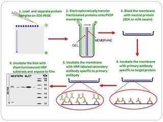
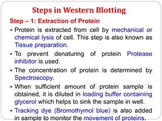
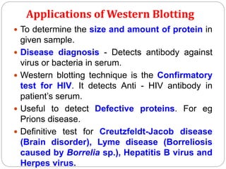
Dr. J. P. Saranraj
IMMUNOBLOTTING –
WESTERN BLOTTING
WESTERN BLOTTING
 Western blotting, also known as
Immunoblotting or Protein blotting, is a core
technique in cell and molecular biology.
 The term “blotting” refers to the transfer of
biological samples from a Gel to a Membrane
and their subsequent detection on the surface of
the membrane.
 It was first described by Towbin et al. (1979).
 It is used to detect the presence of a specific
protein in a complex mixture extracted from
cells or tissue.
Steps in Western Blotting
Step – 1: Extraction of Protein
 Protein is extracted from cell by mechanical or
chemical lysis of cell. This step is also known as
Tissue preparation.
 To prevent denaturing of protein Protease
inhibitor is used.
 The concentration of protein is determined by
Spectroscopy.
 When sufficient amount of protein sample is
obtained, it is diluted in loading buffer containing
glycerol which helps to sink the sample in well.
 Tracking dye (Bromothymol blue) is also added
in sample to monitor the movement of proteins.
Step – 2: Gel Electrophoresis
 The sample is loaded in well of SDS-PAGE.
 The proteins are separated on the basis of
electric charge, isoelectric point, molecular
weight, or combination of these all.
 The small size protein moves faster than large
size protein.
 Protein are negatively charged, so they move
toward positive (Anode) pole as electric current
is applied.
Step – 3: Blotting
 The Nitrocellulose membrane is placed on the
gel. The separated protein from gel get
transferred to nitrocellulose paper by capillary
action. This type of blotting is time consuming
and may take 1-2 days.
 For fast and more efficient transfer of desired
protein from the gel to nitrocellulose paper
electro-blotting can be used.
 In electro-blotting nitrocellulose membrane is
sandwich between gel and cassette of Filter
paper and then electric current is passed through
the gel causing transfer of protein to the
membrane.
Step – 4: Blocking
 Blocking is very important step in Western
blotting.
 Antibodies are also protein so they are likely to
bind the nitrocellulose paper. So before adding
the Primary antibody the membrane is non -
specifically saturated or masked by using casein
or Bovine serum albumin (BSA).
Step – 5: Treatment with Primary Antibody
 The Primary antibody (1° Ab) is specific to
desired protein so it form Ag-Ab complex.
Step – 6: Treatment with Secondary Antibody
 The Secondary antibody is enzyme labelled. For
eg. Alkaline phosphatase or Horseradish
peroxidase is labelled with Secondary antibody.
 Secondary antibody (2° Ab) is antibody against
primary antibody (anti-antibody) so it can bind
with Ag-Ab complex.
Step – 7: Treatment with suitable Substrate
 To visualize the enzyme action, the reaction
mixture is incubated with specific substrate.
 The enzyme convert the substrate to give visible
colored product, so band of color can be
visualized in the membrane.
Applications of Western Blotting
 To determine the size and amount of protein in
given sample.
 Disease diagnosis - Detects antibody against
virus or bacteria in serum.
 Western blotting technique is the Confirmatory
test for HIV. It detects Anti - HIV antibody in
patient’s serum.
 Useful to detect Defective proteins. For eg
Prions disease.
 Definitive test for Creutzfeldt-Jacob disease
(Brain disorder), Lyme disease (Borreliosis
caused by Borrelia sp.), Hepatitis B virus and
Herpes virus.

Immunoblotting

  • 1.
    Dr. J. P.Saranraj IMMUNOBLOTTING – WESTERN BLOTTING
  • 2.
    WESTERN BLOTTING  Westernblotting, also known as Immunoblotting or Protein blotting, is a core technique in cell and molecular biology.  The term “blotting” refers to the transfer of biological samples from a Gel to a Membrane and their subsequent detection on the surface of the membrane.  It was first described by Towbin et al. (1979).  It is used to detect the presence of a specific protein in a complex mixture extracted from cells or tissue.
  • 4.
    Steps in WesternBlotting Step – 1: Extraction of Protein  Protein is extracted from cell by mechanical or chemical lysis of cell. This step is also known as Tissue preparation.  To prevent denaturing of protein Protease inhibitor is used.  The concentration of protein is determined by Spectroscopy.  When sufficient amount of protein sample is obtained, it is diluted in loading buffer containing glycerol which helps to sink the sample in well.  Tracking dye (Bromothymol blue) is also added in sample to monitor the movement of proteins.
  • 5.
    Step – 2:Gel Electrophoresis  The sample is loaded in well of SDS-PAGE.  The proteins are separated on the basis of electric charge, isoelectric point, molecular weight, or combination of these all.  The small size protein moves faster than large size protein.  Protein are negatively charged, so they move toward positive (Anode) pole as electric current is applied.
  • 6.
    Step – 3:Blotting  The Nitrocellulose membrane is placed on the gel. The separated protein from gel get transferred to nitrocellulose paper by capillary action. This type of blotting is time consuming and may take 1-2 days.  For fast and more efficient transfer of desired protein from the gel to nitrocellulose paper electro-blotting can be used.  In electro-blotting nitrocellulose membrane is sandwich between gel and cassette of Filter paper and then electric current is passed through the gel causing transfer of protein to the membrane.
  • 7.
    Step – 4:Blocking  Blocking is very important step in Western blotting.  Antibodies are also protein so they are likely to bind the nitrocellulose paper. So before adding the Primary antibody the membrane is non - specifically saturated or masked by using casein or Bovine serum albumin (BSA).
  • 8.
    Step – 5:Treatment with Primary Antibody  The Primary antibody (1° Ab) is specific to desired protein so it form Ag-Ab complex. Step – 6: Treatment with Secondary Antibody  The Secondary antibody is enzyme labelled. For eg. Alkaline phosphatase or Horseradish peroxidase is labelled with Secondary antibody.  Secondary antibody (2° Ab) is antibody against primary antibody (anti-antibody) so it can bind with Ag-Ab complex.
  • 9.
    Step – 7:Treatment with suitable Substrate  To visualize the enzyme action, the reaction mixture is incubated with specific substrate.  The enzyme convert the substrate to give visible colored product, so band of color can be visualized in the membrane.
  • 10.
    Applications of WesternBlotting  To determine the size and amount of protein in given sample.  Disease diagnosis - Detects antibody against virus or bacteria in serum.  Western blotting technique is the Confirmatory test for HIV. It detects Anti - HIV antibody in patient’s serum.  Useful to detect Defective proteins. For eg Prions disease.  Definitive test for Creutzfeldt-Jacob disease (Brain disorder), Lyme disease (Borreliosis caused by Borrelia sp.), Hepatitis B virus and Herpes virus.