Saajida Sultaana Mahusook
Rocket Immunoelectrophoresis
• Rocket Immunoelectrophoresis is an adaptation of radial
immunodiffusion developed by Laurell.
• It is also known as electroimmunoassay or
electroimmunodiffusion.
• It is called as “rocket electrophoresis” due to the appearance of
the precipitin bands in the shape of cone-like structures (rocket
appearance) at the end of the reaction.
• In rocket immunoelectrophoresis, antigen migrates in an
electric field in a layer of agarose containing an appropriate
antibody.
• The migration of the antigen toward the anode gives rise to
rocket-shaped patterns of precipitation. The area under the
rocket is proportional to antigen concentration.
Objective
• To detect antigen-antibody complexes.
Principle
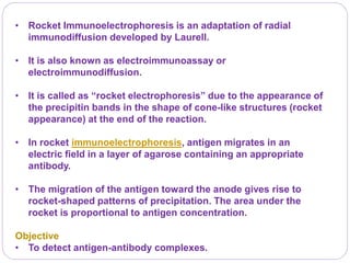
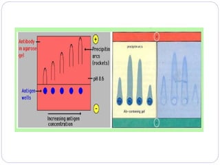
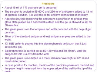
 Rocket immunoelectrophoresis is a quantitative one-dimensional
single electro-immunodiffusion technique. In this method antibody
is incorporated in the gel at a pH value at which the antibodies
remain essentially immobile. Antigen is placed in wells cut in the
gel. Electric current is then passed through the gel, which facilitates
the migration of negatively charged antigens into the agar.
 As the antigen moves out of the well and enters the agarose gel, it
combines with the antibody to form immune complex which
becomes visible. During the initial phase there is considerable
antigen excess over antibody and no visible precipitation occurs.
However, as the antigen sample migrates further through the
agarose gel, more antibody molecules are encountered that
interact with the antigen to form immune complex. This results in
formation of a precipitin line that is conical in shape, resembling a
rocket.
 The greater the amount of antigen loaded in a well, the further the
antigen will have to travel through the gel before it can interact with
sufficient antibody to form a precipitate. Thus, the height of the
Procedure
 About 15 ml of 1 % agarose gel is prepared.
 The solution is cooled to 55-60oC and 250 ml of antiserum added to 13 ml
of agarose solution. It is well mixed for uniform distribution of antibody.
 Agarose solution containing the antiserum is poured on to grease free
glass plate placed on a horizontal surface and the gel is allowed to set for
30 minutes.
 The glass plate is on the template and wells punched with the help of gel
puncher.
 10 ml of the standard antigen and test antigen samples are added to the
wells.
 1X TBE buffer is poured into the electrophoresis tank such that it just
covers the gel.
 Electrophoresis is carried out at 80-120 volts and 60-70 mA, until the
antigen travels 3-4 cms from the well.
 The glass plate is incubated in a moist chamber overnight at 37o C and
results interpreted.
 In case positive for reaction, the tips of the precipitin peaks are marked and
the peak height measured from the upper edge of the well to the tip of the
Results
 A precipitation ‘rocket’ spreading out from the loading well
indicate positive reaction or specific antigen-antibody
reaction due to the presence of antibody specific to the
antigen.
 The absence of the precipitation indicates no reaction or the
absence of any corresponding antibody – antigen.
 The height of the rocket, and its area are directly proportional
to the amount of antigen in the sample, that is, the height of
the precipitin peak depends on the concentration of antigens
loaded in the corresponding wells.
Applications
 Rocket electrophoresis is used mainly for quantitative
estimation of antigen in the serum.
 The method has been used for quantization of human
serum proteins before automated methods became
available.
 Determining the concentration of a specific protein in
a protein mixture.
 In estimation of immunoglobulin protease activity.
 Studies dealing with antigenic relationships between
organisms.
Reference
 https://microbenotes.com/immunoelectrophoresis-
principle-procedure-results-and-applications-
advantages-and-limitations/
 https://microbenotes.com/rocket-
immunoelectrophoresis/
THANK YOU

Rocket immunoelectrophoresis

  • 1.
    Saajida Sultaana Mahusook RocketImmunoelectrophoresis
  • 2.
    • Rocket Immunoelectrophoresisis an adaptation of radial immunodiffusion developed by Laurell. • It is also known as electroimmunoassay or electroimmunodiffusion. • It is called as “rocket electrophoresis” due to the appearance of the precipitin bands in the shape of cone-like structures (rocket appearance) at the end of the reaction. • In rocket immunoelectrophoresis, antigen migrates in an electric field in a layer of agarose containing an appropriate antibody. • The migration of the antigen toward the anode gives rise to rocket-shaped patterns of precipitation. The area under the rocket is proportional to antigen concentration. Objective • To detect antigen-antibody complexes.
  • 3.
    Principle  Rocket immunoelectrophoresisis a quantitative one-dimensional single electro-immunodiffusion technique. In this method antibody is incorporated in the gel at a pH value at which the antibodies remain essentially immobile. Antigen is placed in wells cut in the gel. Electric current is then passed through the gel, which facilitates the migration of negatively charged antigens into the agar.  As the antigen moves out of the well and enters the agarose gel, it combines with the antibody to form immune complex which becomes visible. During the initial phase there is considerable antigen excess over antibody and no visible precipitation occurs. However, as the antigen sample migrates further through the agarose gel, more antibody molecules are encountered that interact with the antigen to form immune complex. This results in formation of a precipitin line that is conical in shape, resembling a rocket.  The greater the amount of antigen loaded in a well, the further the antigen will have to travel through the gel before it can interact with sufficient antibody to form a precipitate. Thus, the height of the
  • 5.
    Procedure  About 15ml of 1 % agarose gel is prepared.  The solution is cooled to 55-60oC and 250 ml of antiserum added to 13 ml of agarose solution. It is well mixed for uniform distribution of antibody.  Agarose solution containing the antiserum is poured on to grease free glass plate placed on a horizontal surface and the gel is allowed to set for 30 minutes.  The glass plate is on the template and wells punched with the help of gel puncher.  10 ml of the standard antigen and test antigen samples are added to the wells.  1X TBE buffer is poured into the electrophoresis tank such that it just covers the gel.  Electrophoresis is carried out at 80-120 volts and 60-70 mA, until the antigen travels 3-4 cms from the well.  The glass plate is incubated in a moist chamber overnight at 37o C and results interpreted.  In case positive for reaction, the tips of the precipitin peaks are marked and the peak height measured from the upper edge of the well to the tip of the
  • 6.
    Results  A precipitation‘rocket’ spreading out from the loading well indicate positive reaction or specific antigen-antibody reaction due to the presence of antibody specific to the antigen.  The absence of the precipitation indicates no reaction or the absence of any corresponding antibody – antigen.  The height of the rocket, and its area are directly proportional to the amount of antigen in the sample, that is, the height of the precipitin peak depends on the concentration of antigens loaded in the corresponding wells.
  • 7.
    Applications  Rocket electrophoresisis used mainly for quantitative estimation of antigen in the serum.  The method has been used for quantization of human serum proteins before automated methods became available.  Determining the concentration of a specific protein in a protein mixture.  In estimation of immunoglobulin protease activity.  Studies dealing with antigenic relationships between organisms.
  • 8.