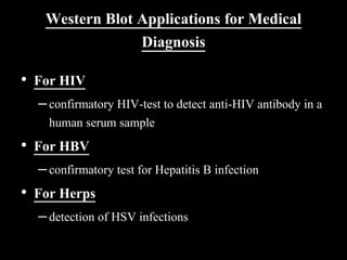
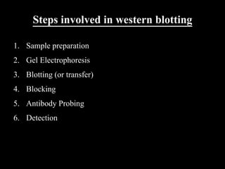
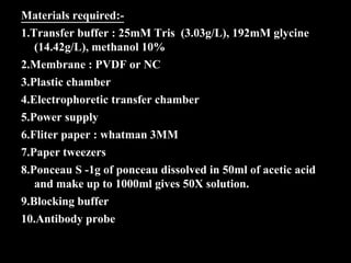
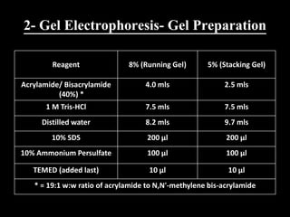
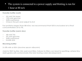
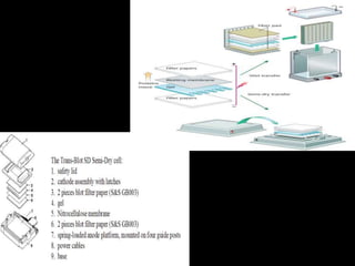
Western blotting is a technique used to detect specific proteins in a complex protein mixture. It involves separating proteins by gel electrophoresis, transferring them to a membrane, and using antibodies to detect the target protein as a band on the membrane. The key steps are sample preparation, gel electrophoresis, blotting, blocking, antibody probing, and detection with a substrate. Western blotting is commonly used for medical diagnosis of conditions like HIV and hepatitis B.
In this slide contains principle, types, methods and application of Western Blotting Technique.
Presented by: T.NIRANJAN REDDY (Department of pharmacology).
RIPER, anantapur
In this slide contains principle, types, methods and application of Western Blotting Technique.
Presented by: T.NIRANJAN REDDY (Department of pharmacology).
RIPER, anantapur
Introduction, the principle of immunofluorescence, Technique, Fluorescent microscope and its components, Application and types of immunofluorescence, Direct and indirect immunofluorescence, FACS (Fluorescence-activated cell sorting), Uses and limitations of Immunofluorescence
Creative Bioarray provides Cell Apoptosis Assays to all of our customers. The process of programmed cell death, or apoptosis, is generally characterized by distinct morphological characteristics and energy-dependent biochemical mechanisms.
https://www.creative-bioarray.com/cell-apoptosis-assays.htm
Western blot is a commonly used method for protein analysis. It can be used for qualitative and semi-quantitative protein analysis. For the accomplishment of the western blot, there are three elements, separation of proteins by size, transferring proteins to a solid support, and marking proteins by primary and secondary antibodies for visualization.
It is the transfer of molecules like proteins or nucleic acids from the gel onto the surface of an immobilizing membrane in the same relative positions as they occur in gel.
Pharmaceutical biotechnology ..in that different blotting techniques such as ELISA , western blotting and southern blotting.There applications and their advantage and disadvantage with their diagrams
Protein Immunoblotting- An Introduction to Western BlottingFatemeh Barantalab
Western blotting is an important technique used in cell and molecular biology. The specificity of the antibody-antigen interaction enables a target protein to be identified from a complex protein mixture. Western blotting can produce qualitative and semi-quantitative data about that protein. This powerpoint will attempt to explain the technique and theory behind western blot.
Cancer cell metabolism: special Reference to Lactate PathwayAADYARAJPANDEY1
Normal Cell Metabolism:
Cellular respiration describes the series of steps that cells use to break down sugar and other chemicals to get the energy we need to function.
Energy is stored in the bonds of glucose and when glucose is broken down, much of that energy is released.
Cell utilize energy in the form of ATP.
The first step of respiration is called glycolysis. In a series of steps, glycolysis breaks glucose into two smaller molecules - a chemical called pyruvate. A small amount of ATP is formed during this process.
Most healthy cells continue the breakdown in a second process, called the Kreb's cycle. The Kreb's cycle allows cells to “burn” the pyruvates made in glycolysis to get more ATP.
The last step in the breakdown of glucose is called oxidative phosphorylation (Ox-Phos).
It takes place in specialized cell structures called mitochondria. This process produces a large amount of ATP. Importantly, cells need oxygen to complete oxidative phosphorylation.
If a cell completes only glycolysis, only 2 molecules of ATP are made per glucose. However, if the cell completes the entire respiration process (glycolysis - Kreb's - oxidative phosphorylation), about 36 molecules of ATP are created, giving it much more energy to use.
IN CANCER CELL:
Unlike healthy cells that "burn" the entire molecule of sugar to capture a large amount of energy as ATP, cancer cells are wasteful.
Cancer cells only partially break down sugar molecules. They overuse the first step of respiration, glycolysis. They frequently do not complete the second step, oxidative phosphorylation.
This results in only 2 molecules of ATP per each glucose molecule instead of the 36 or so ATPs healthy cells gain. As a result, cancer cells need to use a lot more sugar molecules to get enough energy to survive.
introduction to WARBERG PHENOMENA:
WARBURG EFFECT Usually, cancer cells are highly glycolytic (glucose addiction) and take up more glucose than do normal cells from outside.
Otto Heinrich Warburg (; 8 October 1883 – 1 August 1970) In 1931 was awarded the Nobel Prize in Physiology for his "discovery of the nature and mode of action of the respiratory enzyme.
WARNBURG EFFECT : cancer cells under aerobic (well-oxygenated) conditions to metabolize glucose to lactate (aerobic glycolysis) is known as the Warburg effect. Warburg made the observation that tumor slices consume glucose and secrete lactate at a higher rate than normal tissues.
What is greenhouse gasses and how many gasses are there to affect the Earth.moosaasad1975
What are greenhouse gasses how they affect the earth and its environment what is the future of the environment and earth how the weather and the climate effects.
(May 29th, 2024) Advancements in Intravital Microscopy- Insights for Preclini...Scintica Instrumentation
Intravital microscopy (IVM) is a powerful tool utilized to study cellular behavior over time and space in vivo. Much of our understanding of cell biology has been accomplished using various in vitro and ex vivo methods; however, these studies do not necessarily reflect the natural dynamics of biological processes. Unlike traditional cell culture or fixed tissue imaging, IVM allows for the ultra-fast high-resolution imaging of cellular processes over time and space and were studied in its natural environment. Real-time visualization of biological processes in the context of an intact organism helps maintain physiological relevance and provide insights into the progression of disease, response to treatments or developmental processes.
In this webinar we give an overview of advanced applications of the IVM system in preclinical research. IVIM technology is a provider of all-in-one intravital microscopy systems and solutions optimized for in vivo imaging of live animal models at sub-micron resolution. The system’s unique features and user-friendly software enables researchers to probe fast dynamic biological processes such as immune cell tracking, cell-cell interaction as well as vascularization and tumor metastasis with exceptional detail. This webinar will also give an overview of IVM being utilized in drug development, offering a view into the intricate interaction between drugs/nanoparticles and tissues in vivo and allows for the evaluation of therapeutic intervention in a variety of tissues and organs. This interdisciplinary collaboration continues to drive the advancements of novel therapeutic strategies.
Comparing Evolved Extractive Text Summary Scores of Bidirectional Encoder Rep...University of Maribor
Slides from:
11th International Conference on Electrical, Electronics and Computer Engineering (IcETRAN), Niš, 3-6 June 2024
Track: Artificial Intelligence
https://www.etran.rs/2024/en/home-english/
A brief information about the SCOP protein database used in bioinformatics.
The Structural Classification of Proteins (SCOP) database is a comprehensive and authoritative resource for the structural and evolutionary relationships of proteins. It provides a detailed and curated classification of protein structures, grouping them into families, superfamilies, and folds based on their structural and sequence similarities.
2. Introduction
• Blotting is a technique by which a macromolecule such as
DNA, RNA, or protein is resolved in a gel matrix, transferred
to a solid support, and detected with a specific probe
• To identify and characterize specific molecules in a complex
mixture of related molecules
• Some of the more common techniques include:
– Southern blotting (DNA)
– Northern blotting (RNA)
– and Western blotting (for protein)
3. • Western blotting ( immunoblotting or protein blotting) is a
technique used to detect the presence of a specific protein
in a complex protein mixture .
• Western blots have become the analytical tools for the
– Detection of viral proteins
– characterization of monoclonal and Polyclonal antibody
preparations
– and in determining the specificity of the immune response to
viral antigens
4. Western Blot Applications for Medical
Diagnosis
• For HIV
–confirmatory HIV-test to detect anti-HIV antibody in a
human serum sample
• For HBV
–confirmatory test for Hepatitis B infection
• For Herps
–detection of HSV infections
5. • The Western blotting procedure relies upon three key
elements to accomplish this task:
– The separation of protein mixtures by size using gel
electrophoresis
– The efficient transfer of separated proteins to a solid support;
– and the specific detection of a target protein by appropriately
matched antibodies
• The target protein will be visualized as a band on a
blotting membrane, X-ray film, or an imaging system
6. Steps involved in western blotting
1. Sample preparation
2. Gel Electrophoresis
3. Blotting (or transfer)
4. Blocking
5. Antibody Probing
6. Detection
7. Materials required:-
1.Transfer buffer : 25mM Tris (3.03g/L), 192mM glycine
(14.42g/L), methanol 10%
2.Membrane : PVDF or NC
3.Plastic chamber
4.Electrophoretic transfer chamber
5.Power supply
6.Fliter paper : whatman 3MM
7.Paper tweezers
8.Ponceau S -1g of ponceau dissolved in 50ml of acetic acid
and make up to 1000ml gives 50X solution.
9.Blocking buffer
10.Antibody probe
8. 1- Sample Preparation
• All sources of protein, from single cells to whole tissues,
biological fluids and proteins secreted in vitro, are open to analysis
by Western blotting
• In most cases, the cells are harvested, washed, and lysed to release
the target protein
• For best results, all these steps should be carried out on ice
• This will minimize proteolysis, dephosphorylation, and
denaturation, since all begin to occur once the cells are disrupted
9. • The choice of extraction method depends primarily
on the sample and whether the analysis is targeting
all the proteins in a cell or only a component from a
particular subcellular fraction
• The endogenous proteases may be liberated upon
cell disruption and may degrade the target
molecule,
–the sample should be protected during cell disruption
and subsequent purification by the use of a cocktail of
protease inhibitors to avoid uncontrolled protein losses
10. • Numerous methods are available for disrupting cells and
preparing their contents for analysis by Western blotting
Detergent lysis The membranes are solubilized, lysing cells and liberating
their contents
Ultrasonication The sound waves generate a region of low pressure, causing
disruption of the membranes of cells
Freeze/thaw lysis Cells are disrupted by the repeated formation of
ice crystals and the method is usually combined with
enzymatic lysis
Enzymatic digestion The enzymes dissolve cell walls, coats, capsules, capsids, or
other structures
• To ensure that samples are in the proper range of detection for the
assay, and so they can be compared on an equivalent basis, it is
important to know the concentration of total protein in each sample
11. 2- Gel Electrophoresis- Gel Preparation
Reagent 8% (Running Gel) 5% (Stacking Gel)
Acrylamide/ Bisacrylamide
(40%) *
4.0 mls 2.5 mls
1 M Tris-HCl 7.5 mls 7.5 mls
Distilled water 8.2 mls 9.7 mls
10% SDS 200 µl 200 µl
10% Ammonium Persulfate 100 µl 100 µl
TEMED (added last) 10 µl 10 µl
* = 19:1 w:w ratio of acrylamide to N,N'-methylene bis-acrylamide
12. 3- Blotting
• Following gel electrophoresis, the separated protein mixtures are
transferred to a solid support for further analysis
• Transfer can be done in wet or semi-dry conditions
• Semi-dry transfer is generally faster
• Wet transfer is recommended for large proteins, >100 kD
• For both kinds of transfer, the membrane is placed next to the gel
• The two are sandwiched between absorbent materials, and the
sandwich is clamped between solid supports to maintain tight
contact between the gel and membrane
13. • The system is connected to a power supply and blotting is run for
1 hour at 80 mA
14.
15. Blotting Membranes
• The solid support onto which the separated proteins are
transferred is usually of two types, both of which bind
proteins with high affinity:
– Nitrocellulose membrane
• has excellent protein binding and retention capabilities
• is brittle and thus it is usually less effective when blots need to be reused
– Polyvinylidene fluoride (PVDF) membrane
• PVDF demonstrates superior mechanical strength making it suitable for
stripping/reprobing
16. Visualization of proteins in membranes:
Ponceau Red stain
• Ponceau Red is a reversible stain with poor sensitivity
• Ponceau S is compatible with both nitrocellulose and
PVDF membranes
• This is a quick and easy way to visualize proteins
transferred to membranes
• Ponceau S is easily removed with water and is regarded
as a “gentle” treatment that does not interfere with
subsequent immunological detection steps
17. 4- Blocking
• Blocking is a very important step in the immunodetection phase of
Western blotting because it prevents non-specific binding of
antibody to the blotting membrane
• The most commonly used blocking solutions contain 3-5% BSA or
non-fat dried milk in a solution of PBS (phosphate buffered saline)
or TBS (tris buffered saline)
• Often, a small amount of Tween 20 detergent is added to blocking
and washing solutions to reduce background staining, and the
buffer is known as PBST or TBST
18. 5- Antibody Probing
• Usually, Western blotting protocols utilize a non-labeled primary antibody
directed against the target protein
• Wash the membrane several times in TBST while agitating, 5 minutes or
more per wash, to remove residual primary antibody
• A species-specific, labeled secondary antibody directed against the constant
region of the primary antibody is then used
• The secondary antibody serves not only as a carrier of the label but is also a
mechanism to amplify the emitted signals, as many secondary antibodies can
theoretically bind simultaneously to the primary antibody
• Secondary Ab is also diluted according to the manufacturer’s
recommendations and incubated for 1 hour at RT
19. 6- Detection with Substrate
• The most common antibody label used in Western
blots is HRP, a small, stable enzyme with high
specificity and rapid turnover
• The signal is detected when HRP is exposed to a
substrate solution in the final step of the
immunodetection procedure
• Substrate solutions for Western blotting are chemical
reagents that are acted upon by the enzyme to yield a
signal that can be easily measured
• HRP label is typically detected with either
colorimetric or chemiluminescent substrates
20. 6- Detection with Substrate
• Colorimetric substrates for HRP {eg.
Tetramethylbenzidine(TMB)} produce purple/black bands
directly on the surface of the blot
• These substrates are very easy to use and take from a few
minutes to a few hours to produce visible bands
• Detection limits for colorimetric substrates are in the low
nanogram range
21. • More routinely, HRP is used with ECL (enhanced
chemiluminescence) detection
• For ECL detection, the substrate is luminol which is oxidized by
HRP in the presence of H2O2 to produce light
• The emitted light is detected by exposing the Western blot to X-
ray film, or by using a CCD camera for light capture
• The emitted light forms a band on the film, or on the screen of the
imaging system, indicating where the HRP-labeled antibody has
bound to the target protein
• ECL detection of HRP is extraordinarily sensitive, allowing for the
visualization of picogram to femtogram amounts of target protein