PRESENTED BY:
JSP 05903
LT.SHAGUFTA NAZ
Ovulation
Fertilization
Implantation of ovum and
Development of fertilized
zygote
3.
OBJECTIVES
This presentation willenable you to :
Explain the process of ovulation .
Describe the mechanism of fertilization.
Describe the different stages from zygote to
blastocyst
Explain the process of implantation and its normal
site in the uterus.
Describe the stages of zygote development.
4.
CONCEPTION
The term“conception” means to become
pregnant.
Conception or pregnancy occurs when fertilized
ovum embeds in the uterus.
Numerous processes are directly or indirectly
responsible for conception.
GAMETOGENESIS
The process offormation of male and female
gamete.
i. SPERMATOGENESIS-
Process of formation of male gamete
(spermatozoa) in seminiferous tubule of testis.
ii. OOGENESIS-
Process of formation of female gamete
(ovum) in follicles of ovary.
8.
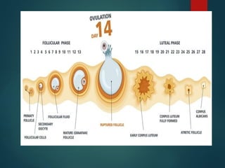
OVULATION:
The processof release of ovum from a mature
Graafian follicle of ovary is called as ovulation.
Fertilisable life span of ovum is 12-24 hours after
ovulation.
A steep increase in the LH levels is known as LH
surge leads to the rupture of the Graafian follicle
to release the secondary oocyte (ovulation).
10.
HORMONAL REGULATION
IN OVULATION:
Follicle Stimulating Hormone(FSH): Stimulates
follicles growth in the ovary.
Luteinising Hormone(LH): LH Surge triggers
ovulation around day 14.
Estrogen: Rises as follicles matures; helps thicken
the endometrium.
Progesterone: Secreted by corpus luteum;
maintains uterine lining post-ovulation.
12.
COPULATION
The processof sexual intercourse which enables
internal fertilization.
The physical act during which the male deposits
sperm into the female reproductive tract.
It is essential for natural conception and
continuation of species.
Fertilisable life span of sperm is 48-72 hrs.
FERTILIZATION
Process offusion of male and female gamete
(sperm and ovum) is called as fertilization.
Site- Normally occurs in the ampulla of uterine
tube within 24 hours after ovulation.
15.
PROCESS OF FERTILIZATION
Ovum is transported to ampulla after ovulation.
Millions of sperms are deposited in vagina during
copulation. Numerous sperms are destroyed in
the acidic medium of vagina. Some of sperms
undergoes “ Capacitation” and reaches vagina.
The acrosomal cap of sperms release “
Hyluronidase enzyme which dissolutes the layer of
corona radiata. Few sperms penetrate the zona
pellucida and only one sperm reaches the
nucleus. After entry of one sperm the membrane
is sealed to avoid further entry of sperm and
hence fertilization occurs and forms single celled
zygote.
17.
DEVELOPMENT OF FERTILIZEDOVUM
Further cell division called “ Cleavage” occurs.
After 30 hours of fertilization 2 celled stage is
reached called as “BLASTOMERE”.
Blastomere continues to divide binary division
through 4, 8, 16, 32 cell stage.
The 16 celled stage resembles a mulberry like ball
or cluster of cells called “ MORULA”.
Morula enters uterine cavity on 4th
day.
Fluid enters into morula and it is now called as “
BLASTOCYST”.
19.
Definition:
Fluid passes intothe morula which separates the
cells of morula and it is called as Blastocyst.
Structure:
it has two parts-
i. Trophoblast (Placenta, Chorion)
ii. Inner Cell Mass (Fetus, Umbilical Cord and Amnion)
BLASTOCYST
20.
Outer layerof blastocyst is called as Trophoblast.
Differentiated into two layers:
i. Syncitiotrophoblast (Outer)
ii. Cytotrophoblast (Inner)
A 3rd
layer called “ Primitive Mesenchyme” is
developed.
Syncitiotrophoblast produces irregular finger like
projections called “ Primary Stem Villi”.
After appearance of primitive mesenchyme , primary
stem villi are renamed as “ Chorionic Villi”.
These villi are differentiated into blood cells and blood
vessels and forms villus .
DIFFERENTIATION OF
TROPHOBLAST
22.
The cellssuspended in the blastocyst is termed as “ Inner Cell
Mass”.
The Inner Cell Mass is differentiated into bilaminar germ layer-
i.Ectoderm
ii.Endoderm
A 3rd
germ layer appears during 3rd
week called mesoderm.
And now bilaminar germ layer becomes trilaminar germ layer.
Two cavities appears one on each side of bilaminar germ
layer-
i. Amniotic Cavity (Filled with amniotic fluid)
ii. Yolk Sac (Incorporated into gut)
DIFFERENTIATION OF INNER CELL
MASS
24.
ECTODERM: Centraland Peripheral Nervous System,
Pituitary Gland, Epidermis of skin with its appendages, Salivary
glands, Mucus lining of mouth, nostril and anus.
MESODERM: Bones, Cartilages, Muscles, Cardiovascular
System , Kidney, Gonads, Suprarenal Glands, Spleen, Most of
genital tracts, lining of Pericardium, Peritoneum, Pleura.
ENDODERM: Liver, Gall Bladder, Pancreas, Epithelial lining of
GI Tract, Respiratory Tract, Mucus lining of Urinary Bladder and
Urethra.
EMBRYO IS DIFFRENTIATED AS HUMAN AT 8TH
WEEK.
GERM LAYER
25.
IMPLANTATION
The blastocystattaching itself to uterine wall or
lining is called as implantation.
It penetrates in the compact layer of decidua
near the fundus.
It occurs on the 6th
day of fertilization and
completed by 10th
to 11th
day.
The deeper penetration of blastocyst into
decidua is called as “ Interstitial implantation”.
DEVELOPMENT OF
FERTILIZED ZYGOTE
WEEK 1-2: Cleavage, blastocyst formation,
implantation.
WEEK 3-8(embryonic stage): Major organs and
systems begin to form.
WEEK 9-Birth (fetal stage): Growth and further
development of organs and body structure.
By the end of the 9th
month, the fetus is ready for
birth.
30.
REFERENCES
Dutta D.C,“Textbook Of Obstetrics”, New Central Book
Agency(P)LTD, 6th
edition, Pg.28-29
Jacob Annamma, “A Comprehensive Textbook of Midwifery”,
Jaypee Brothers Medical Publishers(P)LTD, 2nd
edition, Pg.75-78
Myles, “ Textbook for Midwives”, Churchill Livingstone
Publishers, 13th
edition, Pg.143-147
Editor's Notes
#11 Estrogen in turn stimulates the secretion of GnRH. FSH also stimulates other hormones from the anterior lobe of the pituitary ● GnRH is secreted by the hypothalamus gland. LH stimulates the corpus luteum to secrete progesterone.increasing levels of progesterone inhibit the release of GnRH, which in turn inhibits the release of FSH, LH and progesterone itself
#14 Capacitation- Physiochemical change in sperm by which it becomes hypermotile and able to bind and fertilise ovum.


![4-EMBRYOLOGICAL_DEVELOPMENT_OF_BODY_TISSUES,_ORGANS_AND_SYSTEMS.[1].pptx](https://cdn.slidesharecdn.com/ss_thumbnails/4-embryologicaldevelopmentofbodytissuesorgansandsystems-230811134542-e6d1c32e-thumbnail.jpg?width=640&height=640&fit=bounds)


