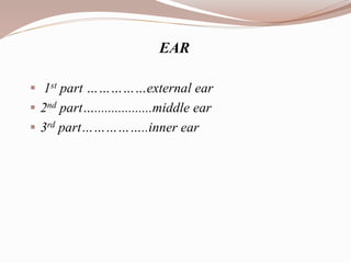
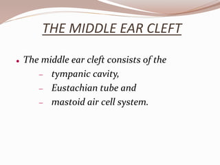
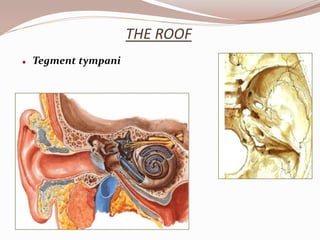
The document describes the anatomy and structures of the human ear. It is divided into three main parts:
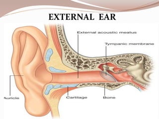
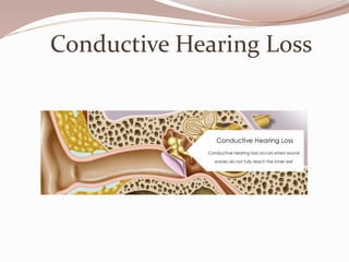
1) The outer ear or external ear collects sound waves and directs them into the middle ear.
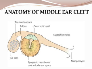
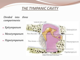
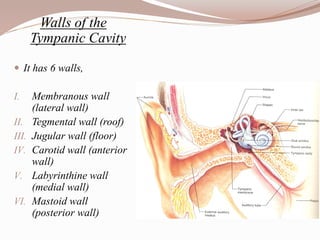
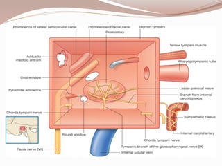
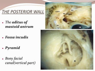
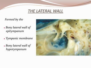
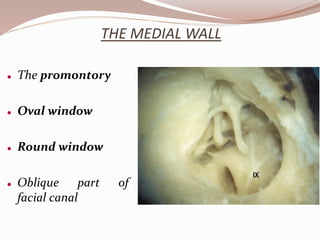
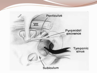
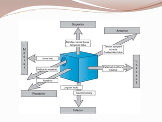
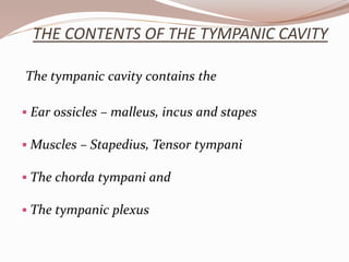
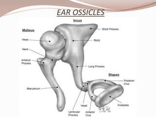
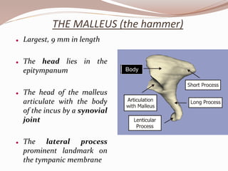
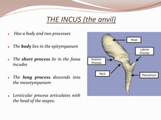
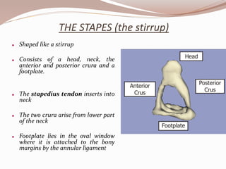
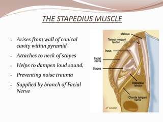
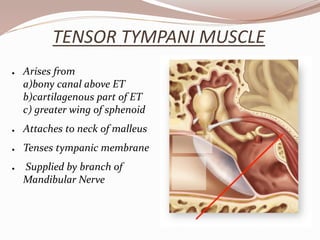
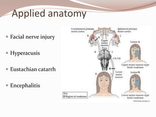
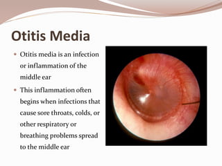
2) The middle ear contains the tympanic cavity with the ossicles (malleus, incus, stapes) that vibrate in response to sound and transmit the vibrations into the inner ear. It also contains muscles and nerves.
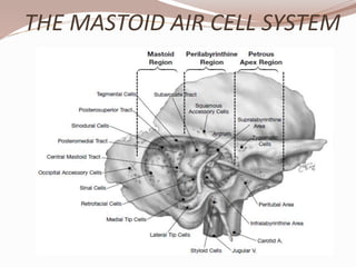
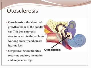
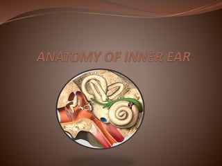
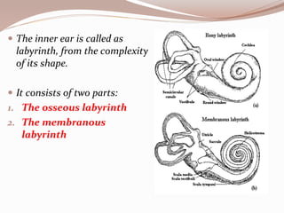
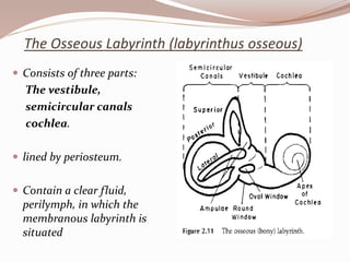
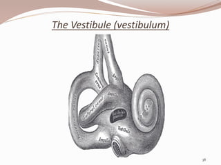
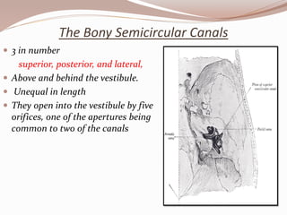
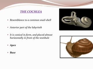
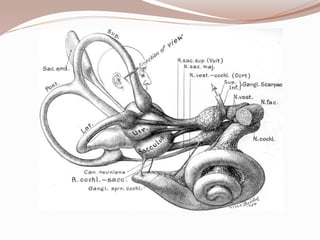
3) The inner ear or labyrinth contains the bony and membranous structures including the cochlea, vestibule and semicircular canals that sense sound and balance. The cochlea converts sound waves into neural signals that


![PERI-PROSTHETIC FRACTURE NAIL-PLATE CONSTRUCT [NPC].pptx](https://cdn.slidesharecdn.com/ss_thumbnails/drarunkumardrmohamedashrafperiprostheticfrasturenail-plateconstructnpc-260209164459-7e9d15a1-thumbnail.jpg?width=640&height=640&fit=bounds)
