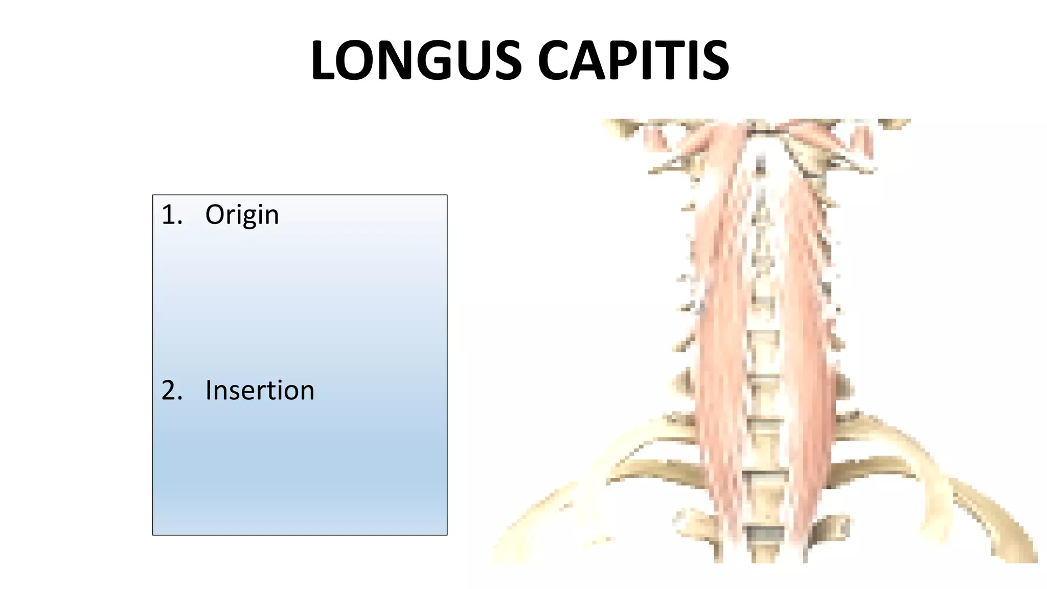
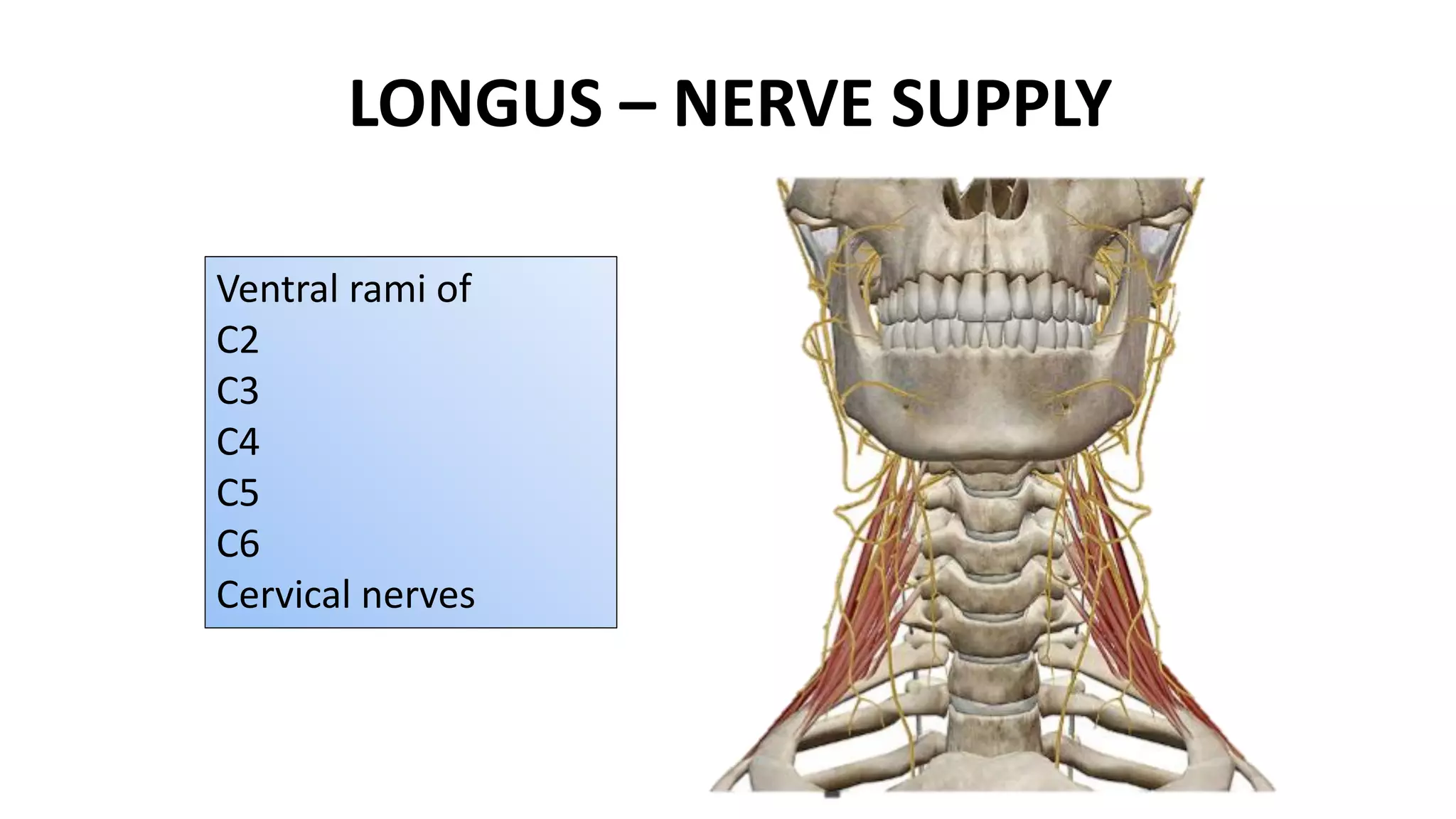
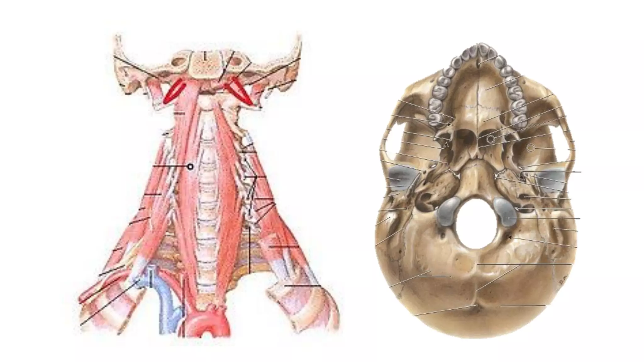
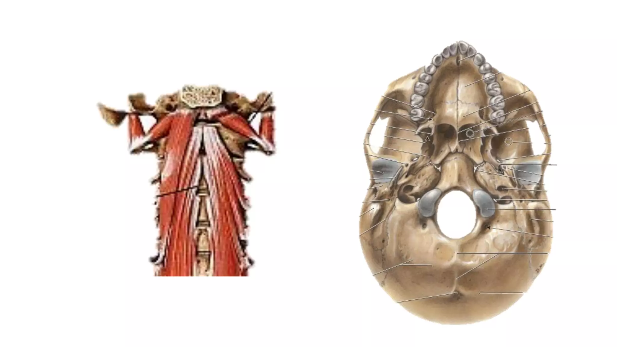
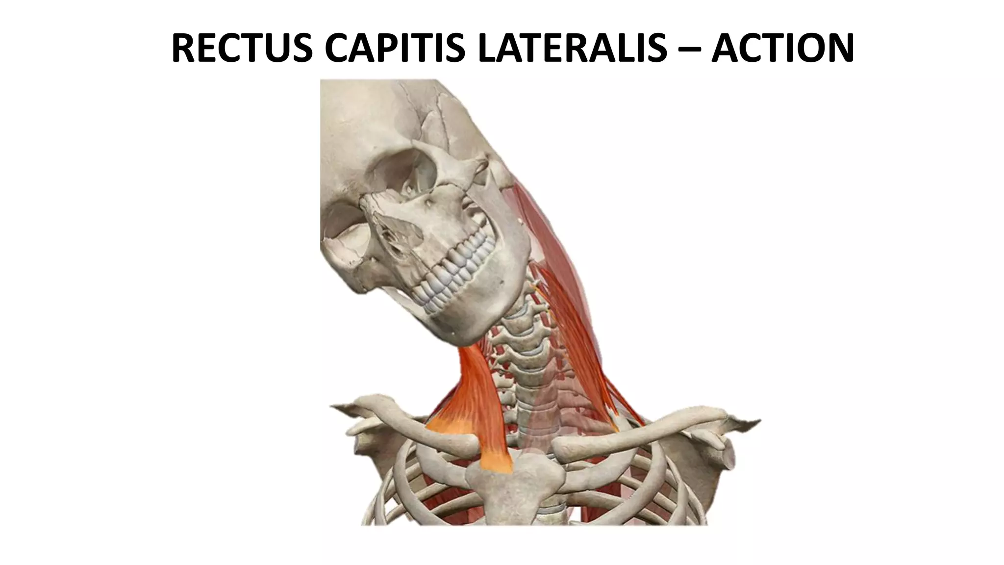
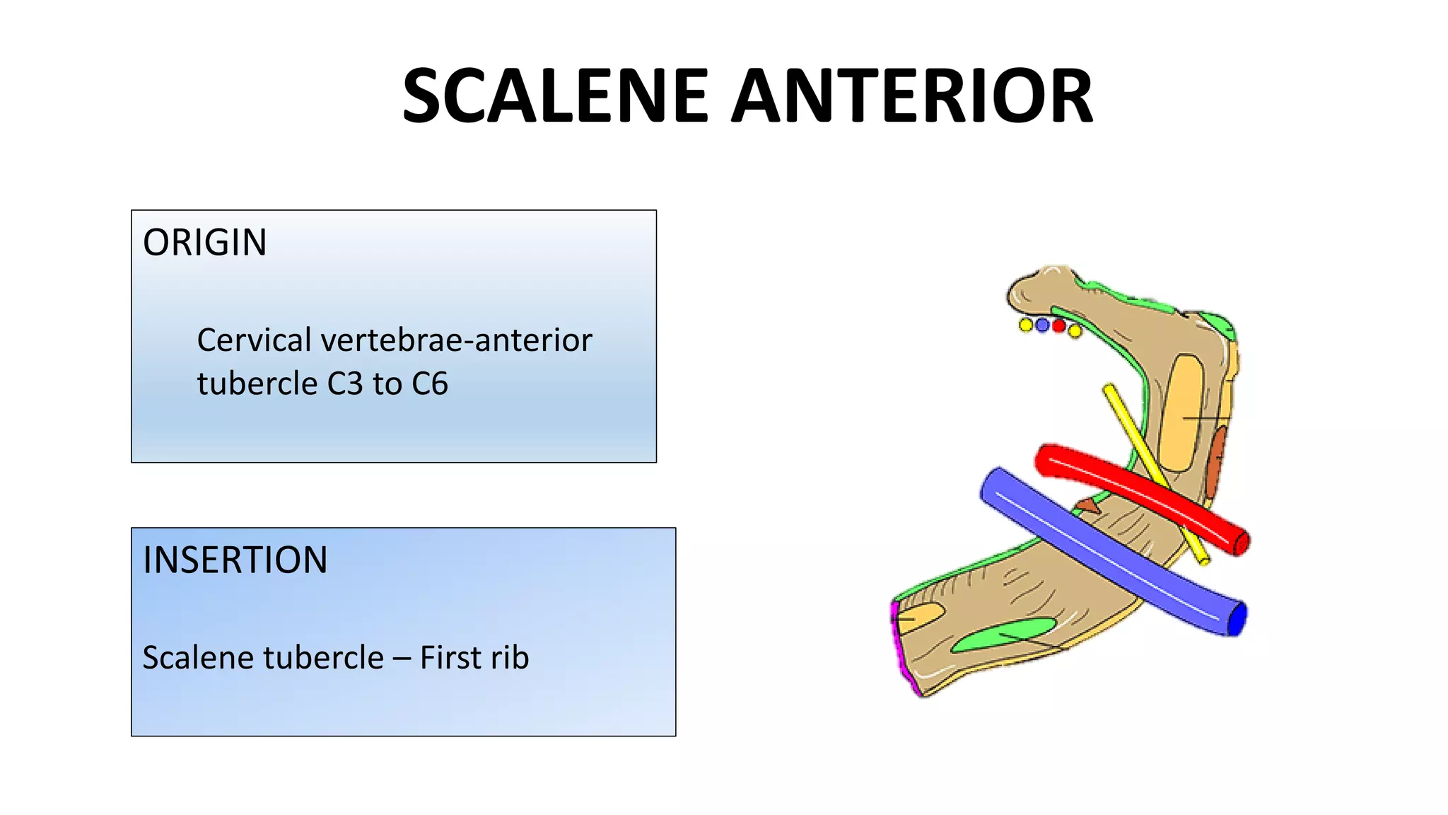
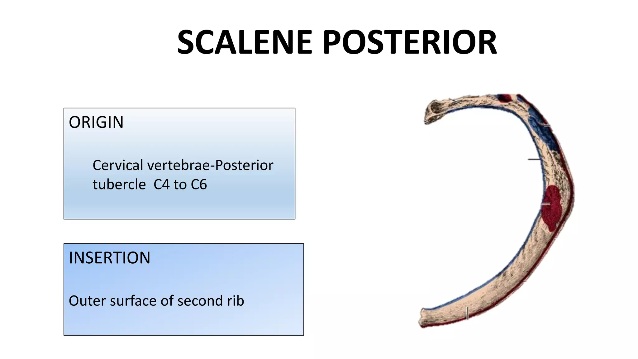
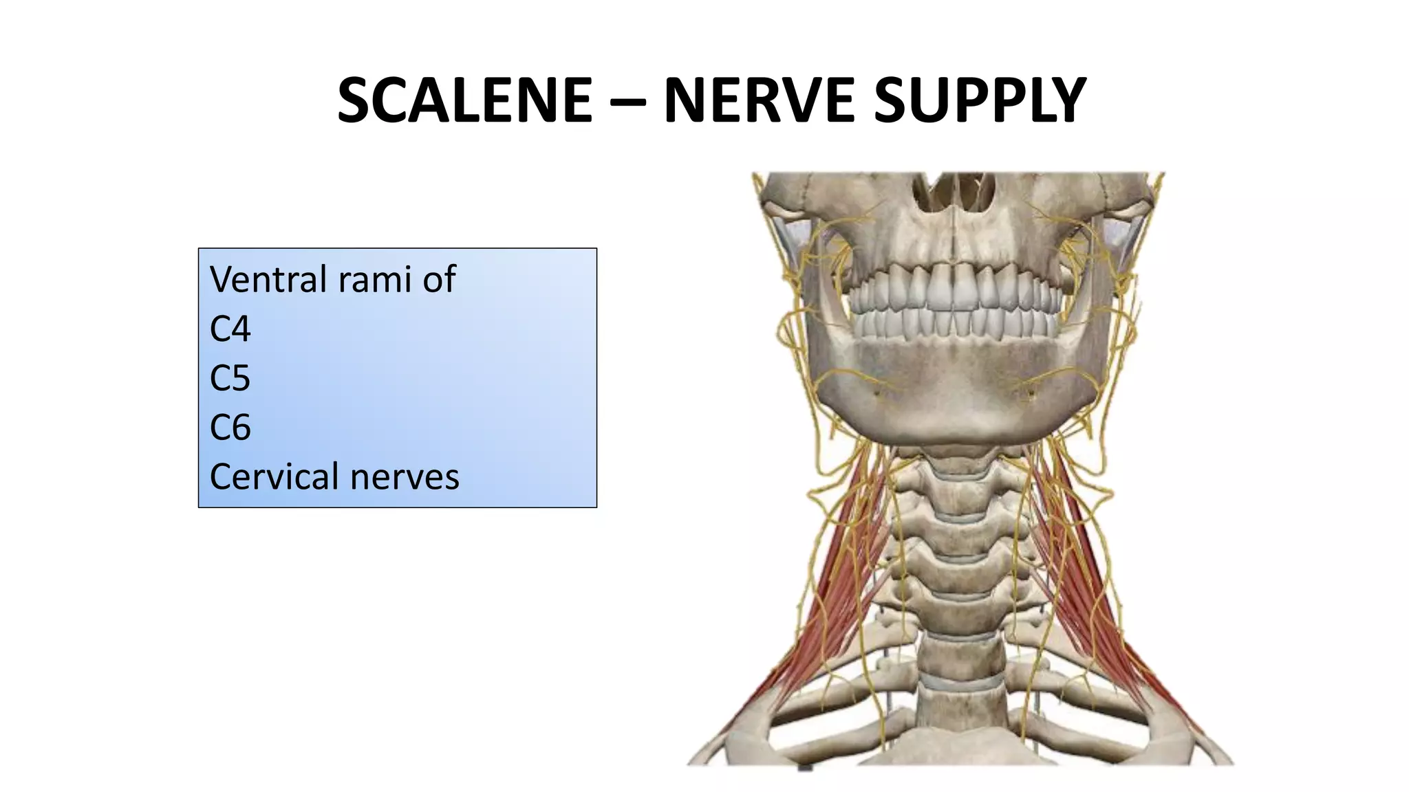
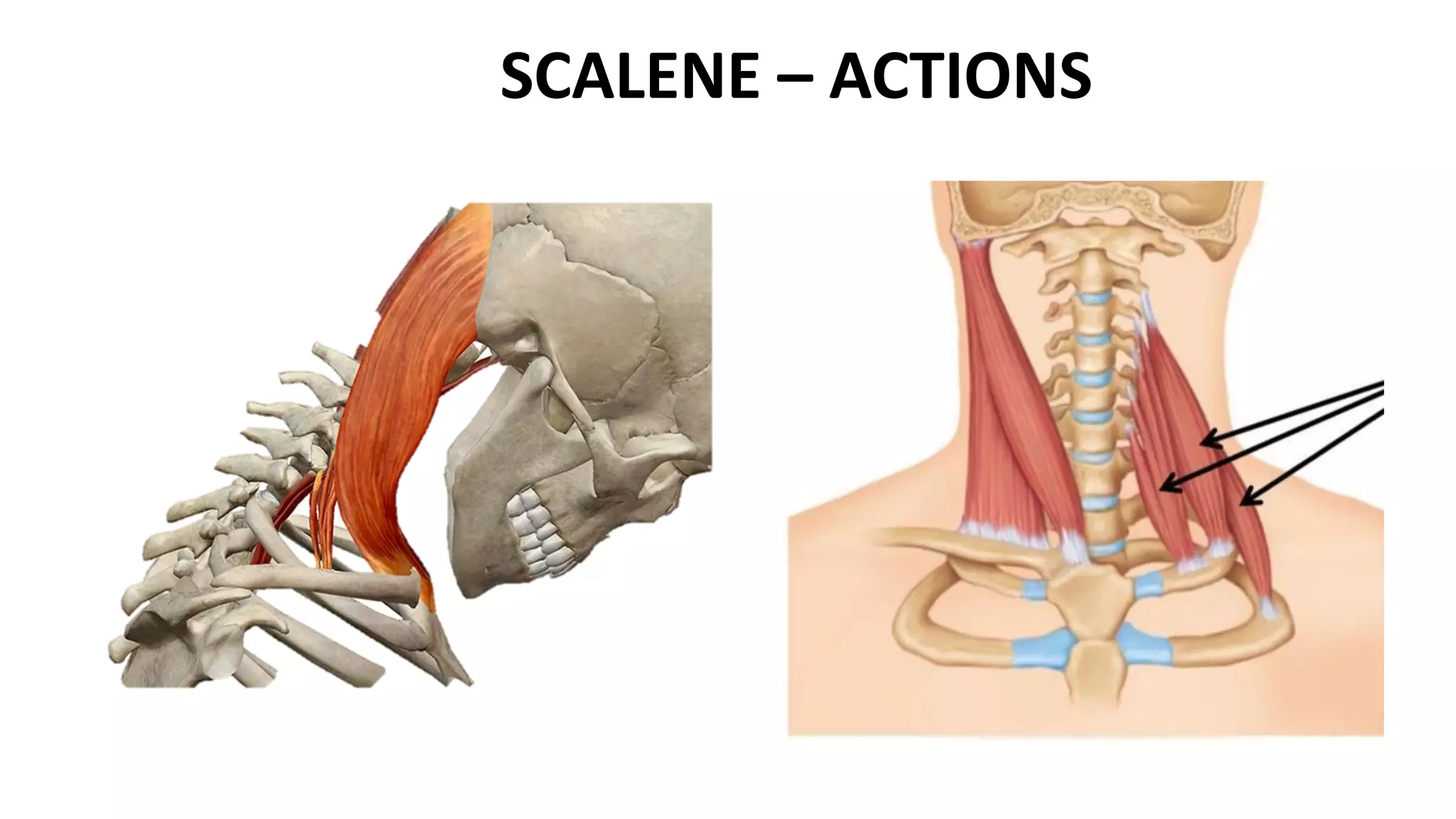
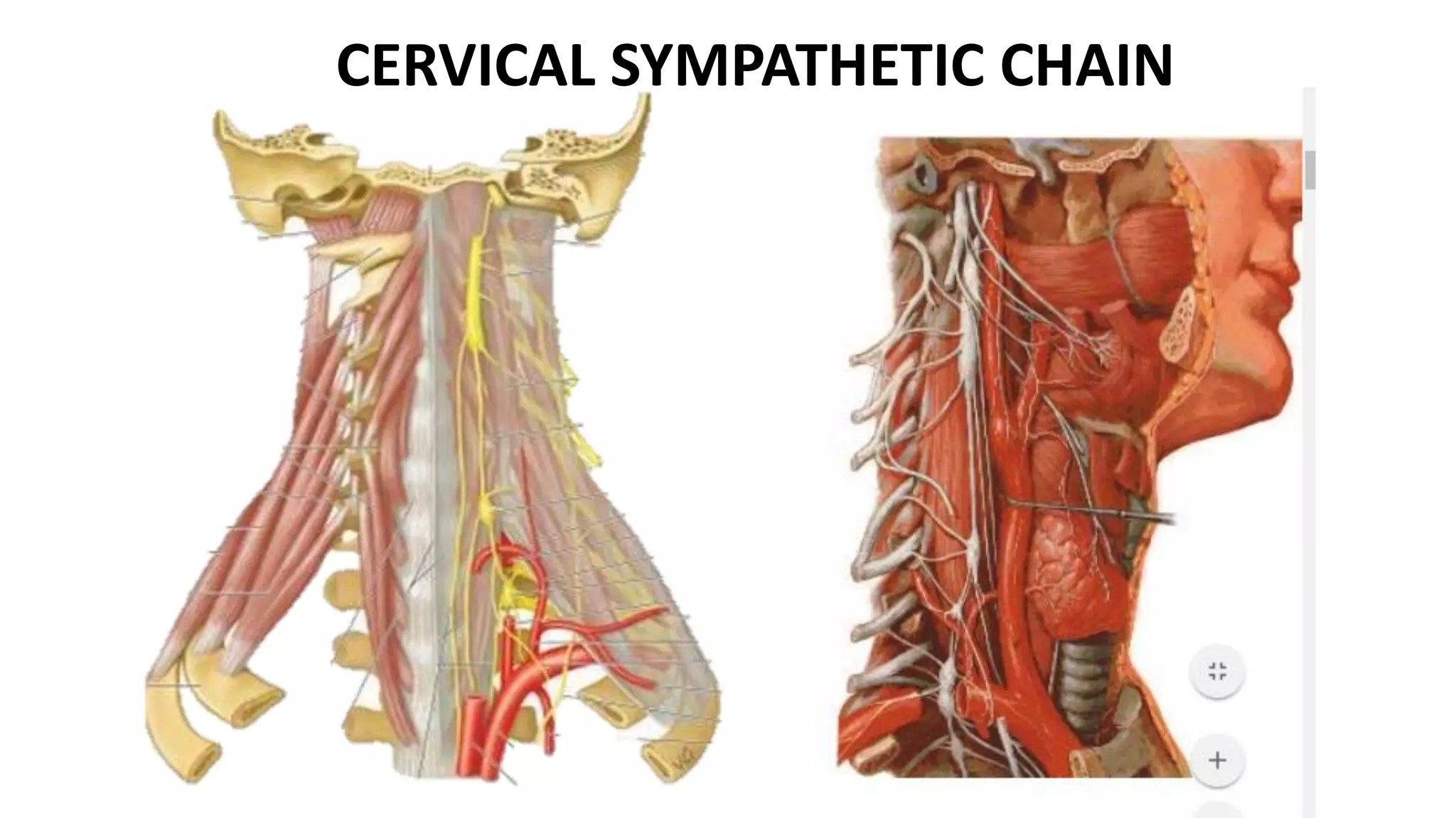
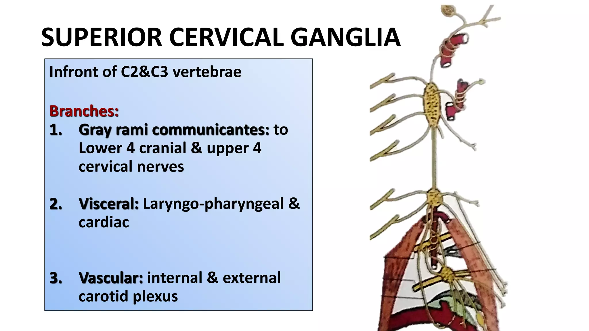
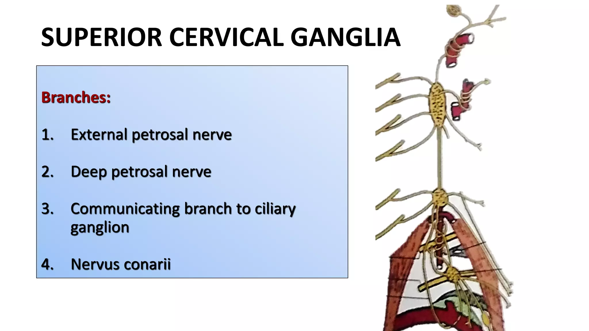
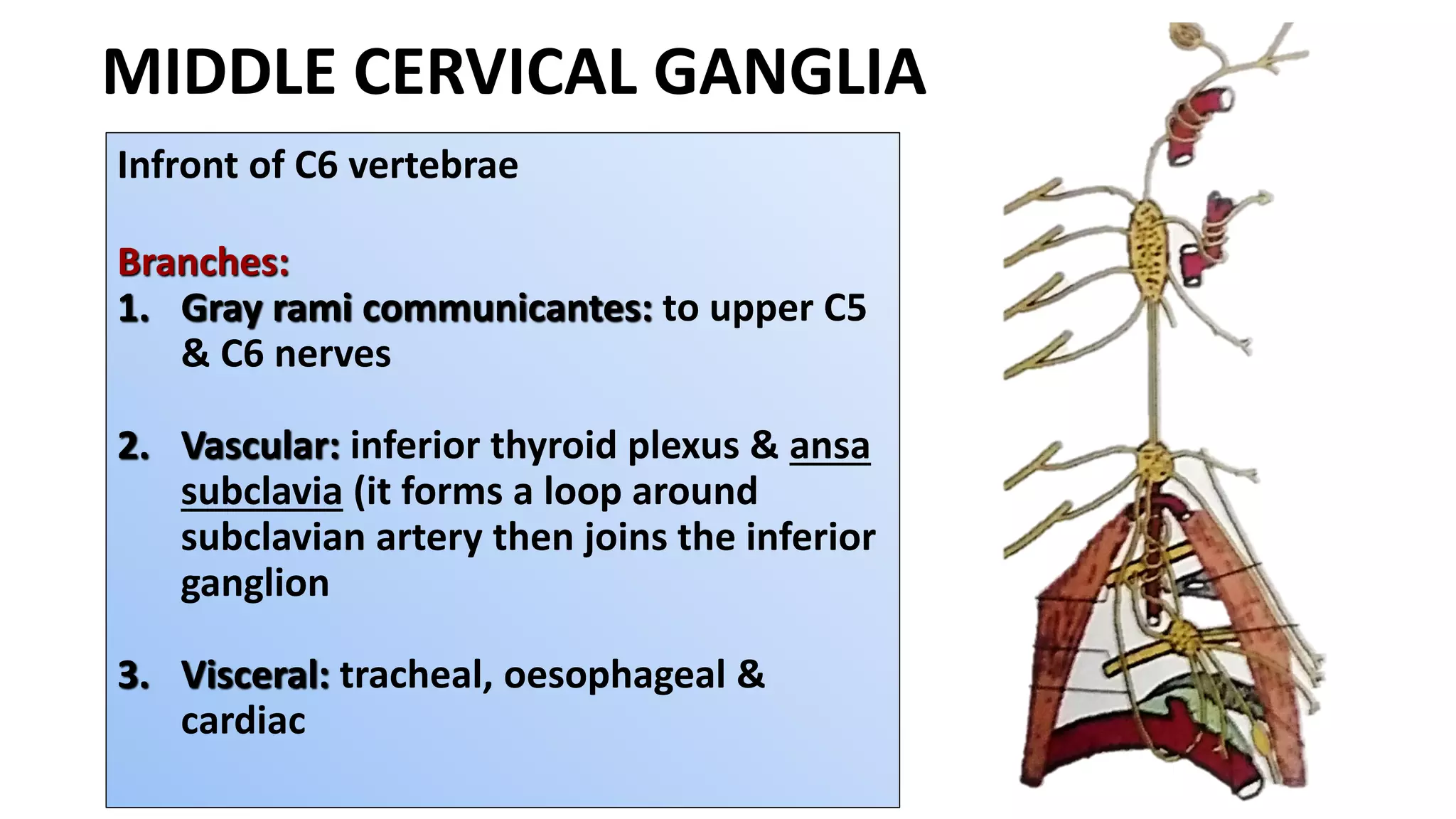
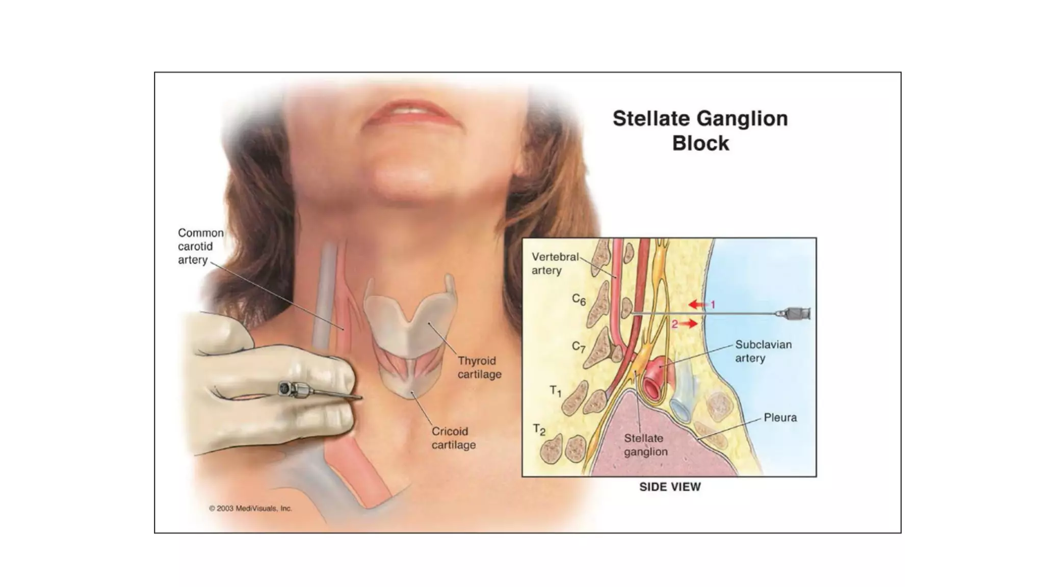
PREVERTEBRAL MUSCLES
SCALENOVERTEBRAL TRIANGLE
CERVICAL SYMPATHETIC CHAIN
Dr.VANI.P.C
ASSISTANT PROFESSOR
DEPARTMENT OF ANATOMY
1. Longus Colli
2. Longus Capitis
3. Rectus Capitis
Anterior
4. Rectus Capitis
Posterior
PREVERTEBRAL MUSCLES
LONGUS COLLI
1. Origin
2. Insertion
Upper part
Anterior tubercles
of transverse
process of C3-C5
vertebrae
Middle part
Anterior surface of
bodies of
C5-C7 &
T1-T3 vertebrae
Lower part
Anterior surface of
bodies of T1—T3
vertebrae
LONGUS COLLI - ORIGIN
LONGUS COLLI - INSERTION
Upper part
Anterior tubercles of
Atlas
Middle part
Anterior surface of
bodies of
C2-C4
vertebrae
Lower part
Anterior tubercle
of transverse
process of C5-C6
vertebrae
LONGUS CAPITIS
1. Origin
2. Insertion
ORIGIN
Anterior tubercles of
transverse process of C3-C6
vertebrae
INSERTION
Inferior surface of basiocciput
on either side of pharyngeal
tubercle
LONGUS CAPITIS
LONGUS – NERVE SUPPLY
Ventral rami of
C2
C3
C4
C5
C6
Cervical nerves
LONGUS – ACTION
RECTUS CAPITIS ANTERIOR
1. Origin
2. Insertion
3. Action
ORIGIN
Anterior Surface Of Lateral
Mass Of Atlas
Root Of Transverse Process Of
Atlas
INSERTION
Inferior Surface Of the basi-occiput
infront of the Occipital condyles
RECTUS CAPITIS ANTERIOR
RECTUS CAPITIS ANTERIOR – ACTION
RECTUS CAPITIS LATERALIS
1. Origin
2. Insertion
3. Action
ORIGIN
Upper Surface Of Transverse
Process Of Atlas
INSERTION
Inferior Surface Of the Jugular
process Of Occipital Bone
RECTUS CAPITIS LATERALIS
RECTUS CAPITIS LATERALIS – ACTION
RECTUS CAPITIS – NERVE SUPPLY
Loop of
communication
between the ventral
rami of C1 & C2
1. Scalene anterior
2. Scalene medius
3. Scalene posterior
PARAVERTEBRAL MUSCLES
ORIGIN
Cervical vertebrae-anterior
tubercle C3 to C6
INSERTION
Scalene tubercle – First rib
SCALENE ANTERIOR
ORIGIN
Cervical vertebrae-Posterior
tubercle
INSERTION
Upper surface of first rib
SCALENE MEDIUS
ORIGIN
Cervical vertebrae-Posterior
tubercle C4 to C6
INSERTION
Outer surface of second rib
SCALENE POSTERIOR
SCALENE – NERVE SUPPLY
Ventral rami of
C4
C5
C6
Cervical nerves
SCALENE – ACTIONS
SCALENOVERTEBRAL TRIANGLE
BOUNDARIES
Medial
Lateral
Apex
Base
Posterior wall
SCALENOVERTEBRAL TRIANGLE
CONTENTS
First part of vertebral artery
Cervical symphathetic chain-
middle & inferior cervical
ganglion
Ansa subclavia
SCALENOVERTEBRAL TRIANGLE
STRUCTURES IN FRONT
Inferior thyroid artery
Thoracic duct on left side only
CERVICAL SYMPATHETIC CHAIN
1. No white rami communicantes
2. Grey rami communicantes to each 8 cervical nerves
3. ORIGIN: Lateral horn cells of T1-T5
CERVICAL SYMPATHETIC CHAIN
SUPERIOR CERVICAL GANGLIA
Infront of C2&C3 vertebrae
Branches:
1. Gray rami communicantes: to
Lower 4 cranial & upper 4
cervical nerves
2. Visceral: Laryngo-pharyngeal &
cardiac
3. Vascular: internal & external
carotid plexus
SUPERIOR CERVICAL GANGLIA
Branches:
1. External petrosal nerve
2. Deep petrosal nerve
3. Communicating branch to ciliary
ganglion
4. Nervus conarii
MIDDLE CERVICAL GANGLIA
Infront of C6 vertebrae
Branches:
1. Gray rami communicantes: to upper C5
& C6 nerves
2. Vascular: inferior thyroid plexus & ansa
subclavia (it forms a loop around
subclavian artery then joins the inferior
ganglion
3. Visceral: tracheal, oesophageal &
cardiac
INFERIOR CERVICAL GANGLIA
Infront of neck of first rib
Stellate ganglion
Branches:
1. Gray rami communicantes: to C7 & C8
nerves
2. Vascular: vertebral and subclavian
plexus
3. Visceral: cardiac
HORNER’S SYNDROME
Prevertebral muscles

Prevertebral muscles