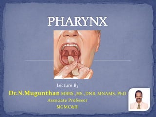
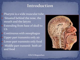
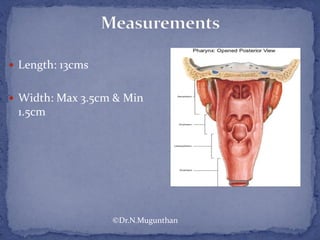
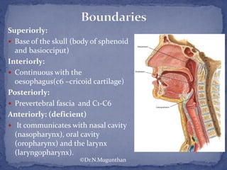
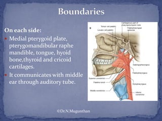
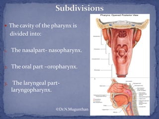
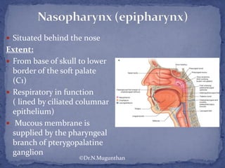
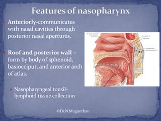
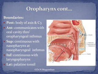
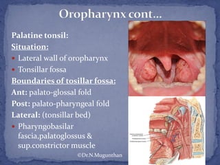
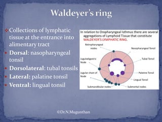
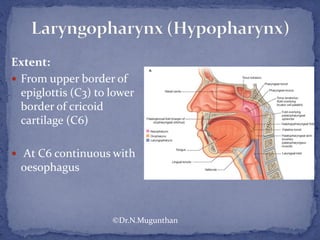
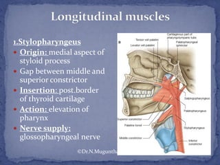
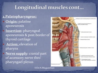
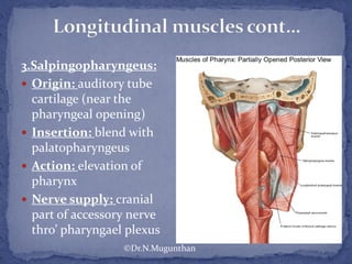
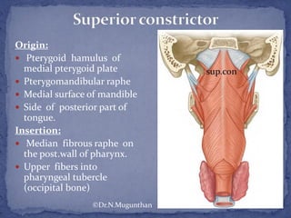
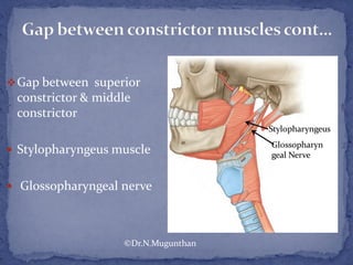
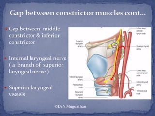
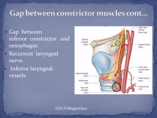
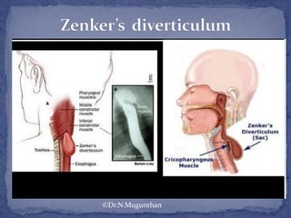
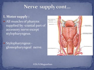
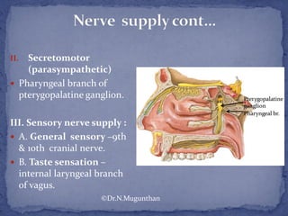
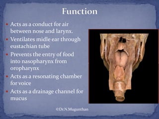
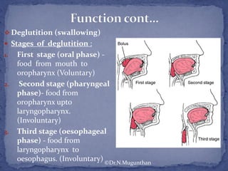
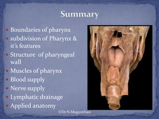
The document outlines the anatomy, structure, and functions of the pharynx, which serves as a muscular tube extending from the base of the skull to the esophagus, dividing into the nasopharynx, oropharynx, and laryngopharynx. It describes various components including muscles, blood supply, nerve supply, and lymphatic drainage, as well as applied anatomy relating to conditions such as tonsillitis and Zenker's diverticulum. The document emphasizes the pharynx's roles in respiration, digestion, and as a conduit for air and food.

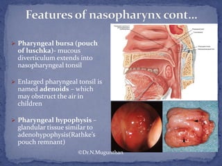


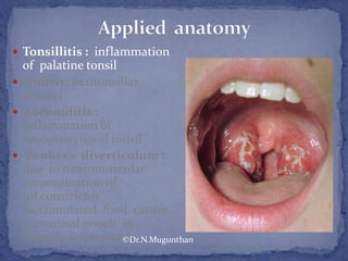
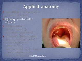
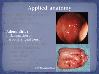
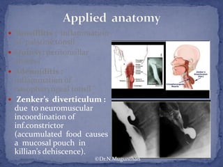
![ Pharyngitis- inflammation
of pharyngeal mucosa -
Nasopharynx -common site
of carcinoma [Fossa of
Rosen muller ].
Infection from
nasopharynx can easily
spread into middle ear
through auditory tube.
Retropharyngeal abscess.
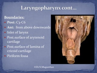
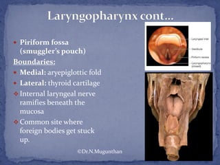
Smuggler's pouch (
common site of foreign
body stuck up)
©Dr.N.Mugunthan](https://image.slidesharecdn.com/pharynx-dr-160406205445/85/Pharynx-Dr-N-Mugunthan-M-S-58-320.jpg)
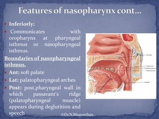
![ Pharyngitis- inflammation
of pharyngeal mucosa .
Nasopharynx -common site
of carcinoma [Fossa of
Rosen muller ].
Infection from
nasopharynx can easily
spread into middle ear
through auditory tube.
Retropharyngeal abscess.
Smuggler's pouch (
common site of foreign
body stuck up)
©Dr.N.Mugunthan](https://image.slidesharecdn.com/pharynx-dr-160406205445/85/Pharynx-Dr-N-Mugunthan-M-S-59-320.jpg)
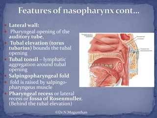
![ Pharyngitis- inflammation
of pharyngeal mucosa .
Nasopharynx -common site
of carcinoma [Fossa of
Rosen muller ].
Infection from
nasopharynx can easily
spread into middle ear
through auditory tube.
Retropharyngeal abscess.
Smuggler's pouch (
common site of foreign
body stuck up)
©Dr.N.Mugunthan](https://image.slidesharecdn.com/pharynx-dr-160406205445/85/Pharynx-Dr-N-Mugunthan-M-S-60-320.jpg)
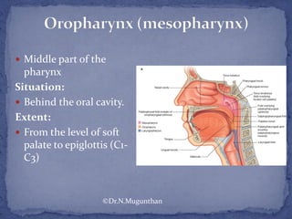
![ Pharyngitis- inflammation
of pharyngeal mucosa .
Nasopharynx -common site
of carcinoma [Fossa of
Rosen muller ].
Infection from
nasopharynx can easily
spread into middle ear
through auditory tube.
Retropharyngeal abscess.
Smuggler's pouch (
common site of foreign
body stuck up)
©Dr.N.Mugunthan](https://image.slidesharecdn.com/pharynx-dr-160406205445/85/Pharynx-Dr-N-Mugunthan-M-S-61-320.jpg)
![ Pharyngitis- inflammation
of pharyngeal mucosa .
Nasopharynx -common site
of carcinoma [Fossa of
Rosen muller ].
Infection from
nasopharynx can easily
spread into middle ear
through auditory tube.
Retropharyngeal abscess.
Smuggler's pouch (
common site of foreign
body stuck up)
©Dr.N.Mugunthan](https://image.slidesharecdn.com/pharynx-dr-160406205445/85/Pharynx-Dr-N-Mugunthan-M-S-62-320.jpg)


![Hypothalamus short ppt by Dr. Neha [PT].pptx](https://cdn.slidesharecdn.com/ss_thumbnails/hypothalamusbydr-260124145759-b9f94a93-thumbnail.jpg?width=640&height=640&fit=bounds)


