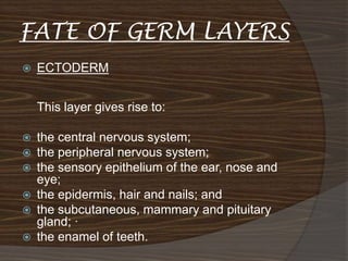
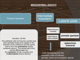
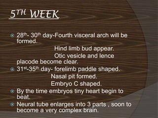
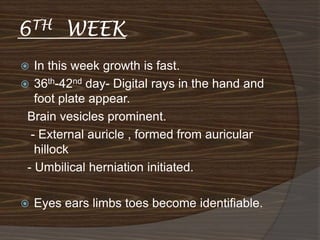
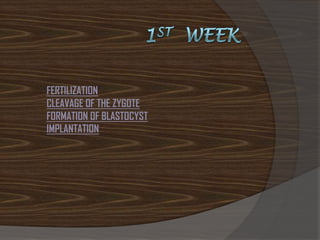
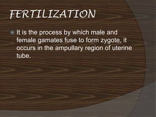
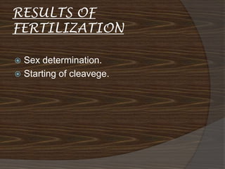
PRESENTED
                 BY

Dr AKHIL.H.S (dept of SHAREERA RACHANA)
 ALVAS AYURVEDIC COLLAGE , MOODBIDRI
                                              :
                                              :
                               (            on . .
     1/1)
-
                               (           .       3)
So                 is a process of the descent of various
     components of the body. This process involves the
     following factors without which a healthy      can’t be
     obtained. It includes spermatogenesis, oogenesis,
     formation of embryo and its complete growth and
ROLE OF
.   PANCHAMAHABHOOTA
    IN GARBHA VRUDHI



           ||
                -SU SA 5-



                  -         -






    ||









    .








    -
   In charaka shareera it is mentioned that the fetus
    gets its nourishment in different ways at different
    stages of growth.

       Either in the initial stages or before
    differentation of various organs it get its
    nourishment from ahara rasa of the mother
    through upa sneha and upa sweda (diffusion and
    osmosis ). After the body parts are formed it gets
    its nutrition through upasneha and nabhi nadi or
    umbilical cord.

      The nabhi nadi is attached to the rasa vaha
    nadi of the mother and this carries ahara rasa
    veerya from the mother to the fetus. The fetus
    grows by the indirect supply of nutrition.
       --
    .
        --

        --

            --   .       .
    .
            --

        --           ...
    .
        --           .
    .
   -




        ||







.        / –                           -
                                             -
                     –
In cha sa 2/ 12 , atreya answer to sishyas regarding the
      .
FERTILIZATION
CLEAVAGE OF THE ZYGOTE
FORMATION OF BLASTOCYST
IMPLANTATION
FERTILIZATION
   It is the process by which male and
    female gamates fuse to form zygote, it
    occurs in the ampullary region of uterine
    tube.
RESULTS OF
FERTILIZATION

 Sex determination.
 Starting of cleavege.
Two days after fertilization, embryo is at
              2celledstage.

Three days after fertilization embryo
  .       becomes morula.

Fourth day -- Blastocyst has formed.
Late implantation/Uteroplacental circulation
Bilaminar disk, amniotic cavity and primary yolk sac
Extra-embryonic mesoderm and coelom
Formation of the chorion and definitive yolk sac
Trophoblast
                             Bilaminar
                           embryonic disc
cytotrophoblast
                                Epiblast
   Syncytiotrophoblast

                               Hypoblast

Cavities
              Amnionic


              Blastocoel
EARLY 2ND WEEK

         Fibrincoagulum

         Lacunar stage

         Exocoelomic memb

         Primary yolk sac
11th 12th day
                Uteroplacental
   Sinusoids   circulation.
                Trophoblast have lacunar
                space in the form of inter
                communicating
                network.More evident at
                embryonic pole.
                The cells of
                synciotropoblast continue
                penetrating deep and erode
                endometrial lining of
                meternal capillaries.
                Lacunae continue with the
                sinusoids.
Extra-embryonic
mesoderm / Extra-
embryonic coelom
Cells derived from the primitive ectoderm fill the space
  between the trophoblast and two cavities

   This loose connective tissue, the extraembryonic
    mesoderm, completely surrounds the amnion and
    primary yolk sac

   Fluid-filled spaces appear in the mesenchyme,
    pushing aside the mesenchyme to form a coelom

   These spaces fuse to form a fluid-filled cavity
    completely surrounding the amnion and yolk sac,
    except at the connecting stalk
   Extra embryonic coelom / Chorionic cavity.

   When EEC enlarges EEM will be restricted
    to lining.

   Extra embryonic somato pleuric mesoderm

   Extra embryonic splanchno pleuric
    mesoderm
Primary villi.

           Secondary yolk sac

           Exocoelomic cyst

           Connecting stalk


13th day
Gastrulation: Primitive Streak and Cell Migrations
Results of Gastrulation: Fate of the Germ Layers
Notochord: the Primary Inducer
Neurulation: Neural Tube Formation
Neural Crest Cells and their Derivatives
GASTRULATION
   Its the process that establishes all 3 germ
    layers. It begins with formation of Primitive
    Streak on surface of epiblast.
   Cephalic end- primitive node.
   Cells of epiblast migrate towards primitive
    streak and it become flask shape.
   It then detach from epiblast and slip beneath it.
    INVAGINATION.
   Once cells invaginated, some displace
    hypoblast and form- embryonic ENDODERM.
   Some lie in b/w epiblast and new embryonic
    endoderm to form MESODERM.
   Remaining cells form embryonic ECTODERM.

   Thus epiblast is source of all 3 layers.
NOTOCHORD
 The pre notochordal cells invaginating in
  the primitive streak, move forward
  cranially, untill they reach prechordal
  plate.
 These cells intercalate in endoderm as
  notochordal plate.
 By further development, the plate
  detaches from endoderm and a solid
  cord is formed.
 This will serve as basis of axial skeleton.
NEURULATION.
   At beginning of 3rd week ectodermal germ layer is
      of disc shape.
   The overlying ectoderm thickens and form NEURAL
    PLATE.
   Cells of the plate make up neuroectoderm.
   Once induction begin the neural plate expands
      towards primitive streak.
   By end of 3rd week,lateral edge of neural plate
     expands to form NEURAL FOLD.
   And mid region – NEURAL GROOVE.
   Gradually Neural folds approaches each other and
    fuse
      in middle ,and continues upward and downwards.
   NEURAL TUBE.
   CRANIAL NEUROPORE & CAUDAL
                                         NEUROPORE.
   Vasculogenesis- EEM – angioblasts-
    angiogenic clustor of cells- endothelial
    cells – this fuse with each side to form
    small blood vessels.

 Hematopoiesis- occur in EEVM –
  angioblasts in angiogenic cell clusters
  give rise to primitive blood cells.
 At 5th week it is taken over by organs.
Somite development
Intraembryonic coelom & body cavities
Head-tail folding
Lateral folding
 In week 4, the embryo undergoes major
  morphological changes as it changes from
  a trilaminar disc-shaped embryo to a
  cylindrical embryo.
 This is also an important week in terms of
  determining placement of future organs.
 Following median and horizontal folding,
  many organs and body cavities will begin
  to form or will be repositioned.
 Embryo begin to fold.
Head-Tail Folding
 Due to the rapid growth in the median
  plane of the brain, amniotic cavity, and
  somites, the embryo elongates, with its
  head and tail ends folding under.
 At the cranial end, the head will be folded
  under, with a very prominent forebrain.
 On the ventral side, the newly positioned
  primitive heart, pericardial cavity, septum
  transversum, and bucco-pharyngeal
  membrane
   At the same time that head-tail folding is
    occurring, lateral folding is also
    occurring to form a cylindrical embryo
   22nd – 23rd day – Fusion of neural fold .
                     Neoropores widly opened.
                   Heart tube begin to fold.

24th -25 th day – Cephalocaudal folding.
                 Cranial neuropore closing.
                  Optic vesicles appear.
                  Otic placcodes appear.

   26th-27th day – Caudal neuropore closing.
                    Upper limbs buds appear.
                    3 pairs of visceral arches.
EMBRYO AT   4TH   WEEK.
Somite Development
 The paraxial mesoderm will develop into paired
  cuboidal bodies, or somites (soma, body).
 These will eventually develop into the bones
  (Sclerotome), Muscles (Myotome), and Dermis
  (Dermatome) of and surrounding the axial skeleton

 Somites appear as bumps on the dorsal surface of the
  embryo.
 At the end of week 3, 4-12 somites are present (visible
  on the dorsal surface of the embryo). By the end of
  week 5, 42-44 can be counted.

   The most appear between days 20-30, giving this
    period the title of the somite period of development.

    Somites appear cranially to caudally, beginning at the
    occipital end. They can be counted and are used to
    roughly estimate the age of the embryo
FATE OF GERM LAYERS
   ECTODERM


    This layer gives rise to:

   the central nervous system;
   the peripheral nervous system;
   the sensory epithelium of the ear, nose and
    eye;
   the epidermis, hair and nails; and
   the subcutaneous, mammary and pituitary
    gland; ·
   the enamel of teeth.
MESODERMAL SHEATH


                                                Intermediate
      Paraxial mesoderm                          mesoderm
                                                                          Lateral plate

                                                         Nephrotomes-
                                                           cranially

                                                         Nephrogenic       somatic (parietal
              Somites ( 42-44)                           cells-caudally
The epithelial cells forming the somites lose
  their epithelial shape and migrate in the                                   splanchnic
 direction of the notochord and the spinal
    cord to form the sclerotome (future
                                                                               (visceral
 vertebral column). The dorsal wall of the
           somite differentiate into
    the myotome (furute muscles) and
      thedermatome (future dermis).                , both developing into the
                                                   excretory units of kidneys,
                                                   gonads, ducts and accessory glands
   ENDODERM

it provide epithelial lining of gastro-
   intestinal tract, respiratory tract, urinary
   tract, urinary bladder, also paranchyma
   of thyroid, parathyroids, liver, pancreas.
5TH    WEEK
 28th- 30th day-Fourth visceral arch will be
  formed.
                 Hind limb bud appear.
                 Otic vesicle and lence
  placode become clear.
 31st-35th day- forelimb paddle shaped.
               Nasal pit formed.
               Embryo C shaped.
 By the time embryos tiny heart begin to
  beat.
 Neural tube enlarges into 3 parts , soon to
  become a very complex brain.
6TH      WEEK
  In this week growth is fast.
 36th-42nd day- Digital rays in the hand and
   foot plate appear.
 Brain vesicles prominent.
  - External auricle , formed from auricular
   hillock
 - Umbilical herniation initiated.

   Eyes ears limbs toes become identifiable.
7TH     WEEK
 43rd-49th day – Pigmentation of retina
  vissible
                - Digitals separating.
                - Nipples and eyes formed.
                 - Maxillary swelling fuse with
  medial nasal swelling and upper lip is
  formed.
              - prominent umbilical herniation.
 Major muscle system is developed.
 The child has its own blood type from
  mothers.
8TH       WEEK

   Limbs long and bend at elbows, knees,
    fingers,toes free.
   Face more human like.
   Tail disppears.
   Umbilical herniation persist end of 3rd week.
   By eighth week all organs develop, and
    embryo is now called foetus.
   Foetus is about half inch long.
   It is protected by amnionic sac, filled with fluid.
Embryonic development

Embryonic development

  • 1.
    PRESENTED BY Dr AKHIL.H.S (dept of SHAREERA RACHANA) ALVAS AYURVEDIC COLLAGE , MOODBIDRI
  • 3.
    : : ( on . . 1/1) - ( . 3) So is a process of the descent of various components of the body. This process involves the following factors without which a healthy can’t be obtained. It includes spermatogenesis, oogenesis, formation of embryo and its complete growth and
  • 4.
    ROLE OF . PANCHAMAHABHOOTA IN GARBHA VRUDHI  || -SU SA 5-  - -
  • 5.
  • 6.
  • 7.
  • 8.
    In charaka shareera it is mentioned that the fetus gets its nourishment in different ways at different stages of growth.  Either in the initial stages or before differentation of various organs it get its nourishment from ahara rasa of the mother through upa sneha and upa sweda (diffusion and osmosis ). After the body parts are formed it gets its nutrition through upasneha and nabhi nadi or umbilical cord.  The nabhi nadi is attached to the rasa vaha nadi of the mother and this carries ahara rasa veerya from the mother to the fetus. The fetus grows by the indirect supply of nutrition.
  • 9.
    -- .  --  --  -- . . .  --  -- ... .  -- . .
  • 10.
    -  || 
  • 11.
  • 12.
    . / – - - – In cha sa 2/ 12 , atreya answer to sishyas regarding the .
  • 15.
    FERTILIZATION CLEAVAGE OF THEZYGOTE FORMATION OF BLASTOCYST IMPLANTATION
  • 16.
    FERTILIZATION  It is the process by which male and female gamates fuse to form zygote, it occurs in the ampullary region of uterine tube.
  • 18.
    RESULTS OF FERTILIZATION  Sexdetermination.  Starting of cleavege.
  • 20.
    Two days afterfertilization, embryo is at 2celledstage. Three days after fertilization embryo . becomes morula. Fourth day -- Blastocyst has formed.
  • 21.
    Late implantation/Uteroplacental circulation Bilaminardisk, amniotic cavity and primary yolk sac Extra-embryonic mesoderm and coelom Formation of the chorion and definitive yolk sac
  • 22.
    Trophoblast Bilaminar embryonic disc cytotrophoblast Epiblast Syncytiotrophoblast Hypoblast Cavities Amnionic Blastocoel
  • 23.
    EARLY 2ND WEEK Fibrincoagulum Lacunar stage Exocoelomic memb Primary yolk sac
  • 24.
    11th 12th day Uteroplacental  Sinusoids circulation. Trophoblast have lacunar space in the form of inter communicating network.More evident at embryonic pole. The cells of synciotropoblast continue penetrating deep and erode endometrial lining of meternal capillaries. Lacunae continue with the sinusoids.
  • 25.
    Extra-embryonic mesoderm / Extra- embryoniccoelom Cells derived from the primitive ectoderm fill the space between the trophoblast and two cavities  This loose connective tissue, the extraembryonic mesoderm, completely surrounds the amnion and primary yolk sac  Fluid-filled spaces appear in the mesenchyme, pushing aside the mesenchyme to form a coelom  These spaces fuse to form a fluid-filled cavity completely surrounding the amnion and yolk sac, except at the connecting stalk
  • 26.
    Extra embryonic coelom / Chorionic cavity.  When EEC enlarges EEM will be restricted to lining.  Extra embryonic somato pleuric mesoderm  Extra embryonic splanchno pleuric mesoderm
  • 28.
    Primary villi. Secondary yolk sac Exocoelomic cyst Connecting stalk 13th day
  • 30.
    Gastrulation: Primitive Streakand Cell Migrations Results of Gastrulation: Fate of the Germ Layers Notochord: the Primary Inducer Neurulation: Neural Tube Formation Neural Crest Cells and their Derivatives
  • 31.
    GASTRULATION  Its the process that establishes all 3 germ layers. It begins with formation of Primitive Streak on surface of epiblast.  Cephalic end- primitive node.  Cells of epiblast migrate towards primitive streak and it become flask shape.  It then detach from epiblast and slip beneath it. INVAGINATION.  Once cells invaginated, some displace hypoblast and form- embryonic ENDODERM.  Some lie in b/w epiblast and new embryonic endoderm to form MESODERM.  Remaining cells form embryonic ECTODERM.  Thus epiblast is source of all 3 layers.
  • 32.
    NOTOCHORD  The prenotochordal cells invaginating in the primitive streak, move forward cranially, untill they reach prechordal plate.  These cells intercalate in endoderm as notochordal plate.  By further development, the plate detaches from endoderm and a solid cord is formed.  This will serve as basis of axial skeleton.
  • 33.
    NEURULATION.  At beginning of 3rd week ectodermal germ layer is of disc shape.  The overlying ectoderm thickens and form NEURAL PLATE.  Cells of the plate make up neuroectoderm.  Once induction begin the neural plate expands towards primitive streak.  By end of 3rd week,lateral edge of neural plate expands to form NEURAL FOLD.  And mid region – NEURAL GROOVE.  Gradually Neural folds approaches each other and fuse in middle ,and continues upward and downwards.  NEURAL TUBE.  CRANIAL NEUROPORE & CAUDAL NEUROPORE.
  • 34.
    Vasculogenesis- EEM – angioblasts- angiogenic clustor of cells- endothelial cells – this fuse with each side to form small blood vessels.  Hematopoiesis- occur in EEVM – angioblasts in angiogenic cell clusters give rise to primitive blood cells.  At 5th week it is taken over by organs.
  • 35.
    Somite development Intraembryonic coelom& body cavities Head-tail folding Lateral folding
  • 36.
     In week4, the embryo undergoes major morphological changes as it changes from a trilaminar disc-shaped embryo to a cylindrical embryo.  This is also an important week in terms of determining placement of future organs.  Following median and horizontal folding, many organs and body cavities will begin to form or will be repositioned.  Embryo begin to fold.
  • 37.
    Head-Tail Folding  Dueto the rapid growth in the median plane of the brain, amniotic cavity, and somites, the embryo elongates, with its head and tail ends folding under.  At the cranial end, the head will be folded under, with a very prominent forebrain.  On the ventral side, the newly positioned primitive heart, pericardial cavity, septum transversum, and bucco-pharyngeal membrane
  • 38.
    At the same time that head-tail folding is occurring, lateral folding is also occurring to form a cylindrical embryo
  • 39.
    22nd – 23rd day – Fusion of neural fold . Neoropores widly opened. Heart tube begin to fold. 24th -25 th day – Cephalocaudal folding. Cranial neuropore closing. Optic vesicles appear. Otic placcodes appear.  26th-27th day – Caudal neuropore closing. Upper limbs buds appear. 3 pairs of visceral arches.
  • 40.
    EMBRYO AT 4TH WEEK.
  • 41.
    Somite Development  Theparaxial mesoderm will develop into paired cuboidal bodies, or somites (soma, body).  These will eventually develop into the bones (Sclerotome), Muscles (Myotome), and Dermis (Dermatome) of and surrounding the axial skeleton  Somites appear as bumps on the dorsal surface of the embryo.  At the end of week 3, 4-12 somites are present (visible on the dorsal surface of the embryo). By the end of week 5, 42-44 can be counted.  The most appear between days 20-30, giving this period the title of the somite period of development.  Somites appear cranially to caudally, beginning at the occipital end. They can be counted and are used to roughly estimate the age of the embryo
  • 42.
    FATE OF GERMLAYERS  ECTODERM This layer gives rise to:  the central nervous system;  the peripheral nervous system;  the sensory epithelium of the ear, nose and eye;  the epidermis, hair and nails; and  the subcutaneous, mammary and pituitary gland; ·  the enamel of teeth.
  • 43.
    MESODERMAL SHEATH Intermediate Paraxial mesoderm mesoderm Lateral plate Nephrotomes- cranially Nephrogenic somatic (parietal Somites ( 42-44) cells-caudally The epithelial cells forming the somites lose their epithelial shape and migrate in the splanchnic direction of the notochord and the spinal cord to form the sclerotome (future (visceral vertebral column). The dorsal wall of the somite differentiate into the myotome (furute muscles) and thedermatome (future dermis). , both developing into the excretory units of kidneys, gonads, ducts and accessory glands
  • 44.
    ENDODERM it provide epithelial lining of gastro- intestinal tract, respiratory tract, urinary tract, urinary bladder, also paranchyma of thyroid, parathyroids, liver, pancreas.
  • 45.
    5TH WEEK  28th- 30th day-Fourth visceral arch will be formed. Hind limb bud appear. Otic vesicle and lence placode become clear.  31st-35th day- forelimb paddle shaped. Nasal pit formed. Embryo C shaped.  By the time embryos tiny heart begin to beat.  Neural tube enlarges into 3 parts , soon to become a very complex brain.
  • 46.
    6TH WEEK  In this week growth is fast.  36th-42nd day- Digital rays in the hand and foot plate appear. Brain vesicles prominent. - External auricle , formed from auricular hillock - Umbilical herniation initiated.  Eyes ears limbs toes become identifiable.
  • 47.
    7TH WEEK  43rd-49th day – Pigmentation of retina vissible - Digitals separating. - Nipples and eyes formed. - Maxillary swelling fuse with medial nasal swelling and upper lip is formed. - prominent umbilical herniation.  Major muscle system is developed.  The child has its own blood type from mothers.
  • 48.
    8TH WEEK  Limbs long and bend at elbows, knees, fingers,toes free.  Face more human like.  Tail disppears.  Umbilical herniation persist end of 3rd week.  By eighth week all organs develop, and embryo is now called foetus.  Foetus is about half inch long.  It is protected by amnionic sac, filled with fluid.