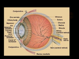
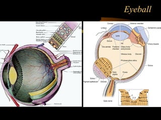
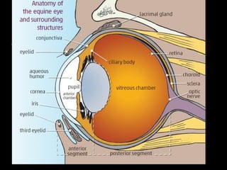
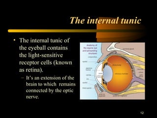
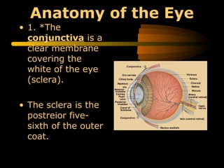
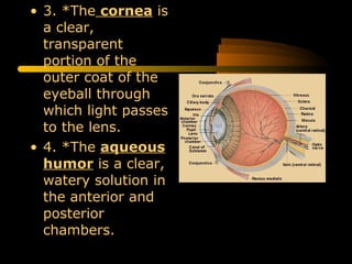
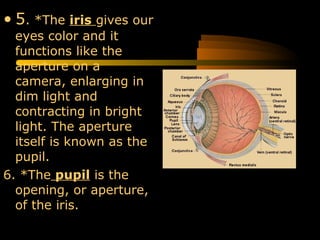
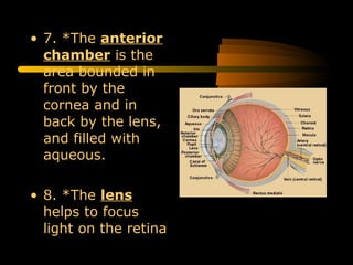
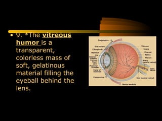
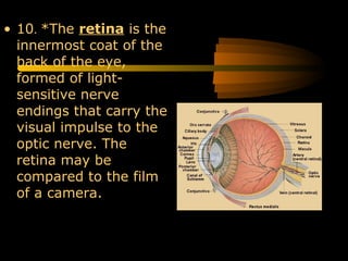
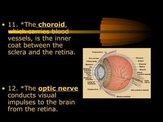
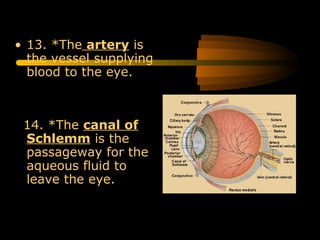
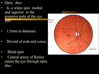
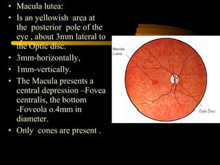
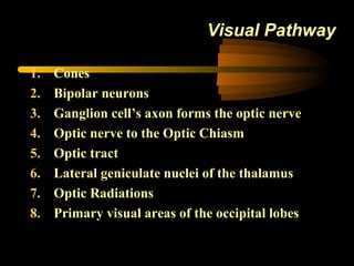
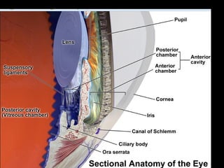
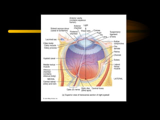
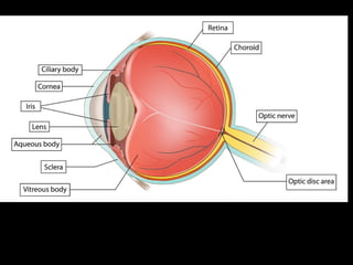
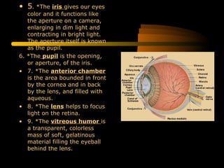
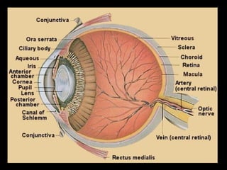
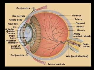
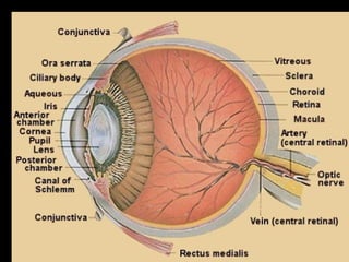
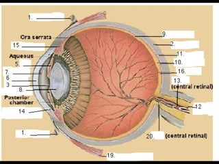
EYE BALL
4
INTRODUCTION
• The Eye is the
organ of vision.
• Composed of :
1. Eyeball.
2. The adnexa.
Eyeball
6
The Eyeball
• Equator :an imaginary line about
the eyeball, which is the
equidistant from the poles.
• Meridian: is one of many lines
passing from pole to pole that
intersects the equator at right
angles.
–Optic nerve :leaves the
eyeball slightly ventral
to the posterior pole
7
Terminology of the eye
• Cornea : the transparent
part of the eyeball .
• Anterior pole: the highest
point on cornea .
• Posterior pole : the
highest point on posterior
surface .
• Optic axis: the straight
line passing through both
poles
9
Eyeball
The three tunics are:
I- An external fibrous tunic-Cornea and Sclera.
II- A middle vascular tunic-Choroid coat,Cilliary body
(cilliary muscle and cilliary process) and Iris.
III- An internal nervous tunic-Retina.
The fibrous tunic
• The cornea forms
about one quarter of
the fibrous tunic and
bulges forward. It is
composed off dense
connective tissue
arranged in lamellar
form .
• The cornea doesn’t
contain blood vessels;
nutrients for its cells
permeate from vessels
in the limbus or are
carried to it its surface
10
11
The vascular Tunic (uvea)
2) The ciliary body :
toward the limbus the choroids
thickness to form it.
3) The Iris: the smallest part of the
vascular tunic, which extends from
the cornea to the lens.
• It attached to sclera and ciliary
body by pectinate ligament.
• the opening in the center is the
pulpi
The internal tunic
• The internal tunic of
the eyeball contains
the light-sensitive
receptor cells (known
as retina).
– It’s an extension of the
brain to which remains
connected by the optic
nerve.
12
Anatomy of the Eye
• 1. *The
conjunctiva is a
clear membrane
covering the
white of the eye
(sclera).
• The sclera is the
postreior five-
sixth of the outer
coat.
• 3. *The cornea is
a clear,
transparent
portion of the
outer coat of the
eyeball through
which light passes
to the lens.
• 4. *The aqueous
humor is a clear,
watery solution in
the anterior and
posterior
chambers.
• 5. *The iris gives our
eyes color and it
functions like the
aperture on a
camera, enlarging in
dim light and
contracting in bright
light. The aperture
itself is known as the
pupil.
6. *The pupil is the
opening, or aperture,
of the iris.
• 7. *The anterior
chamber is the
area bounded in
front by the
cornea and in
back by the lens,
and filled with
aqueous.
• 8. *The lens
helps to focus
light on the retina
• 9. *The vitreous
humor is a
transparent,
colorless mass of
soft, gelatinous
material filling the
eyeball behind the
lens.
• 10. *The retina is the
innermost coat of the
back of the eye,
formed of light-
sensitive nerve
endings that carry the
visual impulse to the
optic nerve. The
retina may be
compared to the film
of a camera.
• 11. *The choroid,
which carries blood
vessels, is the inner
coat between the
sclera and the retina.
• 12. *The optic nerve
conducts visual
impulses to the brain
from the retina.
• 13. *The artery is
the vessel supplying
blood to the eye.
14. *The canal of
Schlemm is the
passageway for the
aqueous fluid to
leave the eye.
• 15. *The ciliary
body is an unseen
part of the iris, and
these together with
the ora serrata form
the uveal tract.
• 16. *The macula is a
small area in the
retina that provides
our most central,
acute vision
• 17. *The ora serrata
and the ciliary body
form the uveal tract,
an unseen part of the
iris.
• 18. *The posterior
chamber is the area
behind the iris, but in
front of the lens, that
is filled with aqueous
• 19. *The rectus
medialis is one of
the six muscles of the
eye.
• 20. *The vein is the
vessel that carries
blood away from the
eye.
• Optic disc:
• Is a white spot medial
and superior to the
posterior pole of the eye
ball
• 1.5mm in diameter.
• Devoid of rods and cones.
• Blind spot.
• Central artery of Retina
enters the eye through optic
disc.
• Macula lutea:
• Is an yellowish area at
the posterior pole of the
eye , about 3mm lateral to
the Optic disc.
• 3mm-horizontally,
• 1mm-vertically.
• The Macula presents a
central depression –Fovea
centralis, the bottom
-Foveola o.4mm in
diameter.
• Only cones are present .
Production of Aqueous Humor and
Intraocular pressure
1. Ciliary Process:
Produces Aqueous Humor
2. Posterior Chamber:
Aqueous Humor flows from
this chamber through the
pupil in Anterior Chamber
3. Canal of Schlemm
Reabsorbs Aqueous Humor
Glaucoma:
Increase in intraocular
pressure due to build up of
Aqueous Humor
Opthalmoscopic view of the retina showing
the location of the Macula to the Optic Disc
Visual Pathway
1. Cones
2. Bipolar neurons
3. Ganglion cell’s axon forms the optic nerve
4. Optic nerve to the Optic Chiasm
5. Optic tract
6. Lateral geniculate nuclei of the thalamus
7. Optic Radiations
8. Primary visual areas of the occipital lobes
Abnormalities of
The Eye:
1. Myopic -
nearsighted
2. Hypermetropic -
Farsighted
3. Presbyopia -
age-related failure of
lens to accommodate
4. Astigmatism -
Distorted vision due to
irregular-shaped lens
or
cornea
5. Color Blindness -
genetic defect that
causes dysfunction of
cones
Hemianopsia or hemianopia is visual field loss on the left or right
side of the vertical midline. It can affect one eye but usually affects
both eyes. Homonymous hemianopsia, or homonymous
hemianopia, is hemianopic visual field loss on the same side of both
eyes.
Homonymous hemianopsia occurs because the right half of the brain
has visual pathways for the left hemifield of both eyes, and the left half
of the brain has visual pathways for the right hemifield of both eyes.
When one of these pathways is damaged, the corresponding visual field
is lost.
Anatomy of the Eyeball
• Fibrous Tunic:
Cornea
Sclera
• Vascular Tunic
Choroid coat
Ciliary Body (Ciliary muscle, Ciliary process)
Iris
• Nervous Tunic
Retina
• 5. *The iris gives our eyes
color and it functions like
the aperture on a camera,
enlarging in dim light and
contracting in bright light.
The aperture itself is known
as the pupil.
6. *The pupil is the opening,
or aperture, of the iris.
• 7. *The anterior chamber
is the area bounded in front
by the cornea and in back
by the lens, and filled with
aqueous.
• 8. *The lens helps to focus
light on the retina.
• 9. *The vitreous humor is
a transparent, colorless
mass of soft, gelatinous
material filling the eyeball
behind the lens.
• 6. *The pupil is the opening, or aperture, of
the iris.
• 7. *The anterior chamber is the area
bounded in front by the cornea and in back by
the lens, and filled with aqueous.
• 8. *The lens helps to focus light on the
retina.
• 9. *The vitreous humor is a transparent,
colorless mass of soft, gelatinous material
filling the eyeball behind the lens.
• 10. *The retina is the innermost coat of the back of
the eye, formed of light-sensitive nerve endings that
carry the visual impulse to the optic nerve. The retina
may be compared to the film of a camera.
• 11. *The choroid, which carries blood vessels, is the
inner coat between the sclera and the retina.
• 12. *The optic nerve conducts visual impulses to the
brain from the retina.
• 13. *The artery is the vessel supplying blood to the
eye.
• 14. *The canal of Schlemm is the passageway for
the aqueous fluid to leave the eye.
• 15. *The ciliary body is an unseen part of the iris,
and these together with the ora serrata form the
uveal tract.
• 16. *The macula is a small area in the retina that
provides our most central, acute vision.
• 17. *The ora serrata and the ciliary body form the
uveal tract, an unseen part of the iris.
• 18. *The posterior chamber is the area behind the
iris, but in front of the lens, that is filled with aqueous.
• 19. *The rectus medialis is one of the six muscles of
the eye.
• 20. *The vein is the vessel that carries blood away
from the eye.
Eyelect1.html usha kannan

Eyelect1.html usha kannan

  • 2.
  • 4.
    4 INTRODUCTION • The Eyeis the organ of vision. • Composed of : 1. Eyeball. 2. The adnexa.
  • 5.
  • 6.
    6 The Eyeball • Equator:an imaginary line about the eyeball, which is the equidistant from the poles. • Meridian: is one of many lines passing from pole to pole that intersects the equator at right angles. –Optic nerve :leaves the eyeball slightly ventral to the posterior pole
  • 7.
    7 Terminology of theeye • Cornea : the transparent part of the eyeball . • Anterior pole: the highest point on cornea . • Posterior pole : the highest point on posterior surface . • Optic axis: the straight line passing through both poles
  • 9.
    9 Eyeball The three tunicsare: I- An external fibrous tunic-Cornea and Sclera. II- A middle vascular tunic-Choroid coat,Cilliary body (cilliary muscle and cilliary process) and Iris. III- An internal nervous tunic-Retina.
  • 10.
    The fibrous tunic •The cornea forms about one quarter of the fibrous tunic and bulges forward. It is composed off dense connective tissue arranged in lamellar form . • The cornea doesn’t contain blood vessels; nutrients for its cells permeate from vessels in the limbus or are carried to it its surface 10
  • 11.
    11 The vascular Tunic(uvea) 2) The ciliary body : toward the limbus the choroids thickness to form it. 3) The Iris: the smallest part of the vascular tunic, which extends from the cornea to the lens. • It attached to sclera and ciliary body by pectinate ligament. • the opening in the center is the pulpi
  • 12.
    The internal tunic •The internal tunic of the eyeball contains the light-sensitive receptor cells (known as retina). – It’s an extension of the brain to which remains connected by the optic nerve. 12
  • 13.
    Anatomy of theEye • 1. *The conjunctiva is a clear membrane covering the white of the eye (sclera). • The sclera is the postreior five- sixth of the outer coat.
  • 14.
    • 3. *Thecornea is a clear, transparent portion of the outer coat of the eyeball through which light passes to the lens. • 4. *The aqueous humor is a clear, watery solution in the anterior and posterior chambers.
  • 15.
    • 5. *Theiris gives our eyes color and it functions like the aperture on a camera, enlarging in dim light and contracting in bright light. The aperture itself is known as the pupil. 6. *The pupil is the opening, or aperture, of the iris.
  • 16.
    • 7. *Theanterior chamber is the area bounded in front by the cornea and in back by the lens, and filled with aqueous. • 8. *The lens helps to focus light on the retina
  • 17.
    • 9. *Thevitreous humor is a transparent, colorless mass of soft, gelatinous material filling the eyeball behind the lens.
  • 18.
    • 10. *Theretina is the innermost coat of the back of the eye, formed of light- sensitive nerve endings that carry the visual impulse to the optic nerve. The retina may be compared to the film of a camera.
  • 19.
    • 11. *Thechoroid, which carries blood vessels, is the inner coat between the sclera and the retina. • 12. *The optic nerve conducts visual impulses to the brain from the retina.
  • 20.
    • 13. *Theartery is the vessel supplying blood to the eye. 14. *The canal of Schlemm is the passageway for the aqueous fluid to leave the eye.
  • 21.
    • 15. *Theciliary body is an unseen part of the iris, and these together with the ora serrata form the uveal tract. • 16. *The macula is a small area in the retina that provides our most central, acute vision
  • 22.
    • 17. *Theora serrata and the ciliary body form the uveal tract, an unseen part of the iris. • 18. *The posterior chamber is the area behind the iris, but in front of the lens, that is filled with aqueous
  • 23.
    • 19. *Therectus medialis is one of the six muscles of the eye. • 20. *The vein is the vessel that carries blood away from the eye.
  • 24.
    • Optic disc: •Is a white spot medial and superior to the posterior pole of the eye ball • 1.5mm in diameter. • Devoid of rods and cones. • Blind spot. • Central artery of Retina enters the eye through optic disc.
  • 25.
    • Macula lutea: •Is an yellowish area at the posterior pole of the eye , about 3mm lateral to the Optic disc. • 3mm-horizontally, • 1mm-vertically. • The Macula presents a central depression –Fovea centralis, the bottom -Foveola o.4mm in diameter. • Only cones are present .
  • 26.
    Production of AqueousHumor and Intraocular pressure 1. Ciliary Process: Produces Aqueous Humor 2. Posterior Chamber: Aqueous Humor flows from this chamber through the pupil in Anterior Chamber 3. Canal of Schlemm Reabsorbs Aqueous Humor Glaucoma: Increase in intraocular pressure due to build up of Aqueous Humor
  • 27.
    Opthalmoscopic view ofthe retina showing the location of the Macula to the Optic Disc
  • 28.
    Visual Pathway 1. Cones 2.Bipolar neurons 3. Ganglion cell’s axon forms the optic nerve 4. Optic nerve to the Optic Chiasm 5. Optic tract 6. Lateral geniculate nuclei of the thalamus 7. Optic Radiations 8. Primary visual areas of the occipital lobes
  • 29.
    Abnormalities of The Eye: 1.Myopic - nearsighted 2. Hypermetropic - Farsighted 3. Presbyopia - age-related failure of lens to accommodate 4. Astigmatism - Distorted vision due to irregular-shaped lens or cornea 5. Color Blindness - genetic defect that causes dysfunction of cones
  • 30.
    Hemianopsia or hemianopiais visual field loss on the left or right side of the vertical midline. It can affect one eye but usually affects both eyes. Homonymous hemianopsia, or homonymous hemianopia, is hemianopic visual field loss on the same side of both eyes. Homonymous hemianopsia occurs because the right half of the brain has visual pathways for the left hemifield of both eyes, and the left half of the brain has visual pathways for the right hemifield of both eyes. When one of these pathways is damaged, the corresponding visual field is lost.
  • 32.
    Anatomy of theEyeball • Fibrous Tunic: Cornea Sclera • Vascular Tunic Choroid coat Ciliary Body (Ciliary muscle, Ciliary process) Iris • Nervous Tunic Retina
  • 36.
    • 5. *Theiris gives our eyes color and it functions like the aperture on a camera, enlarging in dim light and contracting in bright light. The aperture itself is known as the pupil. 6. *The pupil is the opening, or aperture, of the iris. • 7. *The anterior chamber is the area bounded in front by the cornea and in back by the lens, and filled with aqueous. • 8. *The lens helps to focus light on the retina. • 9. *The vitreous humor is a transparent, colorless mass of soft, gelatinous material filling the eyeball behind the lens.
  • 37.
    • 6. *Thepupil is the opening, or aperture, of the iris. • 7. *The anterior chamber is the area bounded in front by the cornea and in back by the lens, and filled with aqueous. • 8. *The lens helps to focus light on the retina. • 9. *The vitreous humor is a transparent, colorless mass of soft, gelatinous material filling the eyeball behind the lens.
  • 39.
    • 10. *Theretina is the innermost coat of the back of the eye, formed of light-sensitive nerve endings that carry the visual impulse to the optic nerve. The retina may be compared to the film of a camera. • 11. *The choroid, which carries blood vessels, is the inner coat between the sclera and the retina. • 12. *The optic nerve conducts visual impulses to the brain from the retina. • 13. *The artery is the vessel supplying blood to the eye. • 14. *The canal of Schlemm is the passageway for the aqueous fluid to leave the eye. • 15. *The ciliary body is an unseen part of the iris, and these together with the ora serrata form the uveal tract.
  • 41.
    • 16. *Themacula is a small area in the retina that provides our most central, acute vision. • 17. *The ora serrata and the ciliary body form the uveal tract, an unseen part of the iris. • 18. *The posterior chamber is the area behind the iris, but in front of the lens, that is filled with aqueous. • 19. *The rectus medialis is one of the six muscles of the eye. • 20. *The vein is the vessel that carries blood away from the eye.