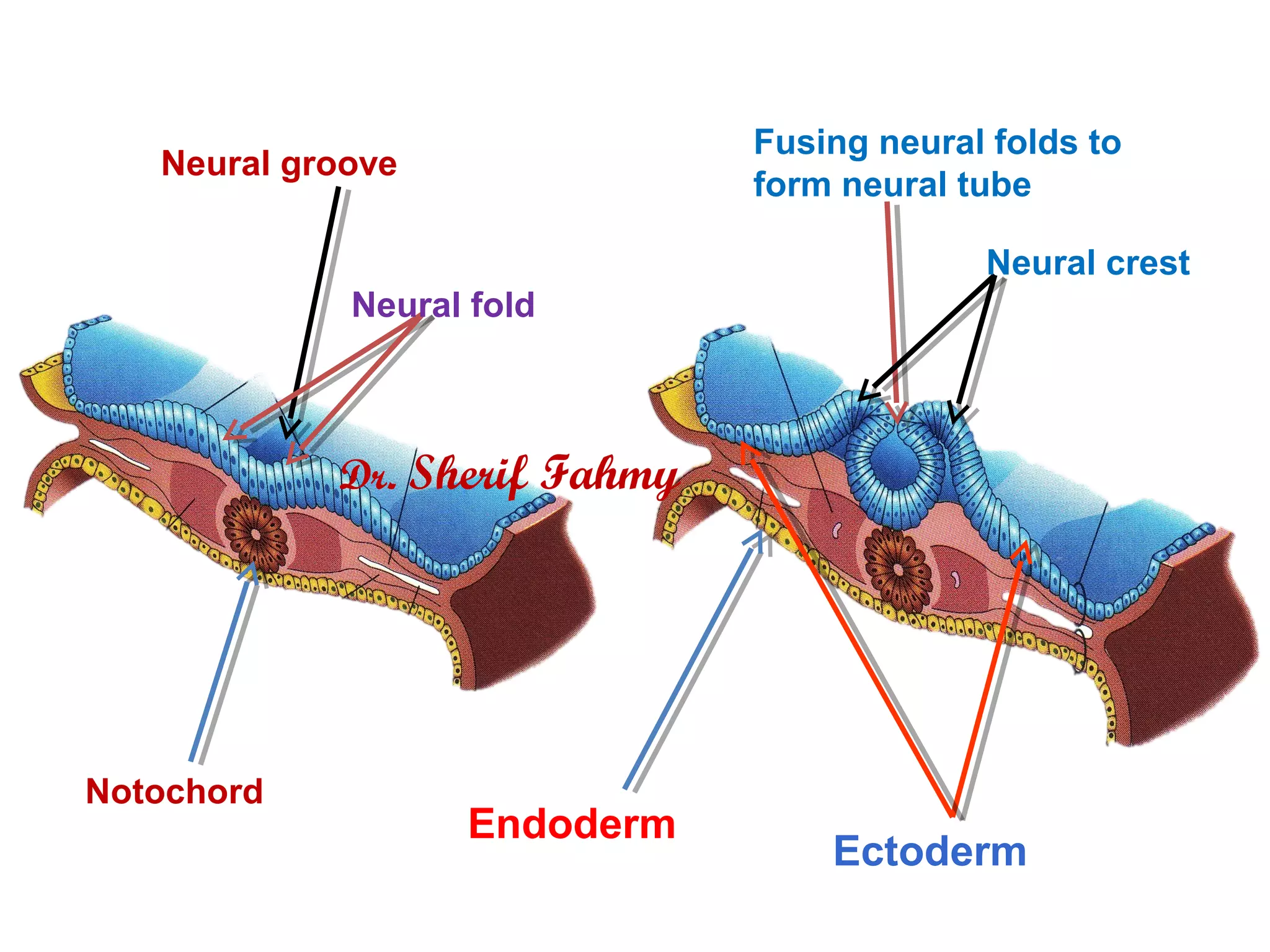
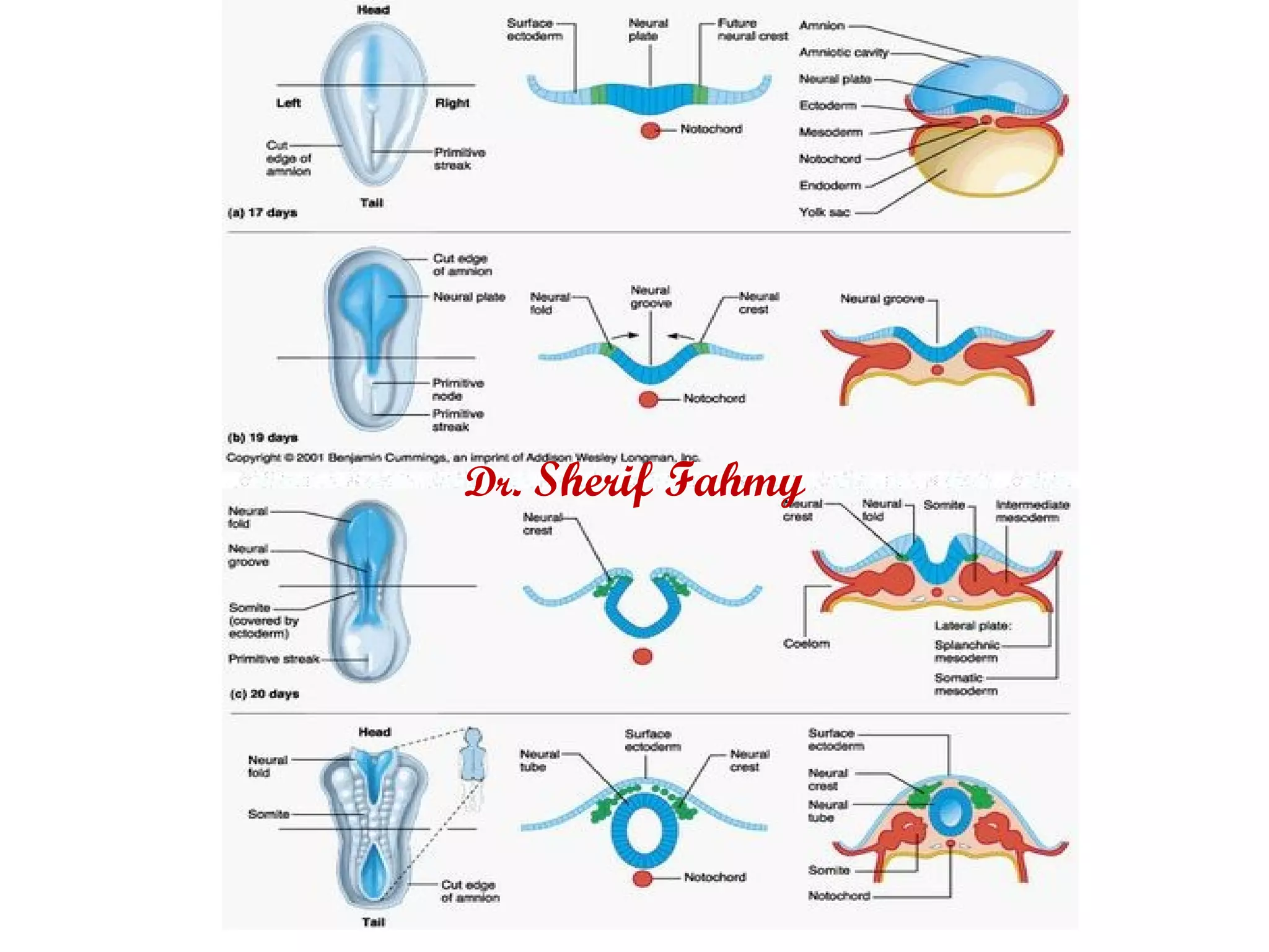
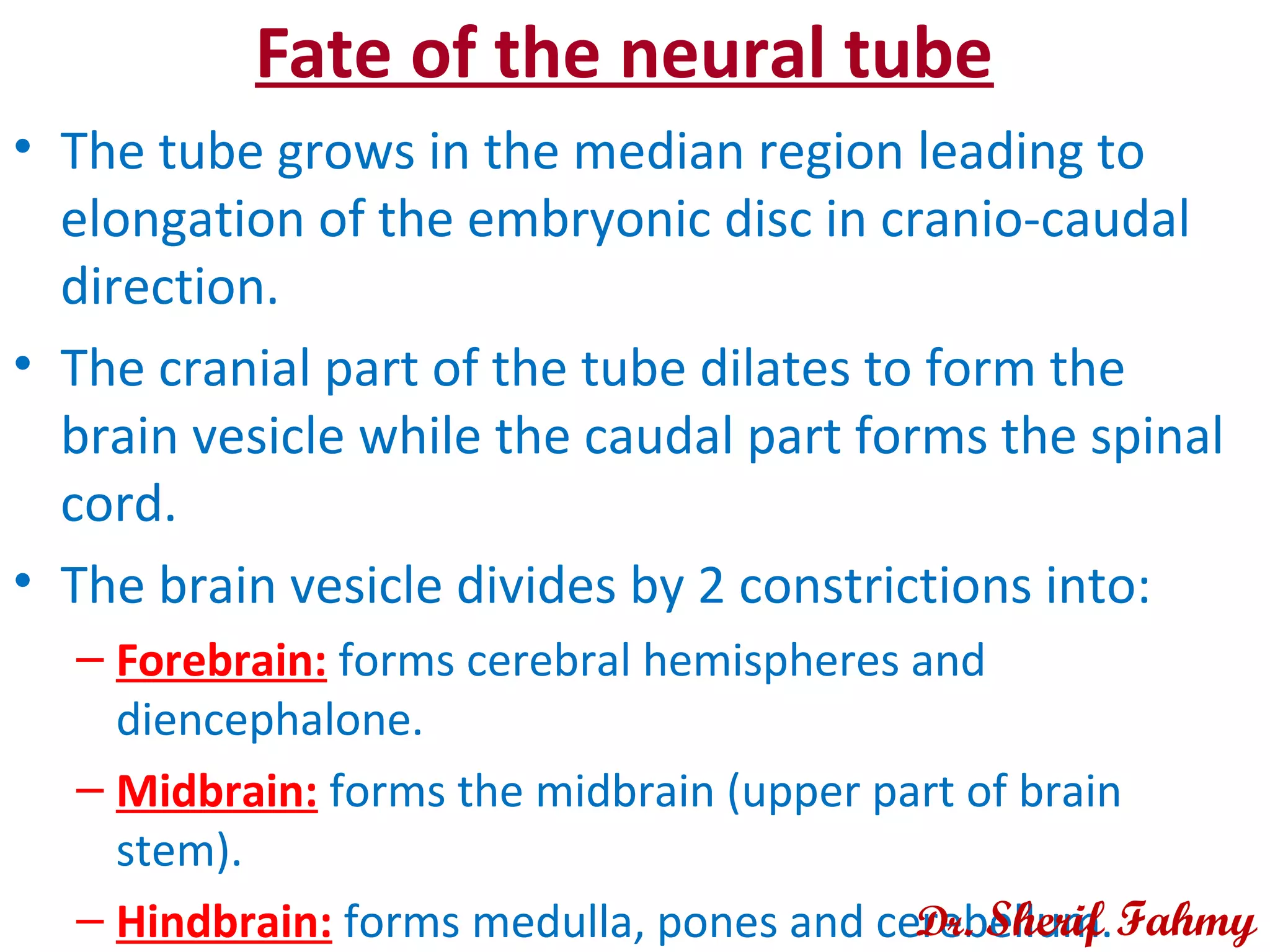
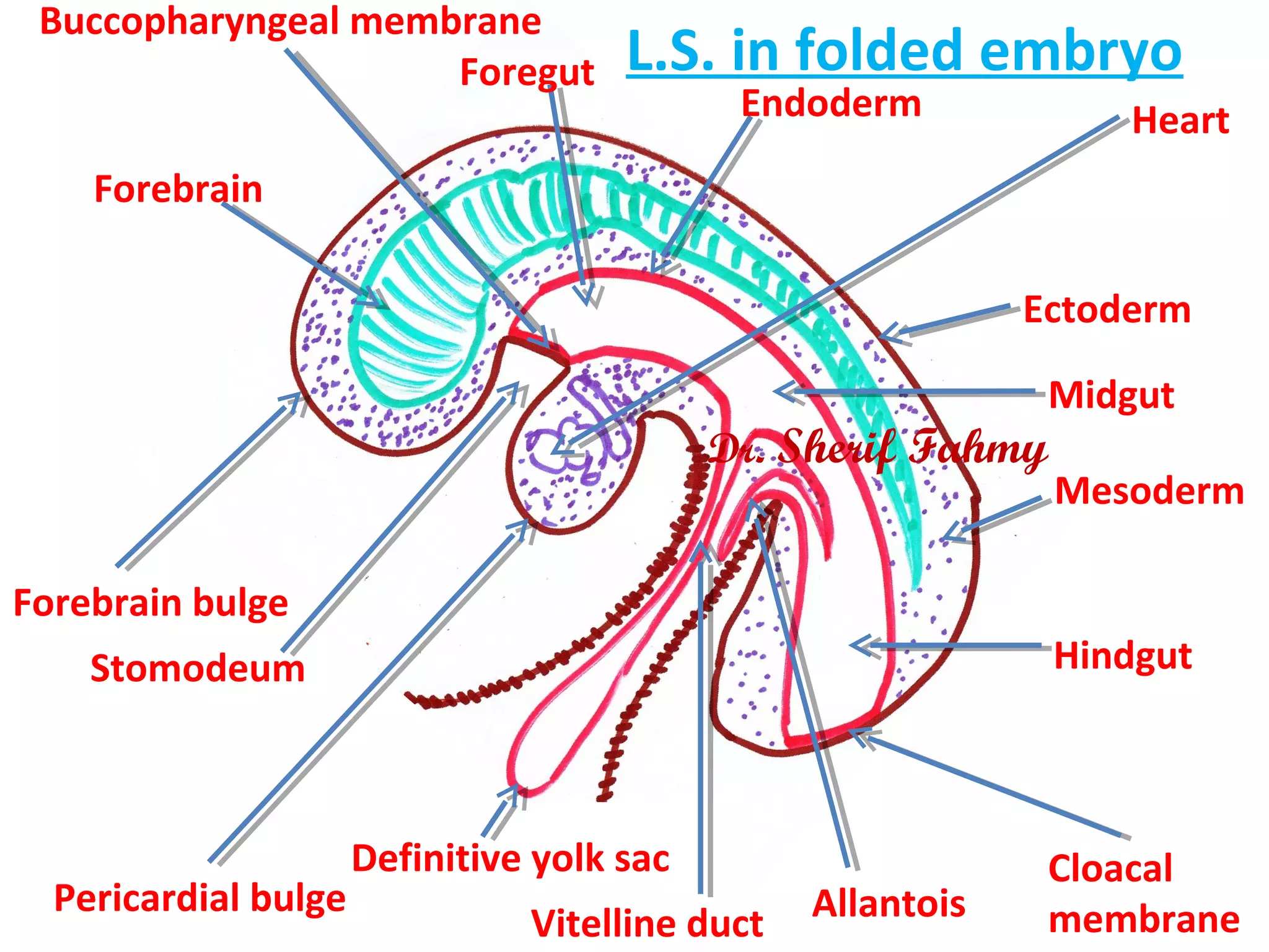
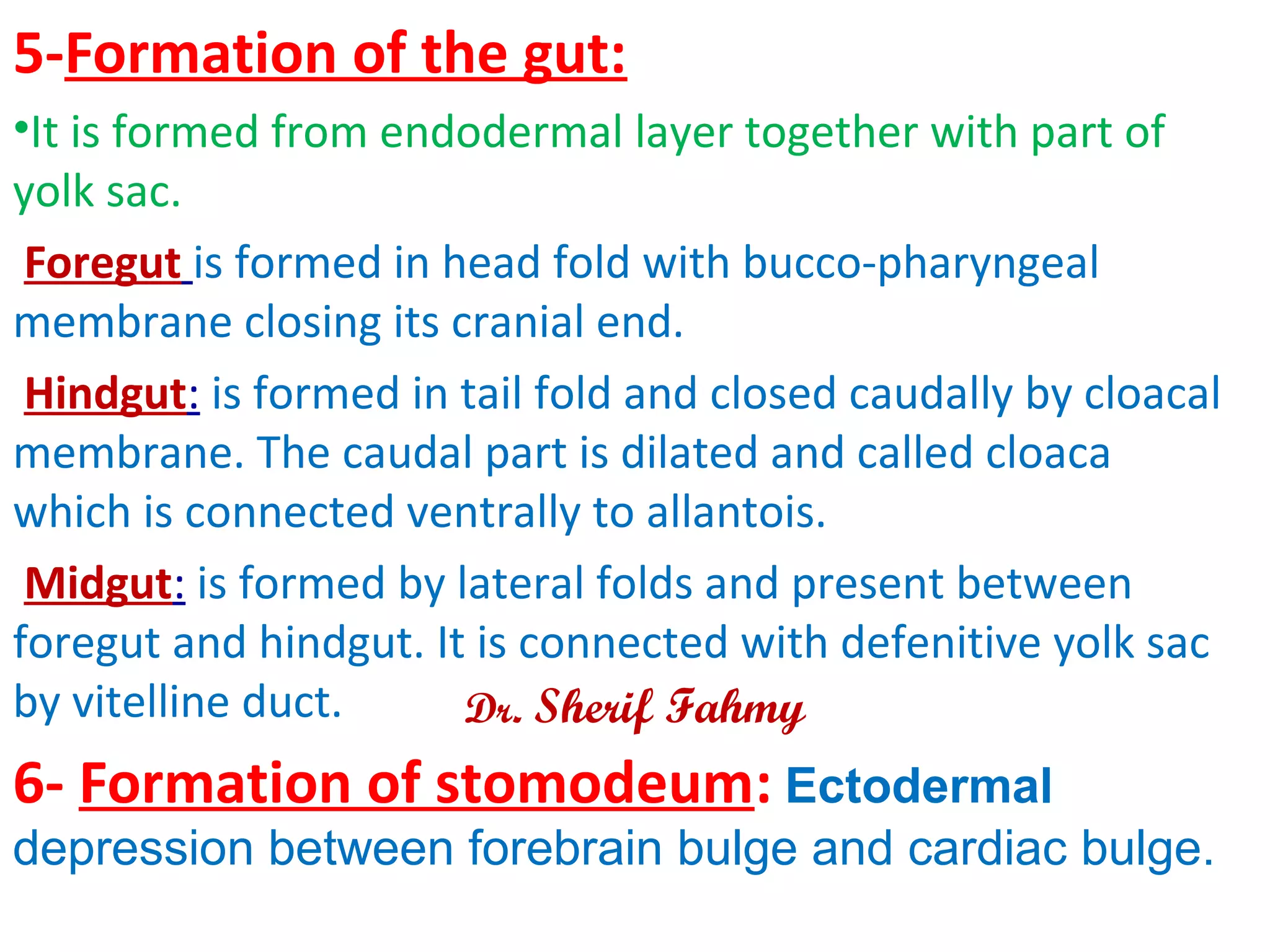
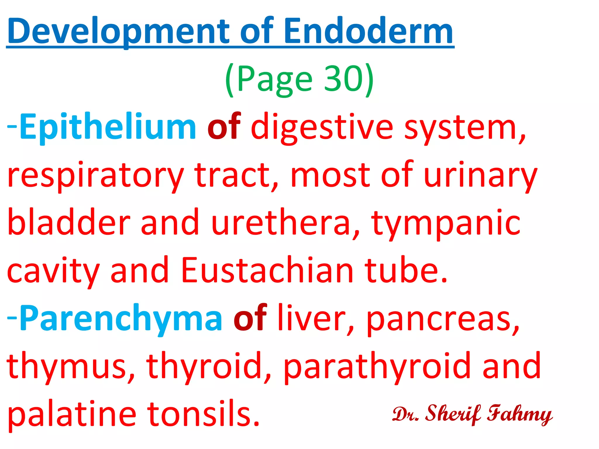
Organogenesis
Embryonic Period (4th
–
8th
week)
Dr. Sherif Fahmy
Fate of Ectoderm
Dr. Sherif Fahmy
DEVELOPMENT OF NEURAL TUBE
• Neural plate is median thickened area between
primitive node and prochordal membrane. Two
strips separate neural plate from the rest of
ectoderm which are called neural crest.
• Neural folds are raised margins of neural plate
while depressed median region is called neural
groove.
• Neural tube is formed by fusion between two
neural folds in its middle and extends cranio-
caudally. Cranial and caudal ends (neuropores) are
the last to be closed. Dr. Sherif Fahmy
Neural groove
Neural fold
Notochord
Fusing neural folds to
form neural tube
Neural crest
Ectoderm
Endoderm
Dr. Sherif Fahmy
Dr. Sherif Fahmy
Dr. Sherif Fahmy
Dr. Sherif Fahmy
Dr. Sherif Fahmy
Dr. Sherif Fahmy
Dr. Sherif Fahmy
Fate of the neural tube
• The tube grows in the median region leading to
elongation of the embryonic disc in cranio-caudal
direction.
• The cranial part of the tube dilates to form the
brain vesicle while the caudal part forms the spinal
cord.
• The brain vesicle divides by 2 constrictions into:
– Forebrain: forms cerebral hemispheres and
diencephalone.
– Midbrain: forms the midbrain (upper part of brain
stem).
– Hindbrain: forms medulla, pones and cerebellum.Dr. Sherif Fahmy
Dr. Sherif Fahmy
Fate of neural crest
• Ganglia: Sensory (of cranial and spinal
nerves), sympathetic and
parasympathetic.
• Cells: Chromaffin cells of supra-renal
medulla, Schwann cells and
melanoblasts.
• Others: Pia mater, arachnoid mater,
enamel of teeth, septa of the heart and
some bones of the skull. Dr. Sherif Fahmy
Other derivatives of ectoderm
- Otic placodes form internal ear.
- Lens placodes form lens of the eye.
- Peripheral nerves.
- Sensory epithelium in ear, nose, eye and
epidermis of skin.
- Pituitary gland.
- Anterior part of oral cavity and lower ½
of anal canal.
Dr. Sherif Fahmy
Folding
Dr. Sherif Fahmy
FOLDING OF THE EMBRYO
• It is the process by which the embryo becomes folded upon
itself.
Time of folding:
• At the end of 3rd
week and completed at the end of 4th
week.
Causes of folding:
• Rapid increase of cranio-caudal length due to rapid growth
of neural tube and somites.
• Rapid expansion of amniotic cavity.
Types of folding:
• Head and tail folds are folding of cranial and caudal parts
of the disc.
• Lateral folds are folding of lateral parts of the disc.Dr. Sherif Fahmy
Dr. Sherif Fahmy
Dr. Sherif Fahmy
Dr. Sherif Fahmy
Results of Folding
Dr. Sherif Fahmy
Dr. Sherif Fahmy
Dr. Sherif Fahmy
Embryonic disc with removed
ectoderm
Cloacal
membrane
Notochord
Paraxial
mesoderm
(somites)
Bucco-
pharyngeal
membrane
Cardiogenic
area
Septum
transversum
Peritoneal canal
Pericardium
Dr. Sherif Fahmy
Ectoderm
Mesoderm
Endoderm
Buccopharyngeal membrane
Cloacal
membrane
Hindgut
Midgut
Foregut
Forebrain
Forebrain bulge
Pericardial bulge
Vitelline duct Allantois
Definitive yolk sac
Stomodeum
L.S. in folded embryo
Heart
Dr. Sherif Fahmy
Peritoneal
canals
Gut
Ventral mersentry
Dorsal mesentry
Dr. Sherif Fahmy
RESULTS OF FOLDING
1-Cylindrical appearance: Transformation of
emryonic disc to cylindrical shape.
2- Amniotic cavity: Before folding it lies dorsal to
embryonic disc, after folding, it surrounds all aspects of
the embryo.
3- Formation of definitive yolk sac: It is the part
of yolk sac outside the embryo in the umbilical cord.
4- Formation of primitive umbilical ring: It is a
ventral defect in anterior abdominal wall that contains
connecting stalk, allantois and vitello-intestinal duct
Dr. Sherif Fahmy
5-Formation of the gut:
•It is formed from endodermal layer together with part of
yolk sac.
Foregut is formed in head fold with bucco-pharyngeal
membrane closing its cranial end.
Hindgut: is formed in tail fold and closed caudally by cloacal
membrane. The caudal part is dilated and called cloaca
which is connected ventrally to allantois.
Midgut: is formed by lateral folds and present between
foregut and hindgut. It is connected with defenitive yolk sac
by vitelline duct.
6- Formation of stomodeum: Ectodermal
depression between forebrain bulge and cardiac bulge.
Dr. Sherif Fahmy
7- Formation of mesenteries: Ventral and
dorsal mesenteries are formed around gut.
8- Reversal of positions:
-Heart and pericardium become cranial to septum
transversum (before folding septum transversum is
most cranial).
-Connecting stalk becomes ventral and more cranial
inspite of being most caudal.
Dr. Sherif Fahmy
Somites After Folding
Dr. Sherif Fahmy
Dr. Sherif Fahmy
Dr. Sherif Fahmy
Dr. Sherif Fahmy
Development of Endoderm
(Page 30)
-Epithelium of digestive system,
respiratory tract, most of urinary
bladder and urethera, tympanic
cavity and Eustachian tube.
-Parenchyma of liver, pancreas,
thymus, thyroid, parathyroid and
palatine tonsils. Dr. Sherif Fahmy
Fetal Membranes
Dr. Sherif Fahmy
Fetal membranes:
1- Chorion
2- Placenta.
2- Amnion.
3- Umbilical cord.
4- Yolk sac.
Dr. Sherif Fahmy
Chorion
Dr. Sherif Fahmy
It is the wall of chorionic vesicle.
Time: Chorionic vesicle is formed at the 12th
day
by the formation of extra-embryonic mesoderm.
Structure of chorion:
1- Syncytiotrophoblast.
2- Cytotrophoblast.
3- Somatic extra-embryonic mesoderm.
Chorionic velli:
1- Primary.
2- Secondary.
3- Tertiary.
Dr. Sherif Fahmy
Connecting stalk
Somatic mesoderm
Syncytio-
trophoblast
Cyto-
trophoblast
Chorion
Chorionic Vesicle
Dr. Sherif Fahmy
Dr. Sherif Fahmy
Dr. Sherif Fahmy
Dr. Sherif Fahmy
Primary
chorionic
villus
Cyto-
trophoblast
Syncytio-
trophoblast
Dr. Sherif Fahmy
Dr. Sherif Fahmy
Syncytio-
trophoblast
Cyto-
trophoblast
Somatic
mesoderm
Secondary
chorionic villus
Dr. Sherif Fahmy
Dr. Sherif Fahmy
Syncytio-
trophoblast
Cyto-
trophoblast
Mesoderm
Fetal blood
vessels
Tertiary
chorioniv villus
Dr. Sherif Fahmy
Decidua
basalis
Chorion frondosum
Chorionic
plate
Chorion leave
Dr. Sherif Fahmy
Dr. Sherif Fahmy
1- Primary chorionic velli (start of 3rd
week):
cyncytiotropholblasts and cytotrophoblast.
2- Secondary chorionic velli (middle of 3rd
week):
Cyncytiotrophoblast, cytotrophoblast and
mesoderm (in the central core).
3- Tertiary chorionic velli (end of 3rd
week):
formation of fetal blood vessels in the mesoderm.
-Tertiary velli, opposite decidua basalis form side
branches and called chorion frondosum while under
decidua capsularis it will degenerates to form chorion
leave.
Dr. Sherif Fahmy
Dr. Sherif Fahmy
PLACENTA
(Page 38)
Dr. Sherif Fahmy
Morphology of Placenta
• It is the organ of exchange of materials between fetal
and maternal blood.
• Shape: Disc like.
• Surfaces:
• -Fetal surface: It is covered with amnion and fetal blood
vessels. Umbilical cord is attached near the center of
this surface.
• -Maternal surface: Shows 15 – 20 rounded elevations
(cotyledons) with septa inbetween).
• Diameter: 15 -25 cm.
• Thickness: About 3 cm.
• Weight: About 500 – 600 gm
• Site: At original implantation site which is upper part of
posterior wall of uterus.
Dr. Sherif Fahmy
Dr. Sherif Fahmy
Cotyledon
Groove between
cotyledons
Umbilical cord
Maternal surface
Dr. Sherif Fahmy
Dr. Sherif Fahmy
Fetal surface covered
with amnion
Umbilical cord
Dr. Sherif Fahmy
Dr. Sherif Fahmy
Formation of Placenta
Dr. Sherif Fahmy
Dr. Sherif Fahmy
Dr. Sherif Fahmy
Dr. Sherif Fahmy
Dr. Sherif Fahmy

Embryonic period 4th – 8th week & Folding (General Embryology)