Downloaded 190 times
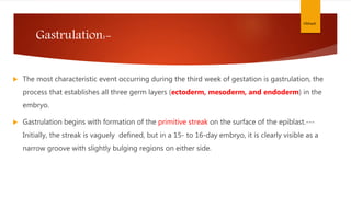

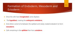
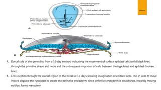
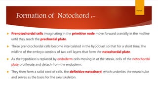
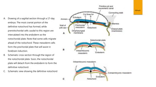


During the third week of development, gastrulation occurs which establishes the three germ layers - ectoderm, mesoderm, and endoderm. Gastrulation begins with the formation of the primitive streak on the surface of the epiblast. Cells migrate through the primitive streak and node, some displacing the hypoblast to form endoderm, while others become mesoderm between the endoderm and remaining ectoderm. This results in the formation of the notochord, and the germ layers differentiate into various tissues and organs.


![3.1_Third_Week_of_Development[1].pptx](https://cdn.slidesharecdn.com/ss_thumbnails/3-221118124853-25c6f4d7-thumbnail.jpg?width=640&height=640&fit=bounds)


