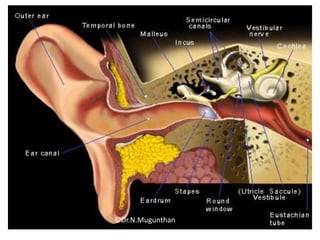
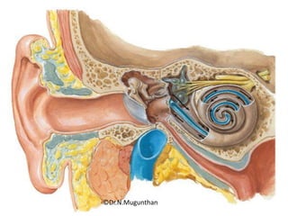
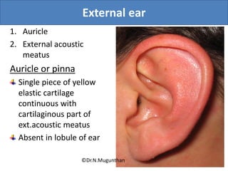
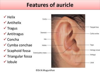
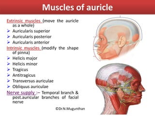
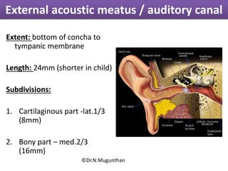
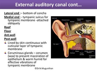
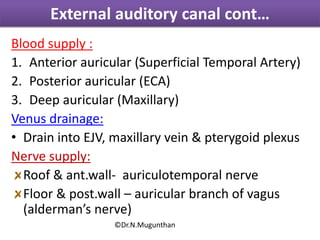
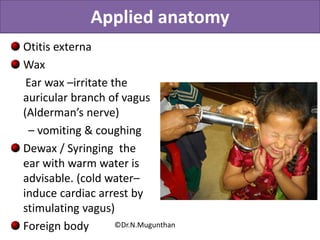
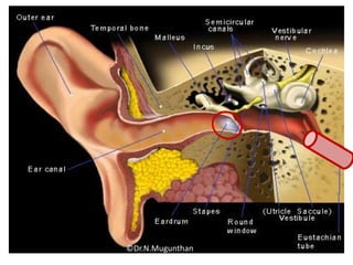
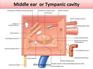
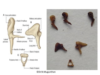
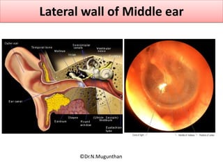
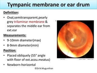
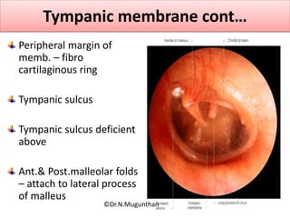
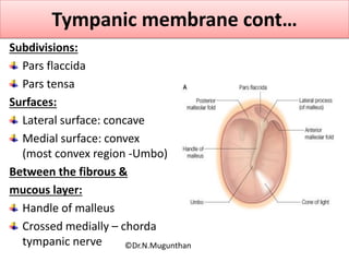
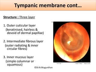
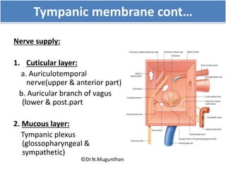
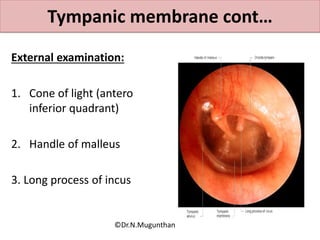
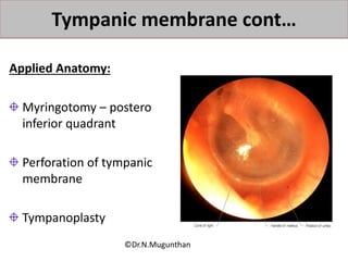
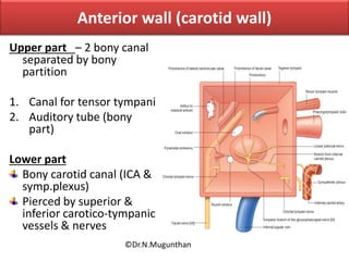
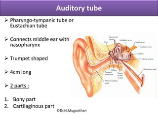
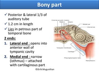
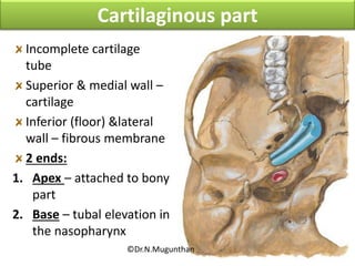
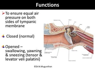
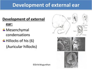
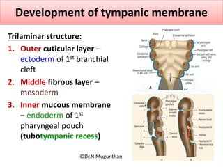
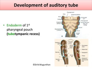
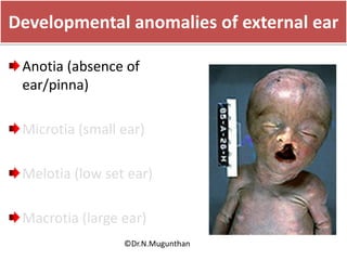
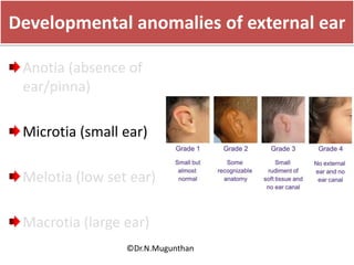
EXTERNAL EAR, TYMPANIC MEMBRANE &
AUDITORY TUBE
Lecture By
Dr.N.Mugunthan. MBBS.,MS.,DNB.,MNAMS.,PhD.,MBA (Hosp.Adm.).,
Associate Professor
MGMC & RI
LEARNING OBJECTIVES
Subdivisions of ear
Concept of hearing
External ear (auricle & external auditory
meatus)
 Tympanic membrane or ear drum
Auditory tube
Development of external ear & it’s anomalies
Applied anatomy
©Dr.N.Mugunthan
Ear
• Ear is an organ of hearing
• Human ear is most sensitive to sound frequency
ranging between 1500 to 3000 Hertz
3 Parts
1. External ear
2. Middle ear
3. Internal ear
©Dr.N.Mugunthan
©Dr.N.Mugunthan
©Dr.N.Mugunthan
External ear
1. Auricle
2. External acoustic
meatus
Auricle or pinna
Single piece of yellow
elastic cartilage
continuous with
cartilaginous part of
ext.acoustic meatus
Absent in lobule of ear
©Dr.N.Mugunthan
Features of auricle
 Helix
 Antihelix
 Tragus
 Antitragus
 Concha
 Cymba conchae
 Scaphoid fossa
 Triangular fossa
 lobule
©Dr.N.Mugunthan
Muscles of auricle
Extrinsic muscles (move the auricle
as a whole)
 Auricularis superior
 Auricularis posterior
 Auricularis anterior
Intrinsic muscles (modify the shape
of pinna)
 Helicis major
 Helicis minor
 Tragicus
 Antitragicus
 Transversus auriculae
 Obliquus auriculae
Nerve supply :– Temporal branch &
post.auricular branches of facial
nerve
©Dr.N.Mugunthan
External acoustic meatus / auditory canal
Extent: bottom of concha to
tympanic membrane
Length: 24mm (shorter in child)
Subdivisions:
1. Cartilaginous part -lat.1/3
(8mm)
2. Bony part – med.2/3
(16mm)
©Dr.N.Mugunthan
Lateral end :– bottom of concha
Medial end :- tympanic sulcus for
tympanic membrane -attached
obliquely
Roof
Floor
Ant.wall
Post.wall
Lined by skin continuous with
cuticular layer of tympanic
membrane
Ceruminous glands – cerumen
(wax) to prevent maceration of
epithelium & warm humid for
effective vibrations of
tympanic membrane
External auditory canal cont…
©Dr.N.Mugunthan
Blood supply :
1. Anterior auricular (Superficial Temporal Artery)
2. Posterior auricular (ECA)
3. Deep auricular (Maxillary)
Venus drainage:
• Drain into EJV, maxillary vein & pterygoid plexus
Nerve supply:
Roof & ant.wall- auriculotemporal nerve
Floor & post.wall – auricular branch of vagus
(alderman’s nerve)
External auditory canal cont…
©Dr.N.Mugunthan
Applied anatomy
Otitis externa
Wax
Ear wax –irritate the
auricular branch of vagus
(Alderman’s nerve)
– vomiting & coughing
Dewax / Syringing the
ear with warm water is
advisable. (cold water–
induce cardiac arrest by
stimulating vagus)
Foreign body ©Dr.N.Mugunthan
©Dr.N.Mugunthan
Middle ear or Tympanic cavity
©Dr.N.Mugunthan
©Dr.N.Mugunthan
Lateral wall of Middle ear
©Dr.N.Mugunthan
Tympanic membrane or ear drum
Definition:
• Oval,semitransparent,pearly
grey trilaminar membrane &
separates the middle ear from
ext.ear
Measurements:
• 9-10mm diameter(max)
• 8-9mm diameter(min)
Position:
• Placed obliquely (55° angle
with floor of ext.acou.meatus)
• Newborn-horizontal
©Dr.N.Mugunthan
Tympanic membrane cont…
Peripheral margin of
memb. – fibro
cartilaginous ring
Tympanic sulcus
Tympanic sulcus deficient
above
Ant.& Post.malleolar folds
– attach to lateral process
of malleus
©Dr.N.Mugunthan
Subdivisions:
Pars flaccida
Pars tensa
Surfaces:
Lateral surface: concave
Medial surface: convex
(most convex region -Umbo)
Between the fibrous &
mucous layer:
Handle of malleus
Crossed medially – chorda
tympanic nerve
Tympanic membrane cont…
©Dr.N.Mugunthan
Structure : Three layer
1. Outer cuticular layer
(keratinised, hairless &
devoid of dermal papillae)
2. Intermediate fibrous layer
(outer radiating & inner
circular fibres)
3. Inner mucous layer
(simple columnar or
squamous)
Tympanic membrane cont…
©Dr.N.Mugunthan
Arterial supply:
Deep auricular artery (Max.Art)
Stylomastoid artery (Post.auric)
Anterior tympanic artery (Max.Art)
Venous drainage:
Outer layer – drain into EJV
Inner layer - drain into transverse sinus &
pterygoid plexus
Tympanic membrane cont…
©Dr.N.Mugunthan
Tympanic membrane cont…
Nerve supply:
1. Cuticular layer:
a. Auriculotemporal
nerve(upper & anterior part)
b. Auricular branch of vagus
(lower & post.part
2. Mucous layer:
Tympanic plexus
(glossopharyngeal &
sympathetic)
©Dr.N.Mugunthan
External examination:
1. Cone of light (antero
inferior quadrant)
2. Handle of malleus
3. Long process of incus
Tympanic membrane cont…
©Dr.N.Mugunthan
Applied Anatomy:
Myringotomy – postero
inferior quadrant
Perforation of tympanic
membrane
Tympanoplasty
Tympanic membrane cont…
©Dr.N.Mugunthan
Anterior wall (carotid wall)
Upper part – 2 bony canal
separated by bony
partition
1. Canal for tensor tympani
2. Auditory tube (bony
part)
Lower part
Bony carotid canal (ICA &
symp.plexus)
Pierced by superior &
inferior carotico-tympanic
vessels & nerves
©Dr.N.Mugunthan
Auditory tube
 Pharyngo-tympanic tube or
Eustachian tube
 Connects middle ear with
nasopharynx
 Trumpet shaped
 4cm long
 2 parts :
1. Bony part
2. Cartilaginous part
©Dr.N.Mugunthan
Bony part
 Posterior & lateral 1/3 of
auditory tube
 1.2 cm in length
 Lies in petrous part of
temporal bone
2 ends:
1. Lateral end : opens into
anterior wall of
tympanic cavity
2. Medial end : narrow
(isthmus) – attached
with cartilaginous part
©Dr.N.Mugunthan
Incomplete cartilage
tube
Superior & medial wall –
cartilage
Inferior (floor) &lateral
wall – fibrous membrane
2 ends:
1. Apex – attached to bony
part
2. Base – tubal elevation in
the nasopharynx
Cartilaginous part
©Dr.N.Mugunthan
Functions
To ensure equal air
pressure on both
sides of tympanic
membrane
Closed (normal)
Opened –
swallowing, yawning
& sneezing (tensor &
levator veli palatini)
©Dr.N.Mugunthan
Applied anatomy
Spread of middle air infection (ASOM &
CSOM)
Eustachian catarrh
Tubal block (enlarged tubal tonsil)– retraction
of tympanic membrane
Tmt
1. Nasal decongestants
2. Inflate the tube
©Dr.N.Mugunthan
Development of external ear
Development of external
ear:
Mesenchymal
condensations
Hillocks of his (6)
(Auricular hillocks)
©Dr.N.Mugunthan
Development of tympanic membrane
Trilaminar structure:
1. Outer cuticular layer –
ectoderm of 1st branchial
cleft
2. Middle fibrous layer –
mesoderm
3. Inner mucous membrane
– endoderm of 1st
pharyngeal pouch
(tubotympanic recess)
©Dr.N.Mugunthan
Development of auditory tube
• Endoderm of 1st
pharyngeal pouch
(tubotympanic recess)
©Dr.N.Mugunthan
Developmental anomalies of external ear
Anotia (absence of
ear/pinna)
Microtia (small ear)
Melotia (low set ear)
Macrotia (large ear)
©Dr.N.Mugunthan
Developmental anomalies of external ear
Anotia (absence of
ear/pinna)
Microtia (small ear)
Melotia (low set ear)
Macrotia (large ear)
©Dr.N.Mugunthan
Developmental anomalies of external ear
Anotia (absence of
ear/pinna)
Microtia (small ear)
Melotia (low set ear)
Macrotia (large ear)
©Dr.N.Mugunthan
Developmental anomalies of external ear
Anotia (absence of
ear/pinna)
Microtia (small ear)
Melotia (low set ear)
Macrotia (large ear)
©Dr.N.Mugunthan
Accessory tragus
©Dr.N.Mugunthan
Summary
Parts of ear : External, middle & internal ear.
External ear: auricle & ext. auditory meatus
Tympanic membrane:
Attachments
3 Layers
Structures in the middle layer
Development
Auditory tube:
Parts
Functions
Applied anatomy:©Dr.N.Mugunthan
External ear,tympanic membrane and auditory tube  Dr.N.Mugunthan.M.S.,

External ear,tympanic membrane and auditory tube Dr.N.Mugunthan.M.S.,

  • 1.
    EXTERNAL EAR, TYMPANICMEMBRANE & AUDITORY TUBE Lecture By Dr.N.Mugunthan. MBBS.,MS.,DNB.,MNAMS.,PhD.,MBA (Hosp.Adm.)., Associate Professor MGMC & RI
  • 2.
    LEARNING OBJECTIVES Subdivisions ofear Concept of hearing External ear (auricle & external auditory meatus)  Tympanic membrane or ear drum Auditory tube Development of external ear & it’s anomalies Applied anatomy ©Dr.N.Mugunthan
  • 3.
    Ear • Ear isan organ of hearing • Human ear is most sensitive to sound frequency ranging between 1500 to 3000 Hertz 3 Parts 1. External ear 2. Middle ear 3. Internal ear ©Dr.N.Mugunthan
  • 4.
  • 5.
  • 6.
    External ear 1. Auricle 2.External acoustic meatus Auricle or pinna Single piece of yellow elastic cartilage continuous with cartilaginous part of ext.acoustic meatus Absent in lobule of ear ©Dr.N.Mugunthan
  • 7.
    Features of auricle Helix  Antihelix  Tragus  Antitragus  Concha  Cymba conchae  Scaphoid fossa  Triangular fossa  lobule ©Dr.N.Mugunthan
  • 8.
    Muscles of auricle Extrinsicmuscles (move the auricle as a whole)  Auricularis superior  Auricularis posterior  Auricularis anterior Intrinsic muscles (modify the shape of pinna)  Helicis major  Helicis minor  Tragicus  Antitragicus  Transversus auriculae  Obliquus auriculae Nerve supply :– Temporal branch & post.auricular branches of facial nerve ©Dr.N.Mugunthan
  • 9.
    External acoustic meatus/ auditory canal Extent: bottom of concha to tympanic membrane Length: 24mm (shorter in child) Subdivisions: 1. Cartilaginous part -lat.1/3 (8mm) 2. Bony part – med.2/3 (16mm) ©Dr.N.Mugunthan
  • 10.
    Lateral end :–bottom of concha Medial end :- tympanic sulcus for tympanic membrane -attached obliquely Roof Floor Ant.wall Post.wall Lined by skin continuous with cuticular layer of tympanic membrane Ceruminous glands – cerumen (wax) to prevent maceration of epithelium & warm humid for effective vibrations of tympanic membrane External auditory canal cont… ©Dr.N.Mugunthan
  • 11.
    Blood supply : 1.Anterior auricular (Superficial Temporal Artery) 2. Posterior auricular (ECA) 3. Deep auricular (Maxillary) Venus drainage: • Drain into EJV, maxillary vein & pterygoid plexus Nerve supply: Roof & ant.wall- auriculotemporal nerve Floor & post.wall – auricular branch of vagus (alderman’s nerve) External auditory canal cont… ©Dr.N.Mugunthan
  • 12.
    Applied anatomy Otitis externa Wax Earwax –irritate the auricular branch of vagus (Alderman’s nerve) – vomiting & coughing Dewax / Syringing the ear with warm water is advisable. (cold water– induce cardiac arrest by stimulating vagus) Foreign body ©Dr.N.Mugunthan
  • 13.
  • 14.
    Middle ear orTympanic cavity ©Dr.N.Mugunthan
  • 15.
  • 16.
    Lateral wall ofMiddle ear ©Dr.N.Mugunthan
  • 17.
    Tympanic membrane orear drum Definition: • Oval,semitransparent,pearly grey trilaminar membrane & separates the middle ear from ext.ear Measurements: • 9-10mm diameter(max) • 8-9mm diameter(min) Position: • Placed obliquely (55° angle with floor of ext.acou.meatus) • Newborn-horizontal ©Dr.N.Mugunthan
  • 18.
    Tympanic membrane cont… Peripheralmargin of memb. – fibro cartilaginous ring Tympanic sulcus Tympanic sulcus deficient above Ant.& Post.malleolar folds – attach to lateral process of malleus ©Dr.N.Mugunthan
  • 19.
    Subdivisions: Pars flaccida Pars tensa Surfaces: Lateralsurface: concave Medial surface: convex (most convex region -Umbo) Between the fibrous & mucous layer: Handle of malleus Crossed medially – chorda tympanic nerve Tympanic membrane cont… ©Dr.N.Mugunthan
  • 20.
    Structure : Threelayer 1. Outer cuticular layer (keratinised, hairless & devoid of dermal papillae) 2. Intermediate fibrous layer (outer radiating & inner circular fibres) 3. Inner mucous layer (simple columnar or squamous) Tympanic membrane cont… ©Dr.N.Mugunthan
  • 21.
    Arterial supply: Deep auricularartery (Max.Art) Stylomastoid artery (Post.auric) Anterior tympanic artery (Max.Art) Venous drainage: Outer layer – drain into EJV Inner layer - drain into transverse sinus & pterygoid plexus Tympanic membrane cont… ©Dr.N.Mugunthan
  • 22.
    Tympanic membrane cont… Nervesupply: 1. Cuticular layer: a. Auriculotemporal nerve(upper & anterior part) b. Auricular branch of vagus (lower & post.part 2. Mucous layer: Tympanic plexus (glossopharyngeal & sympathetic) ©Dr.N.Mugunthan
  • 23.
    External examination: 1. Coneof light (antero inferior quadrant) 2. Handle of malleus 3. Long process of incus Tympanic membrane cont… ©Dr.N.Mugunthan
  • 24.
    Applied Anatomy: Myringotomy –postero inferior quadrant Perforation of tympanic membrane Tympanoplasty Tympanic membrane cont… ©Dr.N.Mugunthan
  • 25.
    Anterior wall (carotidwall) Upper part – 2 bony canal separated by bony partition 1. Canal for tensor tympani 2. Auditory tube (bony part) Lower part Bony carotid canal (ICA & symp.plexus) Pierced by superior & inferior carotico-tympanic vessels & nerves ©Dr.N.Mugunthan
  • 26.
    Auditory tube  Pharyngo-tympanictube or Eustachian tube  Connects middle ear with nasopharynx  Trumpet shaped  4cm long  2 parts : 1. Bony part 2. Cartilaginous part ©Dr.N.Mugunthan
  • 27.
    Bony part  Posterior& lateral 1/3 of auditory tube  1.2 cm in length  Lies in petrous part of temporal bone 2 ends: 1. Lateral end : opens into anterior wall of tympanic cavity 2. Medial end : narrow (isthmus) – attached with cartilaginous part ©Dr.N.Mugunthan
  • 28.
    Incomplete cartilage tube Superior &medial wall – cartilage Inferior (floor) &lateral wall – fibrous membrane 2 ends: 1. Apex – attached to bony part 2. Base – tubal elevation in the nasopharynx Cartilaginous part ©Dr.N.Mugunthan
  • 29.
    Functions To ensure equalair pressure on both sides of tympanic membrane Closed (normal) Opened – swallowing, yawning & sneezing (tensor & levator veli palatini) ©Dr.N.Mugunthan
  • 30.
    Applied anatomy Spread ofmiddle air infection (ASOM & CSOM) Eustachian catarrh Tubal block (enlarged tubal tonsil)– retraction of tympanic membrane Tmt 1. Nasal decongestants 2. Inflate the tube ©Dr.N.Mugunthan
  • 31.
    Development of externalear Development of external ear: Mesenchymal condensations Hillocks of his (6) (Auricular hillocks) ©Dr.N.Mugunthan
  • 32.
    Development of tympanicmembrane Trilaminar structure: 1. Outer cuticular layer – ectoderm of 1st branchial cleft 2. Middle fibrous layer – mesoderm 3. Inner mucous membrane – endoderm of 1st pharyngeal pouch (tubotympanic recess) ©Dr.N.Mugunthan
  • 33.
    Development of auditorytube • Endoderm of 1st pharyngeal pouch (tubotympanic recess) ©Dr.N.Mugunthan
  • 34.
    Developmental anomalies ofexternal ear Anotia (absence of ear/pinna) Microtia (small ear) Melotia (low set ear) Macrotia (large ear) ©Dr.N.Mugunthan
  • 35.
    Developmental anomalies ofexternal ear Anotia (absence of ear/pinna) Microtia (small ear) Melotia (low set ear) Macrotia (large ear) ©Dr.N.Mugunthan
  • 36.
    Developmental anomalies ofexternal ear Anotia (absence of ear/pinna) Microtia (small ear) Melotia (low set ear) Macrotia (large ear) ©Dr.N.Mugunthan
  • 37.
    Developmental anomalies ofexternal ear Anotia (absence of ear/pinna) Microtia (small ear) Melotia (low set ear) Macrotia (large ear) ©Dr.N.Mugunthan
  • 38.
  • 39.
    Summary Parts of ear: External, middle & internal ear. External ear: auricle & ext. auditory meatus Tympanic membrane: Attachments 3 Layers Structures in the middle layer Development Auditory tube: Parts Functions Applied anatomy:©Dr.N.Mugunthan