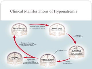
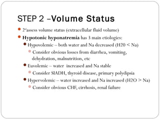
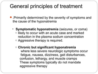
Acute kidney injury (AKI) is a potentially life-threatening
syndrome that occurs primarily in hospitalized patients
and frequently complicates the course of critically ill
patient.
Acute Kidney Injury is is (abrupt) reduction in kidney functions as evidence by changed in laboratory values; serum creatinine, blood urea nitrogen(BUN)and urine output
Interstitial lung disease is a general category that includes many different lung conditions. All interstitial lung diseases affect the interstitium, a part of the lungs' anatomic structure.
Some of the types of interstitial lung disease include:
Interstitial pneumonia: Bacteria, viruses, or fungi may infect the interstitium of the lung. A bacterium called Mycoplasma pneumonia is the most common cause.
Idiopathic pulmonary fibrosis : A chronic, progressive form of fibrosis (scarring) of the interstitium. Its cause is unknown.
Nonspecific interstitial pneumonitis: Interstitial lung disease that's often present with autoimmune conditions (such as rheumatoid arthritis or scleroderma).
ARTIFICIAL INTELLIGENCE IN HEALTHCARE.pdfAnujkumaranit
Artificial intelligence (AI) refers to the simulation of human intelligence processes by machines, especially computer systems. It encompasses tasks such as learning, reasoning, problem-solving, perception, and language understanding. AI technologies are revolutionizing various fields, from healthcare to finance, by enabling machines to perform tasks that typically require human intelligence.
The prostate is an exocrine gland of the male mammalian reproductive system
It is a walnut-sized gland that forms part of the male reproductive system and is located in front of the rectum and just below the urinary bladder
Function is to store and secrete a clear, slightly alkaline fluid that constitutes 10-30% of the volume of the seminal fluid that along with the spermatozoa, constitutes semen
A healthy human prostate measures (4cm-vertical, by 3cm-horizontal, 2cm ant-post ).
It surrounds the urethra just below the urinary bladder. It has anterior, median, posterior and two lateral lobes
It’s work is regulated by androgens which are responsible for male sex characteristics
Generalised disease of the prostate due to hormonal derangement which leads to non malignant enlargement of the gland (increase in the number of epithelial cells and stromal tissue)to cause compression of the urethra leading to symptoms (LUTS
Anti ulcer drugs and their Advance pharmacology ||
Anti-ulcer drugs are medications used to prevent and treat ulcers in the stomach and upper part of the small intestine (duodenal ulcers). These ulcers are often caused by an imbalance between stomach acid and the mucosal lining, which protects the stomach lining.
||Scope: Overview of various classes of anti-ulcer drugs, their mechanisms of action, indications, side effects, and clinical considerations.
Report Back from SGO 2024: What’s the Latest in Cervical Cancer?bkling
Are you curious about what’s new in cervical cancer research or unsure what the findings mean? Join Dr. Emily Ko, a gynecologic oncologist at Penn Medicine, to learn about the latest updates from the Society of Gynecologic Oncology (SGO) 2024 Annual Meeting on Women’s Cancer. Dr. Ko will discuss what the research presented at the conference means for you and answer your questions about the new developments.
TEST BANK for Operations Management, 14th Edition by William J. Stevenson, Ve...kevinkariuki227
TEST BANK for Operations Management, 14th Edition by William J. Stevenson, Verified Chapters 1 - 19, Complete Newest Version.pdf
New Directions in Targeted Therapeutic Approaches for Older Adults With Mantl...i3 Health
i3 Health is pleased to make the speaker slides from this activity available for use as a non-accredited self-study or teaching resource.
This slide deck presented by Dr. Kami Maddocks, Professor-Clinical in the Division of Hematology and
Associate Division Director for Ambulatory Operations
The Ohio State University Comprehensive Cancer Center, will provide insight into new directions in targeted therapeutic approaches for older adults with mantle cell lymphoma.
STATEMENT OF NEED
Mantle cell lymphoma (MCL) is a rare, aggressive B-cell non-Hodgkin lymphoma (NHL) accounting for 5% to 7% of all lymphomas. Its prognosis ranges from indolent disease that does not require treatment for years to very aggressive disease, which is associated with poor survival (Silkenstedt et al, 2021). Typically, MCL is diagnosed at advanced stage and in older patients who cannot tolerate intensive therapy (NCCN, 2022). Although recent advances have slightly increased remission rates, recurrence and relapse remain very common, leading to a median overall survival between 3 and 6 years (LLS, 2021). Though there are several effective options, progress is still needed towards establishing an accepted frontline approach for MCL (Castellino et al, 2022). Treatment selection and management of MCL are complicated by the heterogeneity of prognosis, advanced age and comorbidities of patients, and lack of an established standard approach for treatment, making it vital that clinicians be familiar with the latest research and advances in this area. In this activity chaired by Michael Wang, MD, Professor in the Department of Lymphoma & Myeloma at MD Anderson Cancer Center, expert faculty will discuss prognostic factors informing treatment, the promising results of recent trials in new therapeutic approaches, and the implications of treatment resistance in therapeutic selection for MCL.
Target Audience
Hematology/oncology fellows, attending faculty, and other health care professionals involved in the treatment of patients with mantle cell lymphoma (MCL).
Learning Objectives
1.) Identify clinical and biological prognostic factors that can guide treatment decision making for older adults with MCL
2.) Evaluate emerging data on targeted therapeutic approaches for treatment-naive and relapsed/refractory MCL and their applicability to older adults
3.) Assess mechanisms of resistance to targeted therapies for MCL and their implications for treatment selection
Lung Cancer: Artificial Intelligence, Synergetics, Complex System Analysis, S...Oleg Kshivets
RESULTS: Overall life span (LS) was 2252.1±1742.5 days and cumulative 5-year survival (5YS) reached 73.2%, 10 years – 64.8%, 20 years – 42.5%. 513 LCP lived more than 5 years (LS=3124.6±1525.6 days), 148 LCP – more than 10 years (LS=5054.4±1504.1 days).199 LCP died because of LC (LS=562.7±374.5 days). 5YS of LCP after bi/lobectomies was significantly superior in comparison with LCP after pneumonectomies (78.1% vs.63.7%, P=0.00001 by log-rank test). AT significantly improved 5YS (66.3% vs. 34.8%) (P=0.00000 by log-rank test) only for LCP with N1-2. Cox modeling displayed that 5YS of LCP significantly depended on: phase transition (PT) early-invasive LC in terms of synergetics, PT N0—N12, cell ratio factors (ratio between cancer cells- CC and blood cells subpopulations), G1-3, histology, glucose, AT, blood cell circuit, prothrombin index, heparin tolerance, recalcification time (P=0.000-0.038). Neural networks, genetic algorithm selection and bootstrap simulation revealed relationships between 5YS and PT early-invasive LC (rank=1), PT N0—N12 (rank=2), thrombocytes/CC (3), erythrocytes/CC (4), eosinophils/CC (5), healthy cells/CC (6), lymphocytes/CC (7), segmented neutrophils/CC (8), stick neutrophils/CC (9), monocytes/CC (10); leucocytes/CC (11). Correct prediction of 5YS was 100% by neural networks computing (area under ROC curve=1.0; error=0.0).
CONCLUSIONS: 5YS of LCP after radical procedures significantly depended on: 1) PT early-invasive cancer; 2) PT N0--N12; 3) cell ratio factors; 4) blood cell circuit; 5) biochemical factors; 6) hemostasis system; 7) AT; 8) LC characteristics; 9) LC cell dynamics; 10) surgery type: lobectomy/pneumonectomy; 11) anthropometric data. Optimal diagnosis and treatment strategies for LC are: 1) screening and early detection of LC; 2) availability of experienced thoracic surgeons because of complexity of radical procedures; 3) aggressive en block surgery and adequate lymph node dissection for completeness; 4) precise prediction; 5) adjuvant chemoimmunoradiotherapy for LCP with unfavorable prognosis.
Maxilla, Mandible & Hyoid Bone & Clinical Correlations by Dr. RIG.pptx
Hyponatremia ppt .final
1. Presenter: Dr Arun Karmakar
Moderator : Prof. N. Sharatkumar
Hyponatremia
2. Introduction
Defined as a serum [Na] below 135 mmol/L.
Most common disorder of electrolytes encountered in
clinical practice, occurring in 22% of hospitalized
patients.
3. Important clinically because:
1) acute severe hyponatremia can cause substantial
morbidity and mortality;
2) adverse outcomes are higher in hyponatremic
patients with a wide range of underlying diseases;
3) overly rapid correction of chronic hyponatremia can
cause severe neurological deficits and death.
6. Vomiting or
Diarrhoea
Burns
Peritonitis
Bowel lumen ileus
Loss water &
Na+,
Cl--
free water or hypotonic
fluid intake
Gastrointestinal and Third-Space
Sequestered Losses
Hypovolemic
hyponatremia
Vasopressin
due to volume
contraction
7. Loop diuretics
In TALH
Blocks sodium
reabsorption
interferes with the
generation of a hypertonic
medullary interstitium
Thiazide
diuretics
DCT
interfering with urinary
dilution rather than with
urinary concentration
More free water excretion,
inspite of vasopressin
Limits free water
excretion
Diuretic therapy
8. Is a syndrome described following SAH, head injury, or
neurosurgical procedures, as well in other settings.
Primary defect is salt wasting from the kidneys(?role of
BNP) with subsequent volume contraction, which
stimulates vasopressin release.
Uncommon.
Cerebral Salt Wasting
syndrome
9. Characterized by hyponatremia with ECF volume
contraction (provides the nonosmotic stimulus for
vasopressin release).
Urine [Na+
] above 20 mmol/l, and high serum K+
.
Mineralocorticoid (Aldosterone) Deficiency
11. A defect in osmoregulation causes vasopressin to be
inappropriately stimulated, leading to urinary concentration.
MCC of euvolumic hyponatremia
Excess vasopressin: CNS disturbances such as hemorrhage,
tumors, infections, and trauma.
Ectopic vasopressin: Small cell lung cancers, cancer of the
duodenum and pancreas, and olfactory neuroblastoma.
Idiopathic: seen in elderly(10%).
SIADH (Syndrome of Inappropriate ADH Secretion)
12. Criteria for Diagnosing SIADH
Decreased effective osmolality of the extracellular fluid.
Inappropriate urinary concentration (Uosm >100 mOsmol/kg
H2O) with normal renal function) at some level of plasma hypo-
osmolality.
Clinical euvolemia.
Elevated urinary sodium excretion (>20 mmol/L) while on
normal salt and water intake.
Absence of other potential causes of euvolemic hypo-osmolality
Normal renal function and absence of diuretic use, particularly
thiazide diuretics.
13. Supporting diagnostic criteria for SIADH
Serum uric acid <4 mg/dL
Blood urea nitrogen <10 mg/dL
Fractional sodium excretion >1%; fractional urea
excretion >55%
Failure to improve or worsening of hyponatremia after
0.9% saline infusion
Improvement of hyponatremia with fluid restriction
19. ↑extracellular volume (ascites, edema).
↑ plasma volume (splanchnic venous dilation)
↑plasma renin,
↑ norepinephrine,
↑ vasopressin
↓GFR, ↑free water retention
Dilutional hyponatremia
Cirrhosis
In pts of advanced cirrhosis
20. Advanced Chronic kidney disease
•Urine output is relatively fixed and water intake in
excess of urine output and insensible losses will cause
hyponatremia.
•Edema usually develops when the Na+
ingested exceeds
the kidneys capacity to excrete.
23. STEP 1 – Serum Osmolality
Serum Osmolality: lab value or calculation – in
mosm/kg
=(2 x Na+) + (glucose/18) + (BUN/2.8)
Hypertonic - >295
hyperglycemia, mannitol, glycerol
Isotonic - 280-295
pseudo-hyponatremia from elevated lipids or protein
Hypotonic - <280
excess fluid intake, low solute intake, renal disease, SIADH,
hypothyroidism, adrenal insufficiency, CHF, cirrhosis, etc.
24. STEP 2 –Volume Status
2nd
assess volume status (extracellular fluid volume)
Hypotonic hyponatremia has 3 main etiologies:
Hypovolemic – both water and Na decreased (H20 < Na)
Consider obvious losses from diarrhea, vomiting,
dehydration, malnutrition, etc
Euvolemic – water increased and Na stable
Consider SIADH, thyroid disease, primary polydipsia
Hypervolemic – water increased and Na increased (H2O > Na)
Consider obvious CHF, cirrhosis, renal failure
25. STEP 3 – Urine Studies
For euvolemic hyponatremia, check urine osmolality
Urine osmolality <100 - excess water intake
Primary polydipsia, tap water enemas, post-TURP
Urine osmolality >100 - impaired renal concentration
SIADH, hypothyroidism, cortisol deficiency
Check urine sodium & calculate FeNa %
Low urine sodium (<20) and low FeNa (<1%) implies the
kidneys are appropriately reabsorbing sodium
High urine sodium (>20) and high FeNa (>1%) implies the
kidneys are not functioning properly
26.
27. Treatment
When considering the treatment of patients with
hyponatremia, five issues must be addressed:
• Risk of osmotic demyelination
• Appropriate rate of correction to minimize this risk
•
• Optimal method of raising the plasma sodium
concentration
• Estimation of the sodium deficit if sodium is to be
given
• Management of the patient in whom overly rapid
correction has occurred
28. General principles of treatment
.Primarily determined by the severity of symptoms and
the cause of the hyponatremia
• Symptomatic hyponatremia (seizures, or coma)
o likely to occur with an acute case and marked
reduction in the plasma sodium concentration
o Aggressive therapy is required.
o Chronic but significant hyponatremia
where less severe neurologic symptoms occur
fatigue, nausea, dizziness, gait disturbances,
confusion, lethargy, and muscle cramps
These symptoms typically do not mandate
aggressive therapy
29. Methods of Sodium Correction
• Water restriction
• primary therapy for hyponatremia in edematous states,
SIADH, primary polydipsia, and advanced renal failure.
• Sodium chloride administration
• usually as isotonic saline or increased dietary salt
given to patients with true volume depletion, adrenal
insufficiency, and in some cases of SIADH.
• contraindicated in edematous patients (eg, heart
failure, cirrhosis, renal failure) since it will lead to
exacerbation of the edema
• Hypertonic saline is generally recommended only for
patients with symptomatic or severe hyponatremia.
30. • The increase in plasma Na+
concentration can be highly
unpredictable during treatment with hypertonic saline due
to rapid changes in the underlying physiology.
• Patient should be monitored carefully for changes in
neurologic and pulmonary status, and serum electrolytes
should be checked frequently, every 2 - 4 hours.
31. Goal:
Urgent correction by 1-2 mmol/hr upto 4-6 mmol/L, to
prevent brain herniation and neurological damage from
cerebral ischemia.
Upper limit for correction,10-12 mmol/L in any 24hour
period; 18 mmol/L in any 48-hour period.
Treatment of symptomatic acute hyponatremia
32. how much fluids to give?
Total body water = weight x 0.6 for men / 0.5 for woman
• One liter of NS contains: 154 mmol/L of Na+ Cl−
• One liter of 3% saline contains:513 mmol/L of Na+ Cl−
33. Example: An 60-kg man is having seizure .His s.Na is 110
mmol /L.
Means of correction:
Given the acuity, the patient should be given hypertonic saline,
which has 513 mmol of Na per liter.
{513 - 110} /{60 x 0.6 +1}= 10 mmol/L
One liter of this fluid would increase Na by 10 mmol/L.
Dose of hypertonic saline at 200 mL/hr until symptoms
improve. Maximum 1 litre of 3% NS should be given in 24
hour.
34. Goal:
Minimum correction of serum [Na] by 4-8 mmol/L per
day, with a lower goal of 4-6 mmol/L per day if the risk of
ODS is high.
Limits not to exceed:
• 8-10 mmol/L in any 24-hour period.
Treatment of chronic hyponatremia(Avoiding
ODS)
35. Treatment of hypovolemic hyponatremia
Diuretic related- Discontinuation of thiazides and
correction of volume deficits.
Mineralocorticoid deficiency- Volume repletion with
isotonic saline, Fludrocortisone chronically for
mineralocorticoid replacement.
36. SIADH - For most cases of mild-to moderate SIADH, fluid
restriction represents the cheapest and least toxic therapy. (fluid
restriction 500 mL/d below the 24-hour urine volume.
Failure to water restriction
- Vaptans
- Democlocycline 150- 300 mg PO tid or qid
-Fludrocortisone 0.05-0.2 mg bid
Treatment of euvolemic hyponatremia
37. Glucocorticoid Deficiency-glucocorticoid replacement
at either maintenance or stress doses, depending on
the degree of intercurrent illness.
Severe Hypothyroidism-thyroid hormone replacement at
standard weight-based doses; several days may be needed
to normalize the serum [Na].
38. Heart Failure-for patients with mild to moderate
symptoms, begin with fluid restriction (1 L/d total) and, if
signs of volume overload are present, administer loop
diuretics.
If the serum [Na] does not correct to the desired level, lift the
fluid restriction and start either conivaptan or tolvaptan.
Treatment of hypovolemic hyponatremia
39.
Cirrhosis-Severe daily fluid restriction,
Vaptans an alternative choice if fluid restriction has failed to maintain
a serum [Na] 130 mmol/L; however, tolvaptan use should be
restricted to cases where the potential clinical benefit outweighs the
risk of worsened liver function, such as in patients with end-stage
liver disease and severe hyponatremia who are awaiting imminent
liver transplantation.
40. CKD-Restricting fluid intake. Aquaretics (vaptans)
can be employed{not be expected to cause a
clinically significant aquaresis with severe renal
impairment (ie, serum creatinine >2.5 mg/dL)}.
41. Role of VAPTANS
Vaptans have long been anticipated as a more effective
method to treat hyponatremia by virtue of their unique effect
to selectively increase solute-free water excretion by the
kidneys.
Although not C/I with decreased renal function, these
agents generally will not be effective if S.Cr is >2.5mg%.
42. Conivaptan Tolvaptan Lixivaptan
Receptor V1a/V2 antagonist V2 antagonist V2 antagonist
Route i.v Oral Oral
Urine volume ↑ ↑ ↑
Urine
osmolality
↓ ↓ ↓
Sodium
excretion/d
↔ ↔ ↔ at low dose,
↑at high dose
Status FDA approved FDA & EMA approved Phase 3
completed
Dosage 20mg over 30min f/b cont
inf 20-40mg/d
15mg on D1, then titrate to
30-60mg/d
-
Duration of
treatment
Max 4days (interaction with
CYP3A4)
≤30days(risk of hepatic
injury)
-
Side effects Headache, thirst,
hypokalemia
Drymouth, thirst, dizziness,
hypotension
-
Indications Euvolumic and
hypervolumic hyponatremia
Euvolumic and hypervolumic
hyponatremia
-
44. ODS occurs if chronic hyponatremia is corrected too
rapidly.
Present in a stereotypical biphasic pattern (initially
improve neurologically with correction of hyponatremia,
but then, one to several days later, new, progressive, and
sometimes permanent neurological deficits emerge).
45. • Patients can present para- or quadraparesis, dysphagia,
dysarthria, diplopia, a "locked-in syndrome," and/or loss of
consciousness.
• Most commonly affected area is pons.
• Other regions of the brain affected in ODS: (in order of
frequency) cerebellum, lateral geniculate body, thalamus,
putamen, and cerebral cortex or subcortex.
46. • As these lesions may not appear until 2 weeks after development, a diagnosis
of myelinolysis should not be excluded if the imaging is initially normal.
47. Starting serum [Na] ≥120 mmol/L: Intervention unnecessary.
Starting serum [Na] <120 mmol/L:
Withhold the next dose of vaptan if the correction is >8
mmol/L;
Consider therapeutic re-lowering of serum [Na] if
correction exceeds therapeutic limits;
Consider administration of high-dose glucocorticoids (eg,
dexamethasone, 4 mg every 6 hrs) for 24-48hrs following
the excessive correction.
Managing excessive correction of chronic hyponatremia
48. Re-lowering serum [Na]:
Administer desmopressin to prevent further water losses:
2-4 mg every 8 hours parenterally;
Replace water orally or as 5% dextrose in water
intravenously: 3 mL/kg/h;
Recheck serum [Na] hourly and continue therapy infusion
until serum [Na] is reduced to goal
49. Hypotonic hyponatremia/true
hyponatremia
Pseudohyponatremia/osmotic related
Access renal
status
Primary renal diseaseImpaired renal
function
Access volume
status
Normal
Edema – CHF, cirrhosis, nephrotic syndrome
Volume depletion
Ur, Na+
<20 = diarrhoea,
vomiting, burns, pancreatitis
Ur, Na+
>20 = diuretics, salt
losing nephropathyNormal volume
Adrenal & thyroid
function
Adrenal & thyroid insufficiency
Normal
Access Urine
osmolality(Able to
dilute urine)
>100 mOsmol/kg H2O
Dilute urine Psychogenic polydipsia
NO
YES
Access serum osmolality
low
Normal/high
SIADH
Approach to a case of hyponatremia