This document discusses sodium metabolism and disorders of sodium concentration. It provides details on:
- Water distribution in the body and fluid compartments
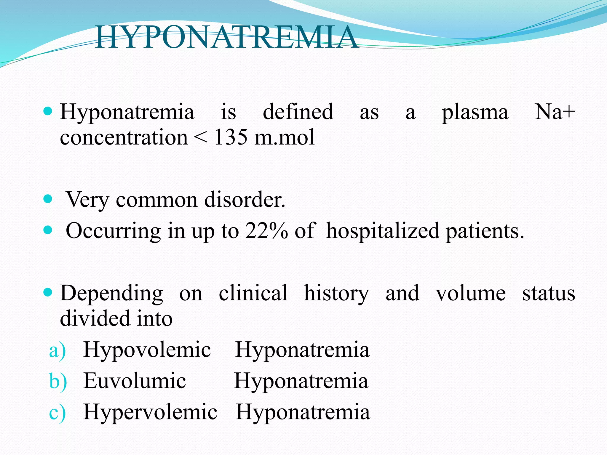
- Causes and types of hyponatremia, including hypovolemic, hypervolemic, and euvolemic hyponatremia
- Evaluation and management of hyponatremia, including treatment based on severity and rate of sodium correction
- Causes and clinical features of hypernatremia
The document is a comprehensive review of sodium disorders and approaches to diagnosis and treatment of hypo- and hypernatremia.

![ Water is the most abundant constituent in the body comprising
approximately 50% of body weight in women and 60% in men.
55-75% is intracellular (intracellular fluid [ICF] ) and 25-45% is
extracellular (extracellular fluid [ECF] ) .
The ECF is further subdivided into intravascular (plasma water) and
extravascular (interstitial) spaces in a ratio of 1 :3.
The solute or particle concentration of a fluid is known as its osmolality
expressed as milliosmoles per kilogram of water (mOsm/kg) .
The major ECF particles are Na+ and its accompanying anions Cl- and
HCO3- whereas K+ and organic phosphate esters (ATP, creatinine
phosphate and phospholipids) are the predominant ICF osmoles.
..](https://image.slidesharecdn.com/sodium-170801182236/75/hyponatremia-2-2048.jpg)


![HYPERVOLUMIC HYPONATREMIA
Patients with hypervolemic hyponatremia develop an increase in total-
body Na+Cl- that is accompanied by a proportionately greater
increase in total body water leading to a reduced plasma Na+
concentration:-
Congestive heart failure [CHF]
Cirrhosis
Nephrotic syndrome
Acute or chronic renal failure,
Characterized by development of pitting edema (about 5 litres of
excess water need to be retained to produce definite pitting edema in
an avereged sized adult)](https://image.slidesharecdn.com/sodium-170801182236/75/hyponatremia-9-2048.jpg)


![LOW SOLUTE INTAKE AND HYPONATREMIA
Alcoholics (beer potomania)
Extreme vegetarian diets
Tea toast diet
PSEUDOHYPONATREMIA
Expansion of extracellular fluid with isotonic fluids that do not contain Na
There is no transcellular shift of water but the [Na+] decreases
e.g. Hypertriglyceridemia
Hyperproteinemia( as in Multiple Myeloma)
Rise in plasma lipids of 4.6 g/L or plasma protein
concentrations greater than 10 g/dL will decrease the sodium
concentration by approximately 1 mEq/L.](https://image.slidesharecdn.com/sodium-170801182236/75/hyponatremia-13-2048.jpg)


![ Water deprivation has long been cornerstone of the therapy of
chronic hyponatremia
The urine-to-plasma electrolyte ratio, (urinary [Na+]+[K+]/plasma
[Na+]) can be exploited as a quick indicator of electrolyte-free water
excretion
Patients with a ratio of >1 should be more aggressively restricted (<500
mL/d), those with a ratio of ~1 should be restricted to 500-700 mL/d
and those with a ratio <1 should be restricted to <1L/d
Hypokalemia should be corrected.](https://image.slidesharecdn.com/sodium-170801182236/75/hyponatremia-28-2048.jpg)

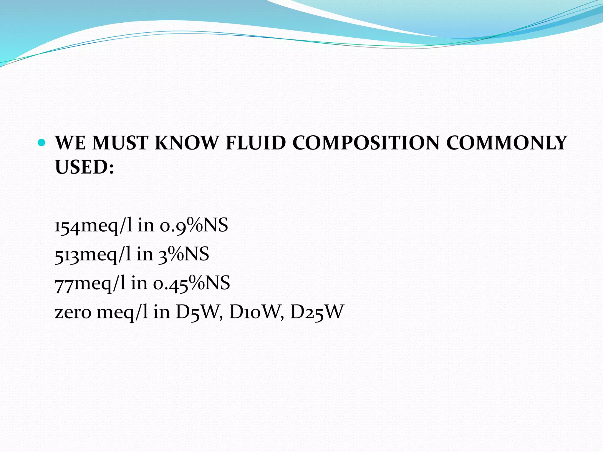
![ Treatment of acute symptomatic hyponatremia should include
hypertonic 3% saline (513 mM ) to acutely increase plasma Na+
concentration by 1-2 mM/h to a total of 4-6 mM.
The traditional approach to calculate Na+ deficit
= 0.6 x body weight x (target plasmaNa+
concentration - starting plasma Na+ concentration)
,
Followed by a calculation of the required rate.
A widely used formula is the Adrogue-Madias formula.
Change in serum Na+ with infusing solution (mM/l/h) =
[infusate (Na + K)]-serum Na/ (total body water +1)](https://image.slidesharecdn.com/sodium-170801182236/75/hyponatremia-30-2048.jpg)