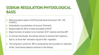
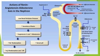
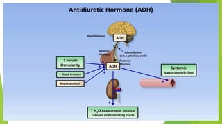
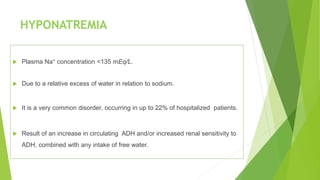
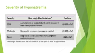
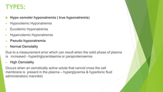
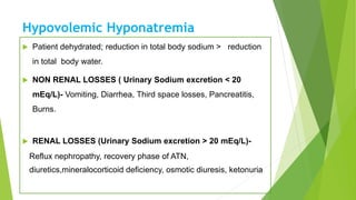
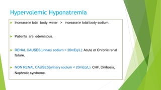
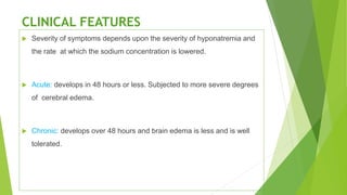
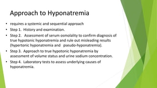
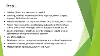
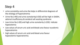
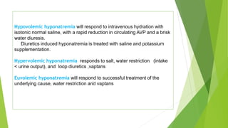
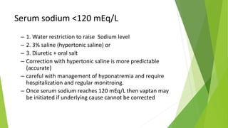
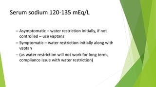
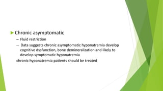
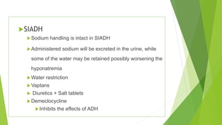
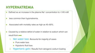
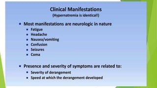
Sodium homeostasis
Candidate : Dr.Ayyanagouda A.G
Guide : Dr.Y.Jamra
SODIUM REGULATION:PHYSIOLOGICAL
BASIS
 Most prevalent cation in ECF(normal level of around 135- 145
mmol/L).
 Intracellular concentration of around 10mmol/L.
 Responsible for 90% of total osmolality of ECF.
 Major function of sodium is to maintain ECF volume and thus BP.
 In normal individuals, the kidney strives to achieve Na+ balance –
that is, to have Na+ excretion equal to Na+ ingestion.
 The long-term control of BP is achieved by the excretion or retention
of Na+ (and hence plasma volume) in the kidney.
Serum sodium concentration regulation
HYPONATREMIA
 Plasma Na+ concentration <135 mEq/L.
 Due to a relative excess of water in relation to sodium.
 It is a very common disorder, occurring in up to 22% of hospitalized patients.
 Result of an increase in circulating ADH and/or increased renal sensitivity to
ADH, combined with any intake of free water.
Severity of hyponatremia
TYPES:
 Hypo osmoler hyponatremia ( true hyponatremia)
 Hypovolemic Hyponatremia
 Euvolemic Hyponatremia
 Hypervolemic Hyponatremia
 Pseudo hyponatremia
 Normal Osmolality
Due to a measurement error which can result when the solid phase of plasma
is increased - hypertriglyceridaemia or paraproteinaemia.
 High Osmolality
Occurs when an osmotically active solute that cannot cross the cell
membrane is present in the plasma – hyperglycemia & hypertonic fluid
administration( mannitol)
Hypovolemic Hyponatremia
 Patient dehydrated; reduction in total body sodium > reduction
in total body water.
 NON RENAL LOSSES ( Urinary Sodium excretion < 20
mEq/L)- Vomiting, Diarrhea, Third space losses, Pancreatitis,
Burns.
 RENAL LOSSES (Urinary Sodium excretion > 20 mEq/L)-
Reflux nephropathy, recovery phase of ATN,
diuretics,mineralocorticoid deficiency, osmotic diuresis, ketonuria.
Euvolemic Hyponatremia
 Patient has a normal store of sodium but an excess of total
body water.
 The syndrome of inappropriate antidiuresis is the most
common condition causing euvolemic hyponatremia.
 Other causes :Glucocorticoid therapy , stress,
hypothyroidism.
SIADH
 Most common cause of Euvolemic hyponatremia.
 The secretion of ADH is not inhibited by either low serum osmolality or
expanded intravascular volume.
 Patient with SIADH is unable to excrete water. This
results in dilution of the serum sodium and hyponatremia.
 Kidney increases sodium excretion in an effort to decrease
intravascular volume to normal; thus, the patient has a mild decrease in
body sodium.
Diagnostic Criteria for SIADH
 Absence of:
 Renal, adrenal, or thyroid insufficiency
 Heart failure, nephrotic syndrome, or cirrhosis
 Diuretic ingestion
 Dehydration
 Urine osmolality >100 mOsm/kg (usually > plasma)
 Serum osmolality <280 mOsm/kg and serum sodium <135 mEq/L
 Urine sodium >30 mEq/L
 Reversal of “sodium wasting” and correction of hyponatremia with
water restriction
Hypervolemic Hyponatremia
 Increase in total body water > increase in total body sodium.
 Patients are edematous.
 RENAL CAUSES(urinary sodium > 20mEq/L): Acute or Chronic renal
failure.
 NON RENAL CAUSES(urinary sodium < 20mEq/L): CHF, Cirrhosis,
Nephrotic syndrome.
CLINICAL FEATURES
 Severity of symptoms depends upon the severity of hyponatremia and
the rate at which the sodium concentration is lowered.
 Acute: develops in 48 hours or less. Subjected to more severe degrees
of cerebral edema.
 Chronic: develops over 48 hours and brain edema is less and is well
tolerated.
SIGNS AND SYMPTOMS OF HYPONATREMIA
Approach to Hyponatremia
• requires a systemic and sequential approach
• Step 1. History and examination.
• Step 2. Assessment of serum osmolality to confirm diagnosis of
true hypotonic hyponatremia and rule out misleading results
(hypertonic hyponatremia and pseudo-hyponatremia).
• Step 3. Approach to true hypotonic hyponatremia by
assessment of volume status and urine sodium concentration.
• Step 4. Laboratory tests to assess underlying causes of
hyponatremia.
Step 1
• detailed history and examination needed
• Vomiting, diarrhea with hypotonic fluid ingestion, recent surgery,
improper IV fluid administration
• Associated diseases (i.e. psychiatric illness, CHF, cirrhosis, renal failure)
• Recent head injury, intracranial surgery, subarachnoid hemorrhage,
stroke, brain tumor, meningitis or brain abscess can cause SIADH.
• Cough, shortness of breath, or pleuritic chest pain should prompt
consideration of respiratory causes of SIADH
• Use of medications
• Skin turgor, mucous membrane appearance and postural hypotension
• Detection of ascites, peripheral edema, pulmonary rales and S 3
• Measuring blood pressure, JVP, CVP and PCWP
Step 2
• Measure plasma osmolality
• 2*Na+ Glucose/18+ BUN/2.8
• osmometer provides actual (correct) osmolality
• Normal plasma osmolality is 280 - 295 mOsm/kg
• low plasma osmolality (POsm < 280 mOsm/kg) confirms
diagnosis
• normal plasma osmolality (POsm 280–295 mOsm/kg) suggests
isotonic pseudo hyponatremia : check for hyperproteinemia,
hyperlipidemia
• high plasma osmolality (POsm > 295 mOsm/kg) suggests
hypertonic hyponatremia : check for hyperglycemia, mannitol
therapy and contrast dyes.
Step 3
• classified into hypovolemic, hypervolemic and
euvolemic
• treatment protocols are absolutely different in
all three categories
• Volume status should be measured
• Urinary sodium should be measured
Step 4
• Check urine Osmolality
• Urine osmolality below 100 mOsm/kg indicates that
antidiuretic hormone (ADH) secretion suppressed
• urine osmolality exceeding 100 mOsm/kg indicates
impaired water excretion which reflects impaired
renal diluting mechanism (SIADH)
Step 4
• urine osmolality and urine Na helps in differential diagnosis of
the etiology of hyponatremia
• Urine Na (>40) and urine osmolarity(>150) will be high in SIADH,
adrenal insufficiency & cerebral salt wasting syndrome
• Low Urine Na (<30) and high urine osmolarity (>150) indicate
hypovolemia
• Low values of serum uric acid and blood urea favour euvolemic
hyponatremia
• high values of serum uric acid and blood urea favour
hypovolemic hyponatremia.
Step 4
• Specific etiologic tests
• Blood sugar: To rule out hyperglycemia
• Serum creatinine: To rule out renal failure
• Serum protein: High in multiple myeloma and low in cirrhosis of liver
• Serum triglycerides: To rule out pseudohyponatremia
• Serum potassium: High in Addison's disease and low in person with diuretics
therapy, diarrhea and vomiting.
• Thyroid function tests: To rule out hypothyroidism
• Adrenal functions: ACTH & ACTH stimulation tests to rule out Addison's disease.
• Acid-base balance: Metabolic alkalosis occurs in diuretic use or vomiting.
Metabolic acidosis occurs in diarrhea or laxative abuse and primary adrenal
insufficiency.
• Head CT scan and Chest X-ray to rule out cerebral salt wasting syndromes
TREATMENT
 Rapid correction is indicated in acute (<48hours) symptomatic or
severe hyponatremia.(serum Na <120 mEq/L).
 In chronic cases patients are at little risk, and rapid correction can
lead to demyelination.
 Total body sodium defict is approximated as :
Na deficit=(target- intial Na+) x TBW(kg)
 Rate of rise of sodium can be predicted as follows:
∆Na =(Inf Na+inf K - plasma Na)/( TBW+ 1)
 TBW = lean weight in kg multiplied by o.6 in male and 0.5 in females.
 Rate of correction should be <8–10 mM in the first 24 h and <18 mM
in the first 48 h .
 0.9% saline (154mEq/L) and 3% NaCl- hypertonic saline (513 mEq/L)
Hypovolemic hyponatremia will respond to intravenous hydration with
isotonic normal saline, with a rapid reduction in circulating AVP and a brisk
water diuresis.
Diuretics induced hyponatremia is treated with saline and potassium
supplementation.
Hypervolemic hyponatremia responds to salt, water restriction (intake
< urine output), and loop diuretics ,vaptans
Euvolemic hyponatremia will respond to successful treatment of the
underlying cause, water restriction and vaptans
Serum sodium <120 mEq/L
– 1. Water restriction to raise Sodium level
– 2. 3% saline (hypertonic saline) or
– 3. Diuretic + oral salt
– Correction with hypertonic saline is more predictable
(accurate)
– careful with management of hyponatremia and require
hospitalization and regular monitroing.
– Once serum sodium reaches 120 mEq/L then vaptan may
be initiated if underlying cause cannot be corrected
Serum sodium 120-135 mEq/L
– Asymptomatic – water restriction initially, if not
controlled – use vaptans
– Symptomatic – water restriction initially along with
vaptan
– (as water restriction will not work for long term,
compliance issue with water restriction)
 Chronic asymptomatic
– Fluid restriction
-- Data suggests chronic asymptomatic hyponatremia develop
cognitive dysfunction, bone demineralization and likely to
develop symptomatic hyponatremia
chronic hyponatremia patients should be treated
SIADH
Sodium handling is intact in SIADH
Administered sodium will be excreted in the urine, while
some of the water may be retained possibly worsening the
hyponatremia
Water restriction
Vaptans
 Diuretics + Salt tablets
Demeclocycline
Inhibits the effects of ADH
Consequence of rapid change of s.sodium
Use of vaptans in hyponatremia
– Euvolemic and hypervolemic hyponatremia
– Chronic symptomatic hyponatremia where underlying cause can not be
corrected and long term treatment is required
– Can be started once serum sodium >120 mEq/L
– augmentation of free-water clearance by specifically antagonising the
V2 vasopressin receptor.
– For initiating vaptan patient should be admitted for 3-4 days, i.e. Vaptan
therapy should be initiated in hospital
– Liver function should be monitored
– Ensure proper intake (good quantity) of water (fluid) while patients are on
vaptan
HYPERNATREMIA
 Defined as an increase in the plasma Na+ concentration to >145 mM.
 less common than hyponatremia.
 Associated with mortality rates as high as 40–60%.
 Caused by a relative deficit of water in relation to sodium which can
result from
1. Net water loss: Accounts for majority of cases
 Pure water loss
 Hypotonic fluid loss
2. Hypertonic gain: Results from iatrogenic sodium loading
Causes of Hypernatremia
Net water loss
Pure water loss
 Unreplaced insensible losses (dermal and respiratory)
 Hypodipsia
 Neurogenic diabetes insipidus
 Congenital nephrogenic diabetes insipidus
 Acquired nephrogenic diabetes insipidus
 Renal disease (e.g. medullary cystic disease)
 Hypercalcemia or hypokalemia
 Drugs (lithium, methoxyflurane, amphotericin B, vasopressin V2-
receptor antagonists)
Hypotonic fluid loss
Renal causes
Loop diuretics
Osmotic diuresis (glucose, urea, mannitol)
Post obstructive diuresis
Polyuric phase of acute tubular necrosis
Gastrointestinal causes
Vomiting
Nasogastric drainage
Entero cutaneous fistula
Diarrhea
Use of osmotic cathartic agents (e.g., lactulose)
Cutaneous causes
Burns
Excessive sweating
Hypertonic sodium gain
Hypertonic sodium bicarbonate infusio
Hypertonic sodium chloride infusion
Primary hyper-aldosteronism
Cushing’s syndrome
Clinical Features
 The symptoms of hypernatremia are predominantly neurologic.
 Altered mental status is the most common manifestation, ranging from
mild confusion and lethargy to deep coma.
 The sudden shrinkage of brain cells in acute hypernatremia may lead to
parenchymal or subarachnoid haemorrhages and/or subdural
hematomas.
 Osmotic damage to muscle membranes also can lead to hypernatremic
rhabdomyolysis.
Administration of Fluids
 Water ideally should be administered by mouth or by nasogastric tube as
the most direct way to provide free water, i.e., water without electrolytes.
 Alternatively, patients can receive free water in dextrose-containing IV
solutions such as 5% dextrose.
 Hypernatremia with ECF volume contraction: Isotonic saline is
given initially till ECF vol is restored. Subsequently water deficit can be
replaced with water by mouth or I.V. 5% dextrose or 0.45% NaCl
 Hypernatremia with increased ECF volume: Since
hypernatremia is secondary to solute administration, it can be rapidly
corrected .
 Patients are volume overloaded- loop diuretic is given along with water
to remove excess sodium
Take home messages..
 Do not get carried away by Na levels
 Asses patient clinically
 Asses volume status
 Correct intravascular volume first
 Correct deficit or excess slowly unless symptomatic
 Rechech response clinically and by lab tests.
THANK YOU

SODIUM HOMEOSTASIS

  • 1.
    Sodium homeostasis Candidate :Dr.Ayyanagouda A.G Guide : Dr.Y.Jamra
  • 2.
    SODIUM REGULATION:PHYSIOLOGICAL BASIS  Mostprevalent cation in ECF(normal level of around 135- 145 mmol/L).  Intracellular concentration of around 10mmol/L.  Responsible for 90% of total osmolality of ECF.  Major function of sodium is to maintain ECF volume and thus BP.  In normal individuals, the kidney strives to achieve Na+ balance – that is, to have Na+ excretion equal to Na+ ingestion.  The long-term control of BP is achieved by the excretion or retention of Na+ (and hence plasma volume) in the kidney.
  • 3.
  • 4.
  • 5.
    HYPONATREMIA  Plasma Na+concentration <135 mEq/L.  Due to a relative excess of water in relation to sodium.  It is a very common disorder, occurring in up to 22% of hospitalized patients.  Result of an increase in circulating ADH and/or increased renal sensitivity to ADH, combined with any intake of free water.
  • 6.
  • 7.
    TYPES:  Hypo osmolerhyponatremia ( true hyponatremia)  Hypovolemic Hyponatremia  Euvolemic Hyponatremia  Hypervolemic Hyponatremia  Pseudo hyponatremia  Normal Osmolality Due to a measurement error which can result when the solid phase of plasma is increased - hypertriglyceridaemia or paraproteinaemia.  High Osmolality Occurs when an osmotically active solute that cannot cross the cell membrane is present in the plasma – hyperglycemia & hypertonic fluid administration( mannitol)
  • 8.
    Hypovolemic Hyponatremia  Patientdehydrated; reduction in total body sodium > reduction in total body water.  NON RENAL LOSSES ( Urinary Sodium excretion < 20 mEq/L)- Vomiting, Diarrhea, Third space losses, Pancreatitis, Burns.  RENAL LOSSES (Urinary Sodium excretion > 20 mEq/L)- Reflux nephropathy, recovery phase of ATN, diuretics,mineralocorticoid deficiency, osmotic diuresis, ketonuria.
  • 9.
    Euvolemic Hyponatremia  Patienthas a normal store of sodium but an excess of total body water.  The syndrome of inappropriate antidiuresis is the most common condition causing euvolemic hyponatremia.  Other causes :Glucocorticoid therapy , stress, hypothyroidism.
  • 10.
    SIADH  Most commoncause of Euvolemic hyponatremia.  The secretion of ADH is not inhibited by either low serum osmolality or expanded intravascular volume.  Patient with SIADH is unable to excrete water. This results in dilution of the serum sodium and hyponatremia.  Kidney increases sodium excretion in an effort to decrease intravascular volume to normal; thus, the patient has a mild decrease in body sodium.
  • 11.
    Diagnostic Criteria forSIADH  Absence of:  Renal, adrenal, or thyroid insufficiency  Heart failure, nephrotic syndrome, or cirrhosis  Diuretic ingestion  Dehydration  Urine osmolality >100 mOsm/kg (usually > plasma)  Serum osmolality <280 mOsm/kg and serum sodium <135 mEq/L  Urine sodium >30 mEq/L  Reversal of “sodium wasting” and correction of hyponatremia with water restriction
  • 13.
    Hypervolemic Hyponatremia  Increasein total body water > increase in total body sodium.  Patients are edematous.  RENAL CAUSES(urinary sodium > 20mEq/L): Acute or Chronic renal failure.  NON RENAL CAUSES(urinary sodium < 20mEq/L): CHF, Cirrhosis, Nephrotic syndrome.
  • 14.
    CLINICAL FEATURES  Severityof symptoms depends upon the severity of hyponatremia and the rate at which the sodium concentration is lowered.  Acute: develops in 48 hours or less. Subjected to more severe degrees of cerebral edema.  Chronic: develops over 48 hours and brain edema is less and is well tolerated.
  • 15.
    SIGNS AND SYMPTOMSOF HYPONATREMIA
  • 16.
    Approach to Hyponatremia •requires a systemic and sequential approach • Step 1. History and examination. • Step 2. Assessment of serum osmolality to confirm diagnosis of true hypotonic hyponatremia and rule out misleading results (hypertonic hyponatremia and pseudo-hyponatremia). • Step 3. Approach to true hypotonic hyponatremia by assessment of volume status and urine sodium concentration. • Step 4. Laboratory tests to assess underlying causes of hyponatremia.
  • 17.
    Step 1 • detailedhistory and examination needed • Vomiting, diarrhea with hypotonic fluid ingestion, recent surgery, improper IV fluid administration • Associated diseases (i.e. psychiatric illness, CHF, cirrhosis, renal failure) • Recent head injury, intracranial surgery, subarachnoid hemorrhage, stroke, brain tumor, meningitis or brain abscess can cause SIADH. • Cough, shortness of breath, or pleuritic chest pain should prompt consideration of respiratory causes of SIADH • Use of medications • Skin turgor, mucous membrane appearance and postural hypotension • Detection of ascites, peripheral edema, pulmonary rales and S 3 • Measuring blood pressure, JVP, CVP and PCWP
  • 18.
    Step 2 • Measureplasma osmolality • 2*Na+ Glucose/18+ BUN/2.8 • osmometer provides actual (correct) osmolality • Normal plasma osmolality is 280 - 295 mOsm/kg • low plasma osmolality (POsm < 280 mOsm/kg) confirms diagnosis • normal plasma osmolality (POsm 280–295 mOsm/kg) suggests isotonic pseudo hyponatremia : check for hyperproteinemia, hyperlipidemia • high plasma osmolality (POsm > 295 mOsm/kg) suggests hypertonic hyponatremia : check for hyperglycemia, mannitol therapy and contrast dyes.
  • 19.
    Step 3 • classifiedinto hypovolemic, hypervolemic and euvolemic • treatment protocols are absolutely different in all three categories • Volume status should be measured • Urinary sodium should be measured
  • 21.
    Step 4 • Checkurine Osmolality • Urine osmolality below 100 mOsm/kg indicates that antidiuretic hormone (ADH) secretion suppressed • urine osmolality exceeding 100 mOsm/kg indicates impaired water excretion which reflects impaired renal diluting mechanism (SIADH)
  • 22.
    Step 4 • urineosmolality and urine Na helps in differential diagnosis of the etiology of hyponatremia • Urine Na (>40) and urine osmolarity(>150) will be high in SIADH, adrenal insufficiency & cerebral salt wasting syndrome • Low Urine Na (<30) and high urine osmolarity (>150) indicate hypovolemia • Low values of serum uric acid and blood urea favour euvolemic hyponatremia • high values of serum uric acid and blood urea favour hypovolemic hyponatremia.
  • 23.
    Step 4 • Specificetiologic tests • Blood sugar: To rule out hyperglycemia • Serum creatinine: To rule out renal failure • Serum protein: High in multiple myeloma and low in cirrhosis of liver • Serum triglycerides: To rule out pseudohyponatremia • Serum potassium: High in Addison's disease and low in person with diuretics therapy, diarrhea and vomiting. • Thyroid function tests: To rule out hypothyroidism • Adrenal functions: ACTH & ACTH stimulation tests to rule out Addison's disease. • Acid-base balance: Metabolic alkalosis occurs in diuretic use or vomiting. Metabolic acidosis occurs in diarrhea or laxative abuse and primary adrenal insufficiency. • Head CT scan and Chest X-ray to rule out cerebral salt wasting syndromes
  • 24.
    TREATMENT  Rapid correctionis indicated in acute (<48hours) symptomatic or severe hyponatremia.(serum Na <120 mEq/L).  In chronic cases patients are at little risk, and rapid correction can lead to demyelination.  Total body sodium defict is approximated as : Na deficit=(target- intial Na+) x TBW(kg)  Rate of rise of sodium can be predicted as follows: ∆Na =(Inf Na+inf K - plasma Na)/( TBW+ 1)  TBW = lean weight in kg multiplied by o.6 in male and 0.5 in females.  Rate of correction should be <8–10 mM in the first 24 h and <18 mM in the first 48 h .  0.9% saline (154mEq/L) and 3% NaCl- hypertonic saline (513 mEq/L)
  • 25.
    Hypovolemic hyponatremia willrespond to intravenous hydration with isotonic normal saline, with a rapid reduction in circulating AVP and a brisk water diuresis. Diuretics induced hyponatremia is treated with saline and potassium supplementation. Hypervolemic hyponatremia responds to salt, water restriction (intake < urine output), and loop diuretics ,vaptans Euvolemic hyponatremia will respond to successful treatment of the underlying cause, water restriction and vaptans
  • 26.
    Serum sodium <120mEq/L – 1. Water restriction to raise Sodium level – 2. 3% saline (hypertonic saline) or – 3. Diuretic + oral salt – Correction with hypertonic saline is more predictable (accurate) – careful with management of hyponatremia and require hospitalization and regular monitroing. – Once serum sodium reaches 120 mEq/L then vaptan may be initiated if underlying cause cannot be corrected
  • 27.
    Serum sodium 120-135mEq/L – Asymptomatic – water restriction initially, if not controlled – use vaptans – Symptomatic – water restriction initially along with vaptan – (as water restriction will not work for long term, compliance issue with water restriction)
  • 28.
     Chronic asymptomatic –Fluid restriction -- Data suggests chronic asymptomatic hyponatremia develop cognitive dysfunction, bone demineralization and likely to develop symptomatic hyponatremia chronic hyponatremia patients should be treated
  • 29.
    SIADH Sodium handling isintact in SIADH Administered sodium will be excreted in the urine, while some of the water may be retained possibly worsening the hyponatremia Water restriction Vaptans  Diuretics + Salt tablets Demeclocycline Inhibits the effects of ADH
  • 30.
    Consequence of rapidchange of s.sodium
  • 31.
    Use of vaptansin hyponatremia – Euvolemic and hypervolemic hyponatremia – Chronic symptomatic hyponatremia where underlying cause can not be corrected and long term treatment is required – Can be started once serum sodium >120 mEq/L – augmentation of free-water clearance by specifically antagonising the V2 vasopressin receptor. – For initiating vaptan patient should be admitted for 3-4 days, i.e. Vaptan therapy should be initiated in hospital – Liver function should be monitored – Ensure proper intake (good quantity) of water (fluid) while patients are on vaptan
  • 32.
    HYPERNATREMIA  Defined asan increase in the plasma Na+ concentration to >145 mM.  less common than hyponatremia.  Associated with mortality rates as high as 40–60%.  Caused by a relative deficit of water in relation to sodium which can result from 1. Net water loss: Accounts for majority of cases  Pure water loss  Hypotonic fluid loss 2. Hypertonic gain: Results from iatrogenic sodium loading
  • 33.
    Causes of Hypernatremia Netwater loss Pure water loss  Unreplaced insensible losses (dermal and respiratory)  Hypodipsia  Neurogenic diabetes insipidus  Congenital nephrogenic diabetes insipidus  Acquired nephrogenic diabetes insipidus  Renal disease (e.g. medullary cystic disease)  Hypercalcemia or hypokalemia  Drugs (lithium, methoxyflurane, amphotericin B, vasopressin V2- receptor antagonists)
  • 34.
    Hypotonic fluid loss Renalcauses Loop diuretics Osmotic diuresis (glucose, urea, mannitol) Post obstructive diuresis Polyuric phase of acute tubular necrosis Gastrointestinal causes Vomiting Nasogastric drainage Entero cutaneous fistula Diarrhea Use of osmotic cathartic agents (e.g., lactulose) Cutaneous causes Burns Excessive sweating
  • 35.
    Hypertonic sodium gain Hypertonicsodium bicarbonate infusio Hypertonic sodium chloride infusion Primary hyper-aldosteronism Cushing’s syndrome
  • 37.
    Clinical Features  Thesymptoms of hypernatremia are predominantly neurologic.  Altered mental status is the most common manifestation, ranging from mild confusion and lethargy to deep coma.  The sudden shrinkage of brain cells in acute hypernatremia may lead to parenchymal or subarachnoid haemorrhages and/or subdural hematomas.  Osmotic damage to muscle membranes also can lead to hypernatremic rhabdomyolysis.
  • 39.
    Administration of Fluids Water ideally should be administered by mouth or by nasogastric tube as the most direct way to provide free water, i.e., water without electrolytes.  Alternatively, patients can receive free water in dextrose-containing IV solutions such as 5% dextrose.
  • 40.
     Hypernatremia withECF volume contraction: Isotonic saline is given initially till ECF vol is restored. Subsequently water deficit can be replaced with water by mouth or I.V. 5% dextrose or 0.45% NaCl  Hypernatremia with increased ECF volume: Since hypernatremia is secondary to solute administration, it can be rapidly corrected .  Patients are volume overloaded- loop diuretic is given along with water to remove excess sodium
  • 41.
    Take home messages.. Do not get carried away by Na levels  Asses patient clinically  Asses volume status  Correct intravascular volume first  Correct deficit or excess slowly unless symptomatic  Rechech response clinically and by lab tests.
  • 42.