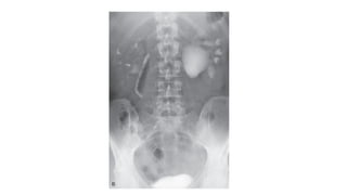
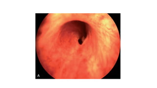
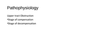
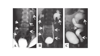
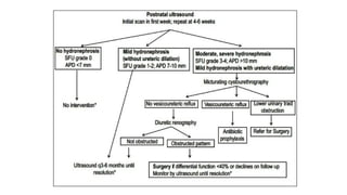
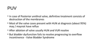
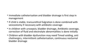
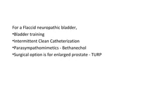
Hydronephrosis
Dr. A. Bharath
Outline
• Definitions
• Etiology
• Pathophysiology
• Clinical features
• Investigations
• Management
Definitions
• Hydronephrosis is the dilation of the renal pelvis or calyces. It
may be associated with obstruction but may be present in the
absence of obstruction.
• Obstructive uropathy refers to the functional or anatomic
obstruction of urinary flow at any level of the urinary tract.
• Obstructive nephropathy is present when the obstruction causes
functional or anatomic renal damage.
Classification
• Cause - Congenital or acquired
• Duration - Acute, Chronic, Intermittent
• Unilateral, Bilateral
• Level - Upper tract, Bladder, Lower tract
Etiology
Congenital
•PUJ obtruction, VUJ abnormalities, Posterior Urethral valve
•Spina bifida, Myelomeningocoele
PUJ Obstruction
• MC congenital abnormality of ureter
• F > M, Left > Right
• Mechanism not clear
• True stenosis is rare; Angulation, kink, high origin of ureter,
hypoplastic ureter etc., are observed
• Abnormal peristalsis interfering with urine flow
VUJ abnormalities
• Obstruction may be similar to PUJ
• Many other structural abnormalities are also noted
- Distal ureteral stenosis
- Ectopic insertion of ureter
- Ureterocoele
- Duplication of ureters
Posterior Urethral Valve
• MC urethral obstructive lesions in newborn males
• MC cause of bilateral hydronephrosis in male fetuses
• Seen in distal prostatic urethra
• Thin membranous folds causing obstruction to unine outflow
• First described by Young
• Assosciated with other congenital abnormalities - Pulmonary
hypoplasia
• Young classification of Posterior Urethral Valves
Etiology
Acquired causes
Upper tract
•Extra mural - Compression by tumor mass, enlarged lymph nodes,
retroperitoneal fibrosis, late stages of pregnancy
•Intramural - Ureteric stricture
•Intra luminal - Ureteric calculus
Mid tract / Bladder
•BPH
•Bladder neck mass
•Bladder neck calculus
Lower tract
•Stricture Urethra
•Calculus in Urethra
Etiology
Functional causes
•Neurogenic bladder
•Vesico Ureteric reflux
VUR
• Intravesical ureter and Bladder trigone act as valve at VUJ and
prevent reflux of vesical urine into ureter
• Any condition that effects - length of intra vesical ureter and tone of
trigone results in deficient valvular function
• High vesical pressures are transmitted into the ureter
• VUR results in Increased workload, High hydrostatic pressure, Weak
ureter musculature
VUR may be caused by
•Congenital weakness of Trigone
•Voiding dysfunction
•Vesical trabeculation
•Edema of vesical wall secondary to cystitis
Neurogenic bladder
• Caused due to lesions along micturition reflex pathway
• Lesions above sacral micturition centre (UMN lesion) - Overactive
bladder with Dyssenrgia
• Overactive bladder with sphincter dyssynergia results in High voiding
pressures, detrusor hypertrophy, VUR / VUJ obstruction
• If lesion is above Pontine micturition centre, detrusor sphincter
dyssnergia doesnot occur
• Lesions at or below sacral micturition centre lead to Flaccid
neuropathic bladder
• Injury to motor nuclei may occur in polio myelitis, herpes zoster, disc
herniation or Cauda equina syndrome
• Injury to afferent pathways result in loss of sensation from bladder
• Seen in Diabetes, Tabes dorsalis and other posterior cord lesions
Spastic Neuropathic Bladder
• Reduced capacity
• Involuntary Detrussor
Contractions
• High voiding pressures
• Marked hypertrophy of bladder
wall
• Spastic external sphincter
Flacid Neuropathic Bladder
• Large capacity
• Lack of voluntary detrussor
contraction
• Low intravesical pressures
• Mild hypertrophy of bladder
• Decreased tone of external
sphincter
Pathophysiology
Upper tract Obstruction
•Stage of compensation
•Stage of decompensation
Stage of Compensation
• Due to increase in resistance to urine flow in the ureter, there is
gradual dilation of ureter and pelvis
• Compensatory hypertrophy occurs and increase in peristaltic activity
occurs resulting in ureter becoming elongated and tortuous
• Secondary fibrous bands may develop resulting in secondary
obstruction
Stage of Decompensation
• As the pressure increases, ureter continues to stretch, it’s wall
becomes weak and attenuated, loses its contractile power
• Some times ureter may be so extremely dilated that it may resemble
a loop of bowel
Changes in Calyces
Renal changes
• Higher the obstruction earlier and greater are the effects on kidney
• As pressure rises initially the pelvis and calyces dilate
• Pelvis may be intra renal or extra renal
• Earlier stages compensatory hypertrophy happens in pelvis
musculature but later it becomes stretched and atonic
• Finally, Compression atrophy and ischemic atrophy occur
• Results in spotty atrophy early on, in advanced stages parenchyma is
thinned out
• In unilateral obstruction these changes are relatively faster as
opposite kidney is functioning and it’s function increases to
compensate for the loss of function in one kidney
• In bilateral obstruction these changes are slow as kidneys attempt to
maintain renal function
• Kidney doesnot exhibit typical response to obstruction and increased
pressure; whereas other glands cease to secrete , kidney continues to
secrete urine
• In the pelvis, water and solutes are reabsorbed via lymphatics
gradually relieving the pressure
• There may be a break in dilated pelvis resulting in extravasation of
urine into renal parenchyma decompressing the pelvis -
Pyelointerstitial back flow
Mid tract / Bladder
• Stage of irritable or hypersensitive bladder
• Stage of compensation
• Stage of decompensation
Stage of irritability
• Occurs in the very initial phases when compensatory hypertrophy
begins, when the detrusor is hypersensitive
• As urine accumulates the urge to micturate cannot be controlled as
the detrusor contraction is so strong it almost goes into a spasm
• Hence first features of BOO are usually Urgency and Frequency
• Can sometimes be severe enough to mimic incontinence
Stage of Compensation
• As the obstruction increases, hypertrophy increases and pressure
required to initiate micturition also increases
• Patient notices Hesitancy to micturate as the bladder needs to
overcome the resistance and generate contraction powerful enough
to initiate micturition
• However the bladder empties completely and no residual urine is
noted in this phase
• Compensatory hypertrophy results in trabeculation of the bladder
• Trigonal area also hypertrophies narrowing ureteric orifices and
thereby resulting in upper tract obstruction
• As intra vesical pressures increase the bladder mucosa in between
trabeculae pouches out to form diverticulae
Stage of Decompensation
• Resistance exceeds the detrusor power, signalled by appearance of
post void residual urine in the bladder
• Can be acute or chronic
• As the volume of residual urine increases the intra vescical pressures
increase simultaneously stretching detrussor resulting in reflux of
urine and transmission of high intra vescical pressures into upper
tract.
• Acute decompensation occurs when muscle tone is impaired due to
rapid filling of bladder or over stretching the detrussor
• Presents as high frequency, very poor stream and low volume voided
in spite of straining with large post void residual urine
• Some times can present as sudden acute retention of urine
• Chronic decompensation occurs as a result of progressive imbalance
between voiding power and resistance to urine flow
• Symptoms of obstruction increase gradually and becomes
increasingly difficult to empty the bladder
• Residual urine increases gradually which stretches the bladder which
loses it’s power, dilates to hold 1000-3000 ml urine
• VUR develops due to weakness of trigone and high intravesical
pressure
• Finally Overflow incontinence results
Lower Tract
• Pathogenesis is similar to bladder outlet obstruction
• In addition, urethra may be dilated and prostatic ducts may also be
dilated
• Diverticulae may be formed and urine infected resulting in
periurethral abscess
Clinical features
• Clinical features are widely variable and mostly depend on cause of
obstruction
• Hydronephrosis per se can be asymptomatic
• Upper tract symptoms are effectively those of ureteric calculus /
stricture
• Pt complains of Loin to groin pain may be accompanied by burning
micturition, colicky pain, vomitings etc
• Bilateral obstruction results in decreased urine output and pt may
end up in uremia
Intermittent obstruction
•Also known as Dietls crisis
•Pt suddenly develops symptoms of upper tract obstruction - pain in
loin, palpable swelling at renal angle especially after intake of fluids
•The symptoms are relieved after sometime with passage of large
amounts of urine
•Could be due to slipping down of ureteric calculus
• Features of Mid tract obstruction are usually those of BOO i.e., LUTS
• Decompensated patients may present with acute urinary retention or
overflow incontinence
• In case of Ca Prostate or Bladder mass causing BOO, there may be
painless hematuria, loss of weight may be present
• In case of lower tract obstruction, pt presents with difficulty in
micturition with features of recurrent uti
Investigations
Imaging studies play an important role, which include
•Plain radiograph KUB
•USG
•CT scan
•IVU
•Retrograde Urography
•MCUG
Plain X ray KUB can give information regarding
•Renal shadow
•Radio opaque calculi
•Any calcifications
IVU aka Excretory Urography is a contrast radiograph of KUB
•Iodine contrast medium is given intra venously
•scout images
•nephrogram (1-2 minutes)
•early and late images of the upper collecting system (>3 minutes)
•supine images of the upper collecting system and proximal ureters
(10-15 minutes)
•Erect films may be taken for ureters
•Post void film
Retrograde Urography
•Rarely used after advent of ct scan
•Can be done if IVU and CT fail to visualise ureteral segment
•Involves ureteroscopy, hence access for simultaneous biopsy or
intervention is available
Retrograde Cystourethrography
•Useful in cases of lower tract obstruction
•Especially helpful to visualise anterior urethra
MCUG
•Done to study bladder, posterior urethra and competence of VUJ
•Film is taken during micturition / when the pt is attempting to void
• Grades of VUR
USG
•Widely used as it is non invasive and helps in identification of wide
variety of conditions
•Helps to establish dilatation of PCS
•Can visualise renal / upper ureteric calculi
•Can visualise any gross structural abnormalities
•Mainstay in prenatal imaging, hence useful for diagnosis of Antenatal
Hydronephrosis
Antenatal Hydronephrosis
•Diagnosed during antenatal usg
•Antero posterior diameter of
renal pelvis is measured
CT scan
•Confirmatory test for most urological pathologies
•Plain ct can visualise anatomy of the kidneys, collecting system,
bladder and urethra
•Contrast can be given antegrade or retrograde for CT urography
Urodynamic study
•Important role in lower tract obstruction, voiding difficulties,
Incontinence and Neuropathic bladder
•Proper interpretation of urodynamics can help us know about Bladder
capacity, Compliance, Bladder sensation, Contractility
•Combined with electromyography it also assesses functioning of
sphinter and pelvic floor muscles
•Videourodynamics is combination of classic urodynamic study with
fluroscopy
Other investigations include
•Complete blood picture
•Routine urine analysis
•Urine examination for malignant cells
•Renal function tests
Treatment
• Treatment as such depends on cause of obstruction
• But main aims of treatment are
- Decompression of urinary tract
- Treating UTI
- Definitive treatment of cause
• In cases of mild to moderate cases of HUN, definitive treatment of
obstruction can be done which simultaneously relieves pressure
• But in case of severe HUN especially where renal function is effected,
urinary tract is decompressed immediately
- Foleys catheterisation
- Supra pubic cystostomy
- Ureteric stenting
- Percutaneous Nephrostomy
ANH
• In case of antenatal hydronephrosis, post natal follow up is advised
• Initial scan done with in 1st week
• Post natal scan done at 48hrs tends to under estimate HUN because
of physiological oliguria in newborn
• Follow up scan can be done after 4-6 weeks
• Postnatally neonates tend to develop VUR, hence if necessary MCUG
is done
PUJ
• MC in such cases is PUJ obstruction
• Definitive treatment includes creation of a dependent, funnel shaped
PUJ of adequate caliber
• No single procedure is considered superior as there are large
differences encountered in anatomy of PUJ
- Dismembered pyeloplasty
- Foleys V-Y pyeloplasty
- Pelvis flap techniques
• Dismembered pyeloplasty
PUV
• In case of Posterior urethral valve, definitive treatment consists of
destruction of the membranes
• Most of the valve cases present with HUN at diagnosis (about 95%)
may / maynot have reflux
• After ablation of valve usually HUN and VUR resolve
• But bladder dysfunction fails to resolve progressing to overflow
incontinence - Valve Bladder Syndrome
• Immediate catheterisation and bladder drainage is first step in
management
• If child is stable, transurethral fulgration is done combined with
vesicostomy if necessary with antibiotic coverage
• In children with urosepsis; bladder drainage, Antibiotic coverage,
correction of fluid and electrolyte abnormalities is done initially
• Children with bladder dysfunction may need Timed voiding, anti
cholinergics, intermittent catheterisation, continuous nocturnal
bladder drainage
VUR
• In cases of VUR, lower grades can be managed with antibiotics to
control uti until reflux resolves or surgically corrected
• When using medical therapy alone, MCUG are repeated every 12-18
months
• Surgical repair is considered if VUR is assosciated with ureteric
anomalies or severe HUN, Pyelonephritis persists despite antibiotics,
Evidence of renal damage on urograms and if pt is not compliant
with medical treatment
• Surgical therapy includes
• Initial temporary urinary diversion - Urethral catheter / Cystostomy /
Ureterostomy / Nephrostomy
• Followed by definitive repair of uretero vesical junction
• Other procedures include
- Nephrectomy
- Heminephrectomy
- Ureteroureterostomy
Neuropathic Bladder
For a Spastic Neuropathic bladder,
•In patients with reasonable capacity, voiding training is given, to void
every 2-3 hrs
•In patients with markedly diminished functional capacity, available
options are
- Muscarinic antagonists
- Indwelling catheter
- Sacral rhizotomy
- Sphincterotomy
For a Flaccid neuropathic bladder,
•Bladder training
•Intermittent Clean Catheterization
•Parasympathomimetics - Bethanechol
•Surgical option is for enlarged prostate - TURP
THANK YOU

Hydronephrosis

  • 1.
  • 2.
    Outline • Definitions • Etiology •Pathophysiology • Clinical features • Investigations • Management
  • 3.
    Definitions • Hydronephrosis isthe dilation of the renal pelvis or calyces. It may be associated with obstruction but may be present in the absence of obstruction. • Obstructive uropathy refers to the functional or anatomic obstruction of urinary flow at any level of the urinary tract. • Obstructive nephropathy is present when the obstruction causes functional or anatomic renal damage.
  • 4.
    Classification • Cause -Congenital or acquired • Duration - Acute, Chronic, Intermittent • Unilateral, Bilateral • Level - Upper tract, Bladder, Lower tract
  • 5.
    Etiology Congenital •PUJ obtruction, VUJabnormalities, Posterior Urethral valve •Spina bifida, Myelomeningocoele
  • 6.
    PUJ Obstruction • MCcongenital abnormality of ureter • F > M, Left > Right • Mechanism not clear • True stenosis is rare; Angulation, kink, high origin of ureter, hypoplastic ureter etc., are observed • Abnormal peristalsis interfering with urine flow
  • 8.
    VUJ abnormalities • Obstructionmay be similar to PUJ • Many other structural abnormalities are also noted - Distal ureteral stenosis - Ectopic insertion of ureter - Ureterocoele - Duplication of ureters
  • 9.
    Posterior Urethral Valve •MC urethral obstructive lesions in newborn males • MC cause of bilateral hydronephrosis in male fetuses • Seen in distal prostatic urethra • Thin membranous folds causing obstruction to unine outflow • First described by Young • Assosciated with other congenital abnormalities - Pulmonary hypoplasia
  • 10.
    • Young classificationof Posterior Urethral Valves
  • 12.
    Etiology Acquired causes Upper tract •Extramural - Compression by tumor mass, enlarged lymph nodes, retroperitoneal fibrosis, late stages of pregnancy •Intramural - Ureteric stricture •Intra luminal - Ureteric calculus
  • 13.
    Mid tract /Bladder •BPH •Bladder neck mass •Bladder neck calculus Lower tract •Stricture Urethra •Calculus in Urethra
  • 14.
  • 15.
    VUR • Intravesical ureterand Bladder trigone act as valve at VUJ and prevent reflux of vesical urine into ureter • Any condition that effects - length of intra vesical ureter and tone of trigone results in deficient valvular function • High vesical pressures are transmitted into the ureter • VUR results in Increased workload, High hydrostatic pressure, Weak ureter musculature
  • 16.
    VUR may becaused by •Congenital weakness of Trigone •Voiding dysfunction •Vesical trabeculation •Edema of vesical wall secondary to cystitis
  • 17.
    Neurogenic bladder • Causeddue to lesions along micturition reflex pathway • Lesions above sacral micturition centre (UMN lesion) - Overactive bladder with Dyssenrgia • Overactive bladder with sphincter dyssynergia results in High voiding pressures, detrusor hypertrophy, VUR / VUJ obstruction • If lesion is above Pontine micturition centre, detrusor sphincter dyssnergia doesnot occur
  • 18.
    • Lesions ator below sacral micturition centre lead to Flaccid neuropathic bladder • Injury to motor nuclei may occur in polio myelitis, herpes zoster, disc herniation or Cauda equina syndrome • Injury to afferent pathways result in loss of sensation from bladder • Seen in Diabetes, Tabes dorsalis and other posterior cord lesions
  • 19.
    Spastic Neuropathic Bladder •Reduced capacity • Involuntary Detrussor Contractions • High voiding pressures • Marked hypertrophy of bladder wall • Spastic external sphincter Flacid Neuropathic Bladder • Large capacity • Lack of voluntary detrussor contraction • Low intravesical pressures • Mild hypertrophy of bladder • Decreased tone of external sphincter
  • 20.
    Pathophysiology Upper tract Obstruction •Stageof compensation •Stage of decompensation
  • 21.
    Stage of Compensation •Due to increase in resistance to urine flow in the ureter, there is gradual dilation of ureter and pelvis • Compensatory hypertrophy occurs and increase in peristaltic activity occurs resulting in ureter becoming elongated and tortuous • Secondary fibrous bands may develop resulting in secondary obstruction
  • 22.
    Stage of Decompensation •As the pressure increases, ureter continues to stretch, it’s wall becomes weak and attenuated, loses its contractile power • Some times ureter may be so extremely dilated that it may resemble a loop of bowel
  • 24.
  • 25.
    Renal changes • Higherthe obstruction earlier and greater are the effects on kidney • As pressure rises initially the pelvis and calyces dilate • Pelvis may be intra renal or extra renal • Earlier stages compensatory hypertrophy happens in pelvis musculature but later it becomes stretched and atonic
  • 26.
    • Finally, Compressionatrophy and ischemic atrophy occur • Results in spotty atrophy early on, in advanced stages parenchyma is thinned out • In unilateral obstruction these changes are relatively faster as opposite kidney is functioning and it’s function increases to compensate for the loss of function in one kidney • In bilateral obstruction these changes are slow as kidneys attempt to maintain renal function
  • 27.
    • Kidney doesnotexhibit typical response to obstruction and increased pressure; whereas other glands cease to secrete , kidney continues to secrete urine • In the pelvis, water and solutes are reabsorbed via lymphatics gradually relieving the pressure • There may be a break in dilated pelvis resulting in extravasation of urine into renal parenchyma decompressing the pelvis - Pyelointerstitial back flow
  • 28.
    Mid tract /Bladder • Stage of irritable or hypersensitive bladder • Stage of compensation • Stage of decompensation
  • 29.
    Stage of irritability •Occurs in the very initial phases when compensatory hypertrophy begins, when the detrusor is hypersensitive • As urine accumulates the urge to micturate cannot be controlled as the detrusor contraction is so strong it almost goes into a spasm • Hence first features of BOO are usually Urgency and Frequency • Can sometimes be severe enough to mimic incontinence
  • 30.
    Stage of Compensation •As the obstruction increases, hypertrophy increases and pressure required to initiate micturition also increases • Patient notices Hesitancy to micturate as the bladder needs to overcome the resistance and generate contraction powerful enough to initiate micturition • However the bladder empties completely and no residual urine is noted in this phase
  • 31.
    • Compensatory hypertrophyresults in trabeculation of the bladder • Trigonal area also hypertrophies narrowing ureteric orifices and thereby resulting in upper tract obstruction • As intra vesical pressures increase the bladder mucosa in between trabeculae pouches out to form diverticulae
  • 32.
    Stage of Decompensation •Resistance exceeds the detrusor power, signalled by appearance of post void residual urine in the bladder • Can be acute or chronic • As the volume of residual urine increases the intra vescical pressures increase simultaneously stretching detrussor resulting in reflux of urine and transmission of high intra vescical pressures into upper tract.
  • 33.
    • Acute decompensationoccurs when muscle tone is impaired due to rapid filling of bladder or over stretching the detrussor • Presents as high frequency, very poor stream and low volume voided in spite of straining with large post void residual urine • Some times can present as sudden acute retention of urine
  • 34.
    • Chronic decompensationoccurs as a result of progressive imbalance between voiding power and resistance to urine flow • Symptoms of obstruction increase gradually and becomes increasingly difficult to empty the bladder • Residual urine increases gradually which stretches the bladder which loses it’s power, dilates to hold 1000-3000 ml urine • VUR develops due to weakness of trigone and high intravesical pressure • Finally Overflow incontinence results
  • 36.
    Lower Tract • Pathogenesisis similar to bladder outlet obstruction • In addition, urethra may be dilated and prostatic ducts may also be dilated • Diverticulae may be formed and urine infected resulting in periurethral abscess
  • 38.
    Clinical features • Clinicalfeatures are widely variable and mostly depend on cause of obstruction • Hydronephrosis per se can be asymptomatic • Upper tract symptoms are effectively those of ureteric calculus / stricture • Pt complains of Loin to groin pain may be accompanied by burning micturition, colicky pain, vomitings etc • Bilateral obstruction results in decreased urine output and pt may end up in uremia
  • 39.
    Intermittent obstruction •Also knownas Dietls crisis •Pt suddenly develops symptoms of upper tract obstruction - pain in loin, palpable swelling at renal angle especially after intake of fluids •The symptoms are relieved after sometime with passage of large amounts of urine •Could be due to slipping down of ureteric calculus
  • 40.
    • Features ofMid tract obstruction are usually those of BOO i.e., LUTS • Decompensated patients may present with acute urinary retention or overflow incontinence • In case of Ca Prostate or Bladder mass causing BOO, there may be painless hematuria, loss of weight may be present • In case of lower tract obstruction, pt presents with difficulty in micturition with features of recurrent uti
  • 41.
    Investigations Imaging studies playan important role, which include •Plain radiograph KUB •USG •CT scan •IVU •Retrograde Urography •MCUG
  • 42.
    Plain X rayKUB can give information regarding •Renal shadow •Radio opaque calculi •Any calcifications
  • 43.
    IVU aka ExcretoryUrography is a contrast radiograph of KUB •Iodine contrast medium is given intra venously •scout images •nephrogram (1-2 minutes) •early and late images of the upper collecting system (>3 minutes) •supine images of the upper collecting system and proximal ureters (10-15 minutes) •Erect films may be taken for ureters •Post void film
  • 44.
    Retrograde Urography •Rarely usedafter advent of ct scan •Can be done if IVU and CT fail to visualise ureteral segment •Involves ureteroscopy, hence access for simultaneous biopsy or intervention is available
  • 45.
    Retrograde Cystourethrography •Useful incases of lower tract obstruction •Especially helpful to visualise anterior urethra MCUG •Done to study bladder, posterior urethra and competence of VUJ •Film is taken during micturition / when the pt is attempting to void
  • 46.
  • 48.
    USG •Widely used asit is non invasive and helps in identification of wide variety of conditions •Helps to establish dilatation of PCS •Can visualise renal / upper ureteric calculi •Can visualise any gross structural abnormalities •Mainstay in prenatal imaging, hence useful for diagnosis of Antenatal Hydronephrosis
  • 49.
    Antenatal Hydronephrosis •Diagnosed duringantenatal usg •Antero posterior diameter of renal pelvis is measured
  • 51.
    CT scan •Confirmatory testfor most urological pathologies •Plain ct can visualise anatomy of the kidneys, collecting system, bladder and urethra •Contrast can be given antegrade or retrograde for CT urography
  • 52.
    Urodynamic study •Important rolein lower tract obstruction, voiding difficulties, Incontinence and Neuropathic bladder •Proper interpretation of urodynamics can help us know about Bladder capacity, Compliance, Bladder sensation, Contractility •Combined with electromyography it also assesses functioning of sphinter and pelvic floor muscles •Videourodynamics is combination of classic urodynamic study with fluroscopy
  • 53.
    Other investigations include •Completeblood picture •Routine urine analysis •Urine examination for malignant cells •Renal function tests
  • 54.
    Treatment • Treatment assuch depends on cause of obstruction • But main aims of treatment are - Decompression of urinary tract - Treating UTI - Definitive treatment of cause
  • 55.
    • In casesof mild to moderate cases of HUN, definitive treatment of obstruction can be done which simultaneously relieves pressure • But in case of severe HUN especially where renal function is effected, urinary tract is decompressed immediately - Foleys catheterisation - Supra pubic cystostomy - Ureteric stenting - Percutaneous Nephrostomy
  • 56.
    ANH • In caseof antenatal hydronephrosis, post natal follow up is advised • Initial scan done with in 1st week • Post natal scan done at 48hrs tends to under estimate HUN because of physiological oliguria in newborn • Follow up scan can be done after 4-6 weeks • Postnatally neonates tend to develop VUR, hence if necessary MCUG is done
  • 58.
    PUJ • MC insuch cases is PUJ obstruction • Definitive treatment includes creation of a dependent, funnel shaped PUJ of adequate caliber • No single procedure is considered superior as there are large differences encountered in anatomy of PUJ - Dismembered pyeloplasty - Foleys V-Y pyeloplasty - Pelvis flap techniques
  • 59.
  • 62.
    PUV • In caseof Posterior urethral valve, definitive treatment consists of destruction of the membranes • Most of the valve cases present with HUN at diagnosis (about 95%) may / maynot have reflux • After ablation of valve usually HUN and VUR resolve • But bladder dysfunction fails to resolve progressing to overflow incontinence - Valve Bladder Syndrome
  • 63.
    • Immediate catheterisationand bladder drainage is first step in management • If child is stable, transurethral fulgration is done combined with vesicostomy if necessary with antibiotic coverage • In children with urosepsis; bladder drainage, Antibiotic coverage, correction of fluid and electrolyte abnormalities is done initially • Children with bladder dysfunction may need Timed voiding, anti cholinergics, intermittent catheterisation, continuous nocturnal bladder drainage
  • 64.
    VUR • In casesof VUR, lower grades can be managed with antibiotics to control uti until reflux resolves or surgically corrected • When using medical therapy alone, MCUG are repeated every 12-18 months • Surgical repair is considered if VUR is assosciated with ureteric anomalies or severe HUN, Pyelonephritis persists despite antibiotics, Evidence of renal damage on urograms and if pt is not compliant with medical treatment
  • 65.
    • Surgical therapyincludes • Initial temporary urinary diversion - Urethral catheter / Cystostomy / Ureterostomy / Nephrostomy • Followed by definitive repair of uretero vesical junction • Other procedures include - Nephrectomy - Heminephrectomy - Ureteroureterostomy
  • 66.
    Neuropathic Bladder For aSpastic Neuropathic bladder, •In patients with reasonable capacity, voiding training is given, to void every 2-3 hrs •In patients with markedly diminished functional capacity, available options are - Muscarinic antagonists - Indwelling catheter - Sacral rhizotomy - Sphincterotomy
  • 67.
    For a Flaccidneuropathic bladder, •Bladder training •Intermittent Clean Catheterization •Parasympathomimetics - Bethanechol •Surgical option is for enlarged prostate - TURP
  • 68.