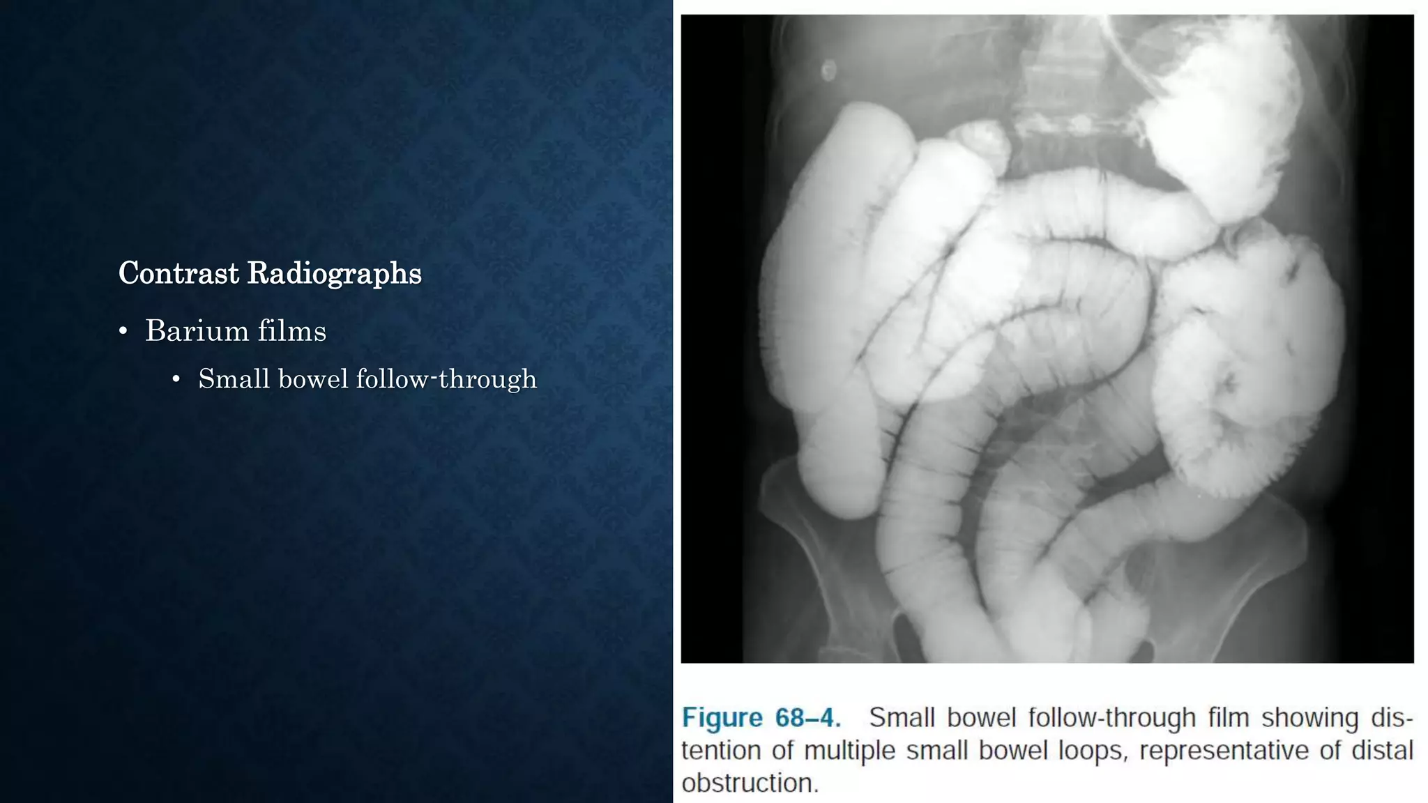
SMALL BOWEL OBSTRUCTION
Yugal Jyoty Nepal
30/12/2074
INTRODUCTION
• Small Bowel Obstruction (SBO) account for 12-16% of surgical admissions for acute
abdominal complaints
• Manifestations can range from
• Slight abdominal discomfort and distension
to
• Hypovolemic or septic shock (or both) – emergency operation
• Decision to treat operatively or nonoperatively – dependent on surgeon’s clinical
experience
• Mortality from SBO
50% in 1900
< 3% today
• Decreased mortality may be due to
• Improved imaging techniques – prompt earlier operative intervention vs
conservative management
• Advanced methods of resuscitation and intensive care
CLASSIFICATION
According to mechanism
• Mechanical – Contents cannot pass through d/t physical blockage
• Extrinsic/ Extraluminal -- eg. Adhesions
• Intrinsic/Mural – eg. Duodenal hematoma
• Intraluminal – eg. Gallstone or Intussusception
• Functional – Dysmotility of bowel without physical obstacle
• Neurogenic disturbances
• Eg. Ileus, Pseudo-obstruction (Ogilvie’s syndrome)
• Partial –
• Passage of gas or liquid stool or both
• Complete –
• No passage of any substance
MOTILITY OF SMALL BOWEL
• During fasting, migrating myoelectric complex (MMC) start in duodenum – progress
to terminal ileum
• Occur every 90-150 min; last 90 min
• In early obstruction, MMC – aggressive – increase intraluminal pressure
• Later, subside and recur episodically
• High obstruction – duration of quiescence shorter
• Low obstruction – duration of quiescence longer
PATHOPHYSIOLOGY OF OBSTRUCTION AND
STRANGULATION
• Method of fluid loss in SBO
• Net secretion into the lumen
• Into wall of bowel
• Small bowel secretes 8.5 L of fluid daily – most reabsorbed in small intestine
• SBO: net flux of fluid
• Due to prostaglandin release – d/t bowel distension
• Manifestation –
• Symptoms of thirst and dry mucus membrane
• Renal failure and shock
• Fluid loss into bowel wall – secondary to venous congestion and edema
• Serosal layer secretes fluid into peritoneum – Ascitis
• Degree of wall edema – corresponds to duration of obstruction
Electrolyte disturbances
• Early obstruction
• Isotonic volume depletion
• Later, hypokalemia d/t vomiting and hyperaldosteronism (response to hypovolemia)
• Loss of bicarbonate – expelled in pancreatic and enteric fluid
• D/t accumulation of air and fluid within lumen,
deterioration of normal absorptive capabilities of gut
• Bacterial colonization d/t stasis
resulting in more gas production from bacteria – worsen distension
• Increased risk of bacteremia, peritonitis and bacterial translocation
• If obstruction not resolved
• Strangulation – necrosis – perforation
CLOSED LOOP OBSTRUCTION
• Caused by obstruction of both afferent and efferent limbs of involved loop of bowel
• Earlier progression to strangulation – d/t inability to decompress proximally or
distally
• Causes:
• Mesenteric torsion
• Adhesive bands
• Hernias
• In colon, any obstructing lesion if IC valve is competent
ETIOLOGY
• Peritoneal adhesion – 75%
• Pelvic or lower abdominal procedures – more risk
• Hernias – 25%
• Inflammatory processes – secondary angulation of bowel
• Diverticulitis and appendicitis
• Crohn’s disease
• Ischemia
• Radiation
• Intussusception
• Volvulus
• Mass lesions – Neoplasms, Gallstones, Bezoars
ETIOLOGY OF POSTOPERATIVE SBO
Adhesions
• Account for majority of early postoperative obstructions
• Develop after peritoneal violation
• Result in inflammatory cascade – activation of complement and coagulation
• Fibrinogen – converted to fibrin by thrombin
• Persistence of fibrin – adherence of injured surfaces
• Peritoneal injury inhibits fibrin degradation by
• Increasing plasminogen activator inhibitors
• Decreasing tissue plasminogen activator levels
Internal herniation
• Through defects formed during surgery
• Mesenteric or omental defects
Inflammatory processes
• Abscesses after bowel surgery
• Form adhesions to nearby loops of bowel – cause partial obstruction
CLINICAL FINDINGS
• Nausea, vomiting
• Crampy abdominal pain
• Distension
• Obstipation
• Mechanical obstruction – pain before vomiting
• Non mechanical obstruction – vomiting before pain
Symptoms according to site of obstruction
• Proximal obstruction – Early and frequent vomiting
• Distal obstruction – Crampy pain and obstipation with delayed vomiting
• Symptoms of postoperative ileus often confused with those of bowel obstruction
• Method to diminish extent of ileus
• Sham feeding: Gum chewing – stimulate GI motility
PHYSICAL EXAMINATION
• Signs of dehydration
• Poor skin turgor
• Dry mucous membrane
• Tachycardia
• Hypotension
• Oliguria
• Mental status changes
• Per abdomen –
• Distension of varying severity
• Palpation: Tenderness, guarding, peritonitis if strangulation, ischemia or perforation
• Percussion: Tympanic if bowel loops filled with gas; dull if filled with fluid
• Auscultation: Rushing of tinkling high pitched bowel sounds OR absent bowel sounds in
advanced stages
• Previous surgical scars – used to predict location and degree of adhesions during
exploration
• Look for hernias
• DRE –
• Rectal masses?
• Hematochezia – more proximal mass, inflammation or strangulation and infarction of
bowel
• Fecal impaction – in elderly; often mimics bowel obstruction
LABORATORY TESTS
• Evidence of dehydration
• Abnormal electrolytes – Hyponatremia, Hypokalemia
• Elevated BUN, Cr and Hct
• Metabolic acidosis d/t dehydration, starvation, ketosis, and loss of alkaline fluid by
secretion
• Metabolic alkalisos – seen occasionally in severe vomiting secondary to vomiting of acidic
juices
• Increased WBCs
• Mild elevation
• Severe leukocytosis – s/o strangulation
• However, bowel ischemia may be present despite normal lab studies
and clinical suspicion should prompt surgical intervention
RADIOLOGIC INVESTIGATION
Plain radiographs
• Abdominal series
• Upright CXR
• Erect and supine abdominal X-Ray
• Goal:
• To rule out free intra-abdominal air
• Delineate severity of bowel distension
• Identify location of obstruction
Disadvantages
• Plain films diagnostic in 50-60% of cases of SBO
• Sensitivity 66%
• Low specificity
Advantages
• Low cost
• Easy availability
• Non-invasive
• Assessment of disease progression
• Patterns suggestive of SBO:
• Multiple loops of small bowel filled with gas or fluid
• Finding of colonic gas indicates partial SBO, early complete SBO or ileus
Contrast Radiographs
• Barium films
• Small bowel follow-through
• Enteroclysis
• Intubation of jejunum and direct infusion of
contrast boluses
• Water soluble contrast
• Diatrizoate meglumine (Gastrografin)
• Therapeutic effect – controversial
• Used as a mode of differentiating partial from complete SBO – predict need for early
surgical intervention
• If gastrografin not passed in to colon after 24 hours, operation is performed
Computed Tomography
• Valuable tool
• Cause of obstruction
• Diagnosis of ischemic bowel, precipitating factors – bowel volvulus, intussusception
• Diagnosis of external as well as internal hernias
• Preexisting pathology
• Abdominal malignancy or inflammatory processes
• Findings of partial SBO on CT
• Mildly dilated small bowel loops (>2.5 cm in diameter) with an ill-defined transition point
• Incompletely collapsed distal bowel
• Moderate amount of gas and fluid in colon
• Closed-loop obstruction
• Involved segment of bowel is nearly completely filled with fluid
• Proximal portion of bowel contain air-fluid levels
• Mesentery may show whirl sign, suggestive of twisted mesenteric vessels
Whirl sign
MEDICAL TREATMENT
• For partial SBO with no evidence of complete obstruction, strangulation, or ischemia
• Adequate intravenous hydration
• Correction of electrolytes
• Close monitoring of urine output with or without a Foley catheter
• Nasogastric tube drainage
• Frequent assessment of patient’s abdominal examination
• Approximately 80% to 90% of partial SBO cases resolve spontaneously with
conservative measures
SURGICAL TREATMENT
• Bowel incarceration, strangulation or ischemia – urgent operation after adequate
resuscitation
• In cases of nonoperative management of partial SBO, factors prompting surgical
intervention include
1. Worsening abdominal pain and distention
2. Findings of peritonitis, fever, and leukocytosis
3. Failure of resolution of complete obstruction within 12 to 24 hours
4. Failure of improvement of partial obstruction after 48 to 72 hours or progression to
complete obstruction
• In case of early postoperative obstruction, initial expectant management upto 4 weeks
• Dense and vascular adhesions – significant morbidity with increased risk for enterotomy and
bleeding
• If patient’s condition worsen during waiting period, surgical intervention implemented in
timely fashion
• Surgical approach depends on suspected cause of obstruction and intra-operative
findings
Prevention of adhesion
• Adhesions after abdominal surgery, esp. colorectal surgery – most common cause of SBO
• Technical efforts
• gentle tissue handling
• Minimal use of foreign materials
• Careful hemostatic measures
• Prevention of infection, ischemia, and desiccation
• Physical barrier
• Sodium hyaluronate–based bioresorbable membrane (Seprafilm)
• Persists in the abdomen for 5 to 7 days
THANK YOU

Small bowel obstruction

  • 1.
    SMALL BOWEL OBSTRUCTION YugalJyoty Nepal 30/12/2074
  • 2.
    INTRODUCTION • Small BowelObstruction (SBO) account for 12-16% of surgical admissions for acute abdominal complaints • Manifestations can range from • Slight abdominal discomfort and distension to • Hypovolemic or septic shock (or both) – emergency operation
  • 3.
    • Decision totreat operatively or nonoperatively – dependent on surgeon’s clinical experience • Mortality from SBO 50% in 1900 < 3% today • Decreased mortality may be due to • Improved imaging techniques – prompt earlier operative intervention vs conservative management • Advanced methods of resuscitation and intensive care
  • 4.
    CLASSIFICATION According to mechanism •Mechanical – Contents cannot pass through d/t physical blockage • Extrinsic/ Extraluminal -- eg. Adhesions • Intrinsic/Mural – eg. Duodenal hematoma • Intraluminal – eg. Gallstone or Intussusception • Functional – Dysmotility of bowel without physical obstacle • Neurogenic disturbances • Eg. Ileus, Pseudo-obstruction (Ogilvie’s syndrome)
  • 5.
    • Partial – •Passage of gas or liquid stool or both • Complete – • No passage of any substance
  • 6.
    MOTILITY OF SMALLBOWEL • During fasting, migrating myoelectric complex (MMC) start in duodenum – progress to terminal ileum • Occur every 90-150 min; last 90 min • In early obstruction, MMC – aggressive – increase intraluminal pressure • Later, subside and recur episodically • High obstruction – duration of quiescence shorter • Low obstruction – duration of quiescence longer
  • 7.
    PATHOPHYSIOLOGY OF OBSTRUCTIONAND STRANGULATION • Method of fluid loss in SBO • Net secretion into the lumen • Into wall of bowel • Small bowel secretes 8.5 L of fluid daily – most reabsorbed in small intestine • SBO: net flux of fluid • Due to prostaglandin release – d/t bowel distension • Manifestation – • Symptoms of thirst and dry mucus membrane • Renal failure and shock
  • 8.
    • Fluid lossinto bowel wall – secondary to venous congestion and edema • Serosal layer secretes fluid into peritoneum – Ascitis • Degree of wall edema – corresponds to duration of obstruction
  • 9.
    Electrolyte disturbances • Earlyobstruction • Isotonic volume depletion • Later, hypokalemia d/t vomiting and hyperaldosteronism (response to hypovolemia) • Loss of bicarbonate – expelled in pancreatic and enteric fluid
  • 10.
    • D/t accumulationof air and fluid within lumen, deterioration of normal absorptive capabilities of gut • Bacterial colonization d/t stasis resulting in more gas production from bacteria – worsen distension • Increased risk of bacteremia, peritonitis and bacterial translocation • If obstruction not resolved • Strangulation – necrosis – perforation
  • 11.
    CLOSED LOOP OBSTRUCTION •Caused by obstruction of both afferent and efferent limbs of involved loop of bowel • Earlier progression to strangulation – d/t inability to decompress proximally or distally • Causes: • Mesenteric torsion • Adhesive bands • Hernias • In colon, any obstructing lesion if IC valve is competent
  • 13.
    ETIOLOGY • Peritoneal adhesion– 75% • Pelvic or lower abdominal procedures – more risk • Hernias – 25% • Inflammatory processes – secondary angulation of bowel • Diverticulitis and appendicitis • Crohn’s disease • Ischemia • Radiation • Intussusception • Volvulus • Mass lesions – Neoplasms, Gallstones, Bezoars
  • 14.
    ETIOLOGY OF POSTOPERATIVESBO Adhesions • Account for majority of early postoperative obstructions • Develop after peritoneal violation • Result in inflammatory cascade – activation of complement and coagulation • Fibrinogen – converted to fibrin by thrombin • Persistence of fibrin – adherence of injured surfaces • Peritoneal injury inhibits fibrin degradation by • Increasing plasminogen activator inhibitors • Decreasing tissue plasminogen activator levels
  • 15.
    Internal herniation • Throughdefects formed during surgery • Mesenteric or omental defects Inflammatory processes • Abscesses after bowel surgery • Form adhesions to nearby loops of bowel – cause partial obstruction
  • 16.
    CLINICAL FINDINGS • Nausea,vomiting • Crampy abdominal pain • Distension • Obstipation • Mechanical obstruction – pain before vomiting • Non mechanical obstruction – vomiting before pain
  • 17.
    Symptoms according tosite of obstruction • Proximal obstruction – Early and frequent vomiting • Distal obstruction – Crampy pain and obstipation with delayed vomiting
  • 19.
    • Symptoms ofpostoperative ileus often confused with those of bowel obstruction • Method to diminish extent of ileus • Sham feeding: Gum chewing – stimulate GI motility
  • 20.
    PHYSICAL EXAMINATION • Signsof dehydration • Poor skin turgor • Dry mucous membrane • Tachycardia • Hypotension • Oliguria • Mental status changes
  • 21.
    • Per abdomen– • Distension of varying severity • Palpation: Tenderness, guarding, peritonitis if strangulation, ischemia or perforation • Percussion: Tympanic if bowel loops filled with gas; dull if filled with fluid • Auscultation: Rushing of tinkling high pitched bowel sounds OR absent bowel sounds in advanced stages • Previous surgical scars – used to predict location and degree of adhesions during exploration • Look for hernias
  • 22.
    • DRE – •Rectal masses? • Hematochezia – more proximal mass, inflammation or strangulation and infarction of bowel • Fecal impaction – in elderly; often mimics bowel obstruction
  • 23.
    LABORATORY TESTS • Evidenceof dehydration • Abnormal electrolytes – Hyponatremia, Hypokalemia • Elevated BUN, Cr and Hct • Metabolic acidosis d/t dehydration, starvation, ketosis, and loss of alkaline fluid by secretion • Metabolic alkalisos – seen occasionally in severe vomiting secondary to vomiting of acidic juices • Increased WBCs • Mild elevation • Severe leukocytosis – s/o strangulation
  • 24.
    • However, bowelischemia may be present despite normal lab studies and clinical suspicion should prompt surgical intervention
  • 25.
    RADIOLOGIC INVESTIGATION Plain radiographs •Abdominal series • Upright CXR • Erect and supine abdominal X-Ray • Goal: • To rule out free intra-abdominal air • Delineate severity of bowel distension • Identify location of obstruction
  • 26.
    Disadvantages • Plain filmsdiagnostic in 50-60% of cases of SBO • Sensitivity 66% • Low specificity Advantages • Low cost • Easy availability • Non-invasive • Assessment of disease progression
  • 27.
    • Patterns suggestiveof SBO: • Multiple loops of small bowel filled with gas or fluid • Finding of colonic gas indicates partial SBO, early complete SBO or ileus
  • 29.
    Contrast Radiographs • Bariumfilms • Small bowel follow-through
  • 30.
    • Enteroclysis • Intubationof jejunum and direct infusion of contrast boluses
  • 31.
    • Water solublecontrast • Diatrizoate meglumine (Gastrografin) • Therapeutic effect – controversial • Used as a mode of differentiating partial from complete SBO – predict need for early surgical intervention • If gastrografin not passed in to colon after 24 hours, operation is performed
  • 32.
    Computed Tomography • Valuabletool • Cause of obstruction • Diagnosis of ischemic bowel, precipitating factors – bowel volvulus, intussusception • Diagnosis of external as well as internal hernias • Preexisting pathology • Abdominal malignancy or inflammatory processes
  • 33.
    • Findings ofpartial SBO on CT • Mildly dilated small bowel loops (>2.5 cm in diameter) with an ill-defined transition point • Incompletely collapsed distal bowel • Moderate amount of gas and fluid in colon • Closed-loop obstruction • Involved segment of bowel is nearly completely filled with fluid • Proximal portion of bowel contain air-fluid levels • Mesentery may show whirl sign, suggestive of twisted mesenteric vessels
  • 34.
  • 35.
    MEDICAL TREATMENT • Forpartial SBO with no evidence of complete obstruction, strangulation, or ischemia • Adequate intravenous hydration • Correction of electrolytes • Close monitoring of urine output with or without a Foley catheter • Nasogastric tube drainage • Frequent assessment of patient’s abdominal examination • Approximately 80% to 90% of partial SBO cases resolve spontaneously with conservative measures
  • 36.
    SURGICAL TREATMENT • Bowelincarceration, strangulation or ischemia – urgent operation after adequate resuscitation • In cases of nonoperative management of partial SBO, factors prompting surgical intervention include 1. Worsening abdominal pain and distention 2. Findings of peritonitis, fever, and leukocytosis 3. Failure of resolution of complete obstruction within 12 to 24 hours 4. Failure of improvement of partial obstruction after 48 to 72 hours or progression to complete obstruction
  • 37.
    • In caseof early postoperative obstruction, initial expectant management upto 4 weeks • Dense and vascular adhesions – significant morbidity with increased risk for enterotomy and bleeding • If patient’s condition worsen during waiting period, surgical intervention implemented in timely fashion • Surgical approach depends on suspected cause of obstruction and intra-operative findings
  • 38.
    Prevention of adhesion •Adhesions after abdominal surgery, esp. colorectal surgery – most common cause of SBO • Technical efforts • gentle tissue handling • Minimal use of foreign materials • Careful hemostatic measures • Prevention of infection, ischemia, and desiccation • Physical barrier • Sodium hyaluronate–based bioresorbable membrane (Seprafilm) • Persists in the abdomen for 5 to 7 days
  • 39.